Exam 4 Stuff: DIGESTION + MALE/FEMALE REPROD + everything else
 Call Kai
 Learn
Learn Practice Test
 Spaced Repetition
 Match
Match Flashcards
 Knowt Play
1/131

There's no tags or description
Looks like no tags are added yet.
Name | Mastery | Learn | Test | Matching | Spaced | Call with Kai | Chat |
|---|
No analytics yet
Send a link to your students to track their progress
132 Terms
Layers of primordial follicle
1 layer of simple squamous epithelium
Layers of primary follicle
Multiple layers of stratified cuboidal epithelium
follicular cells outside, oocyte inside
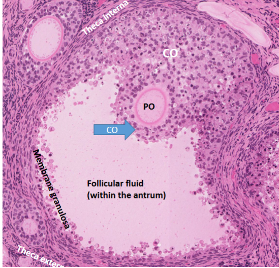
outer ring is theca interna
Graafian follicle: follicular liquid inside, secondary oocyte next to, cumulus oophorus outside both. Membrane granulosa at the end of follicular fluid
Corpus Luteum + hormone
Unfertilized egg, must be shed
LH- is the hormone via granulosa lutein cells
Secreted by granulosum lutein cells
Oviduct/fallopian tube regions
Infundibulum - hold ovary in place (CT)
Ampulla - trumpet shape (SITE OF FERTILIZATION)
Isthmus - straightened out part
Intramural regions - aka fundus
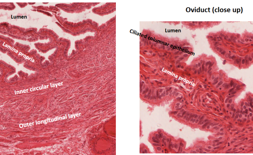
Oviduct cells by the lamina propria are ???
CILIATED columnar epithelium (aid in menstruation)
Oviduct muscular layers
Lamina Propria
Inner circular layer
Outer longitudinal layer
serosa
Middle word describes how the fibers grow across
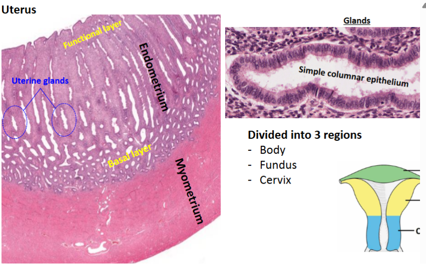
Uterus notes
VERY MUSCULAR
Endometrium and Myometrium
Basal layer inbetween
Endo- dense regular CT
Myo- muscle
Uterine glands (simple columnar epithelium)- secretes hormones for child if present +
Top layer; functional layer
Vagina !!
Very similar to bladder but has SOME keratinized regions, divot at top
Stratified squamous non-keratinized epithelium
NO GLANDS PRESENT
Muscle layers: lamina propria
mucosa
submucosa
inner circular
outer longitudinal
SEVEN UP
S-Seminiferous Tubules
E-Epididymis
V-Vas Deferens
E-Ejaculatory duct
N…..
U-Urethra
P-Penis
Sperm complete maturation where?
Epididymis
Testes : Epidiymis on top
Contains seminiferous tubules
Cells: Leydig cells : secrete testosterone
in stroma surrounding individual tubules
Lining: stratified seminiferous epithelium
Inside: Sperm/spermatozoa
next to sperm: spermatids (small/early)
Outside(larger): spermatogonia
Sertoli cells: Lighter stained than spermatogonia, help develop germ cells.
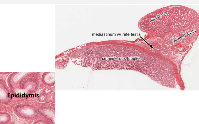
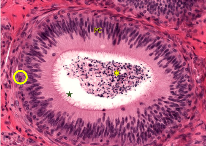
Epididymis
Function: sperm transport and maturation
Histology things:
Principal cells: long, pseudostratified/simple columnar epithelium
basal cells
stereocilia(on principal cells)
Spermatozoa (in middle)
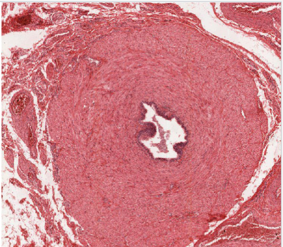
Know layers of vans deferens + why it has so much muscle
Thin lumen pseudostratified ciliated columnar epithelium
thick muscle layers bc it needs to work against gravity
Function: carry sperm from epididymis
Lamina propria
inner longitudinal
middle circular
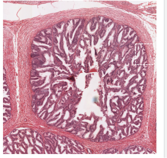
Seminal vesicle webbing cells (type)
Simple columnar and pseudostratified epthelia
Mucous membrane function in seminal vesicles
Liquid IV, keeping sperm moist
Prostatic Concretion function (prostate)
UNKNOWN - but important histological sign of prostate
Pseudostratified columnar and simple cuboidal cells surrounding
Know 5 major components of prostate secretion
Lipids
Proteolytic enzymes
Acid phosphotase
Fibrinolysin
Citric acid
Prostate Gland
secretes fluid that nourishes and protects sperm
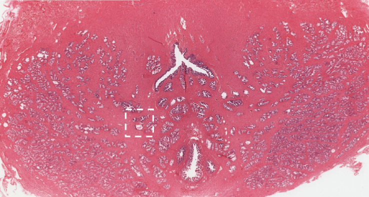
Glands of male reprod. system to know
Seminal Vesicles + prostate gland
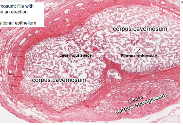
Penis has what structure seen in osteal tissue?
Fibrous trabeculae
Penis corpus cavernosum vs Spongiosum
Stains to know
Pams, pas, mason’s trichrome, ihc, wright, giemsa, acid fast, silver stain, reticulin, elastin, neuro ones
Oral Cavity contains:
Mouth/lips and Tongue
Oral Cavity Glands
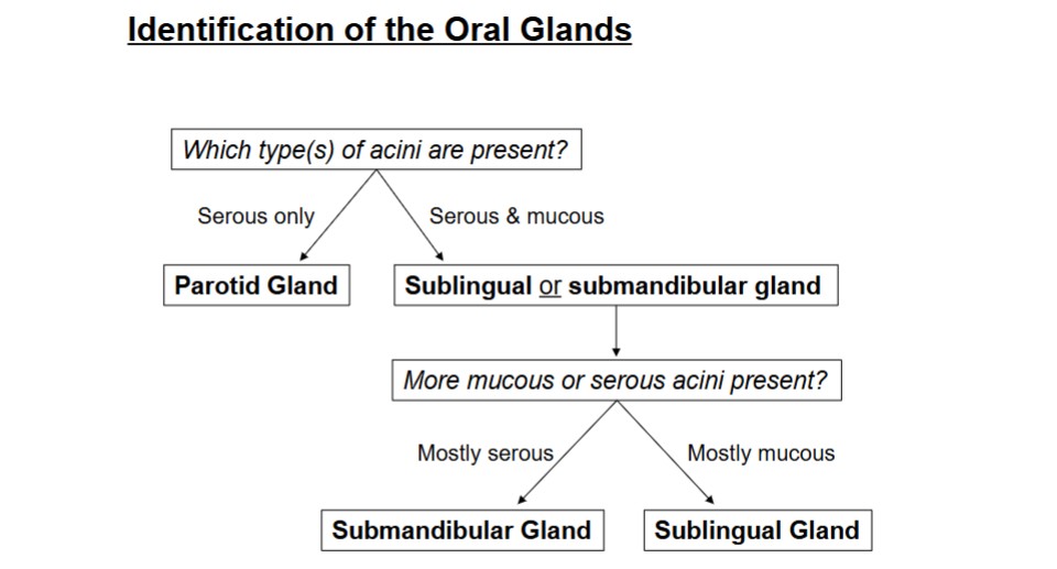
Submandibular (mostly serous mix), Sublingual (mostly mucous mix), and Parotid (serous only)
Alimentary Canal Parts
Esophagus, Stomach, Small and Large Intestines (LESS)
Extramural Glands
Pancreas, Gallbladder, and Liver
Small Intestine Parts
Duodenum, Jejunum, ileum
Large Intestine Parts
Cecum, Colon, Appendix, Rectum, Anal Canal
Lip Sections:
Inner and Outer
Outer lip Hallmarks
Can be keratinized,
non-keratinized, and
para keratinized.
• Vermilion zone
• Sebaceous gland
• Muscular core
• Rete Ridges
![<p>Describe the major sections in this Image of the []</p>](https://assets.knowt.com/user-attachments/3930c397-1d62-40ee-a654-37da73998af4.jpg)
Describe the major sections in this Image of the []
[Outer Lip]
Hf- Hair follicles
Sg: Sebaceous Glands
D: Dermis
E: Epidermis
Keratinization of the Lip
Outer lip (with hair
follicles) is keratinized
2. Vermillion zone is Para
keratinized in the early
transition and becomes
non-keratinized
3. inner lip is non-
keratinized but
epithelium becomes very
thick compared to the
other two.
![<p>Describe the major sections in this Image of the []</p>](https://assets.knowt.com/user-attachments/0c42f100-5ba4-40f2-8717-4abfaf56846a.jpg)
Describe the major sections in this Image of the []
[Inner Lip]
Rete Ridges (RR)
Vascular Supply (BV)
Core (C)
Connective Papillae (CP)
Skeletal Muscle (SM)
Vermilion Zone (VZ)
Tongue Differences from the Mouth
No Vermillion Zone, has wispy taste bud structures on top of itself.
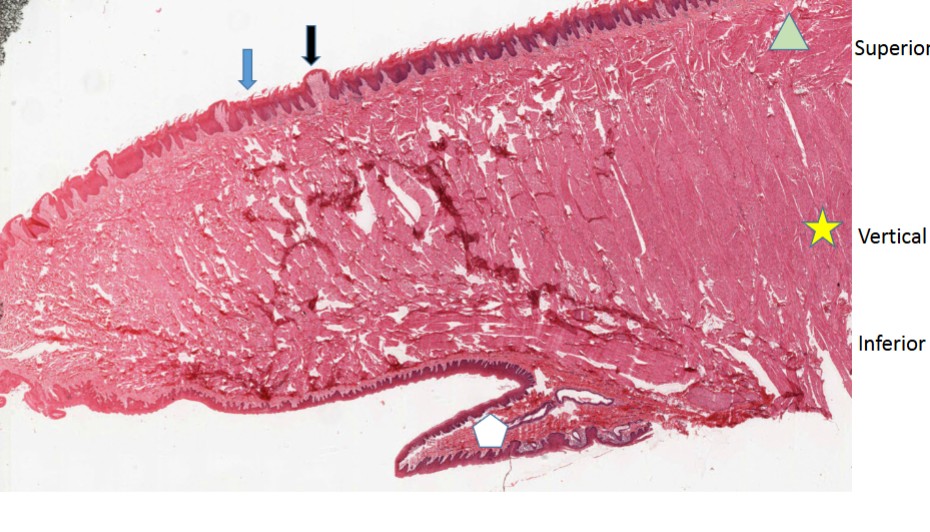
Tongue Overview
Hallmarks
• Intrinsic muscle-
superior, vertical,
inferior and horizontal
(not shown)
• 3 pairs of salivary glands
• Lingual papillae
• Filiform-keratinized
(no taste buds)
• Fungiform-non-
keratinized (few
taste buds,
mushroom shape)
• Circumvallate
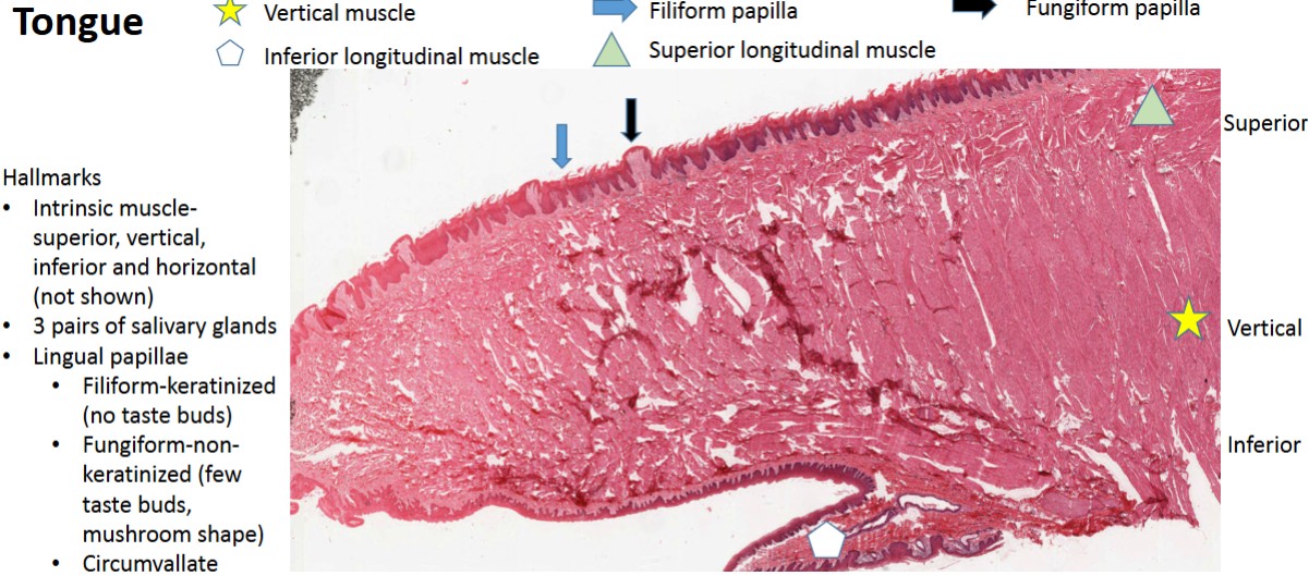
Difference between filiform and fungiform papillae
filiform: keratinized, wispy structure on top
fungiform: non keratinized, thicker dome like shape
Circumvallate Papillae vs Fungiform papillae
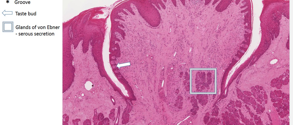
Circumvallate papillae have gaps on sides, flower like shape, and glands of von ebner
Tongue breakdown image
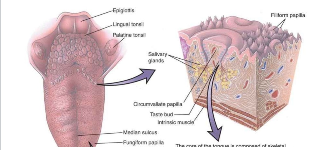
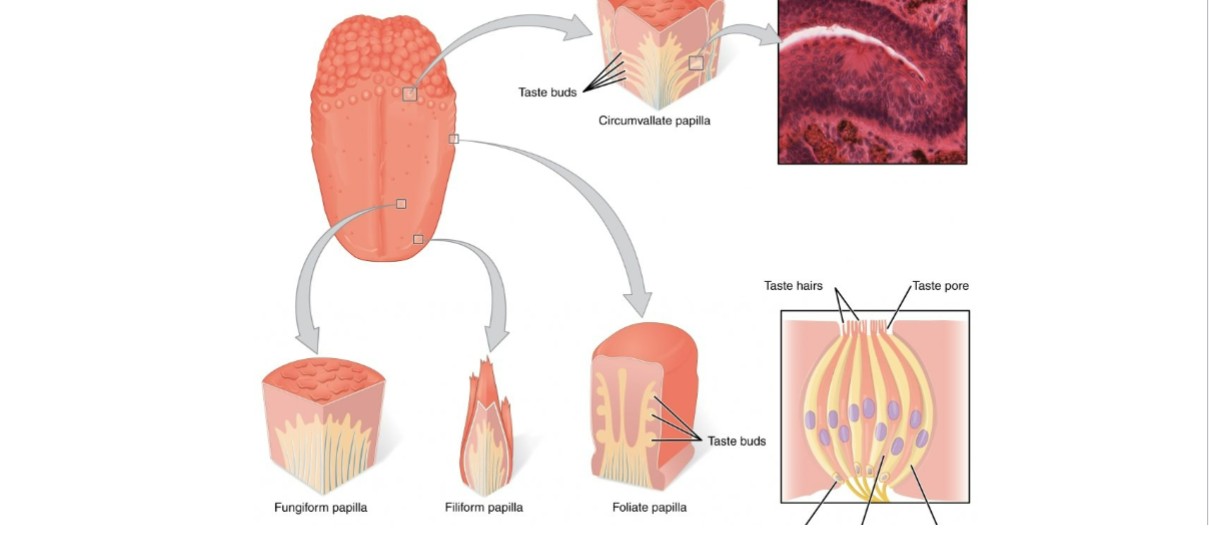
Tongue breakdown of taste buds 2
What kind of secretion is the glands of von ebner?
Serous
What are the “Oral Glands”
Notes: parotid is the darkest, followed by submandibular, and then sublingual. Sublingual has the most fat within it.
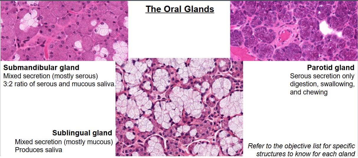
Identification of the Oral Glands Flow Chart
Alimentary Canal Region 1
Esophagus
Esophagus notes
Right next to the lumen of trachea, so will see hyaline cartilage nearby if it is a wider image
Lumen of the esophagus is what shaped
open heart or “m” shaped
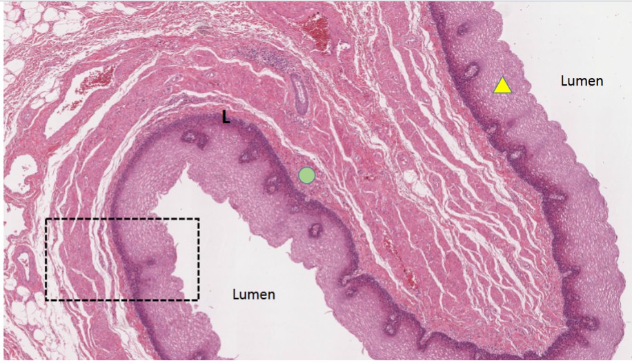
Name the important structures of the Mucosa of the esophagus
Dot: muscularis Mucosae
Triangle: Epithelium
L: lamina propria
What kind of muscle is the muscularis mucosae?
smooth muscle
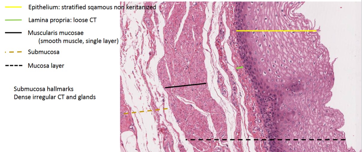
what kind of epithelium lines top of esophageal mucosa?
Stratified Squamous, non-keratinized epithelium
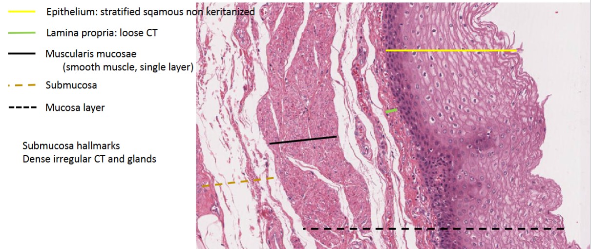
Esophageal lamina propria is what kind of tissue?
Loose CT
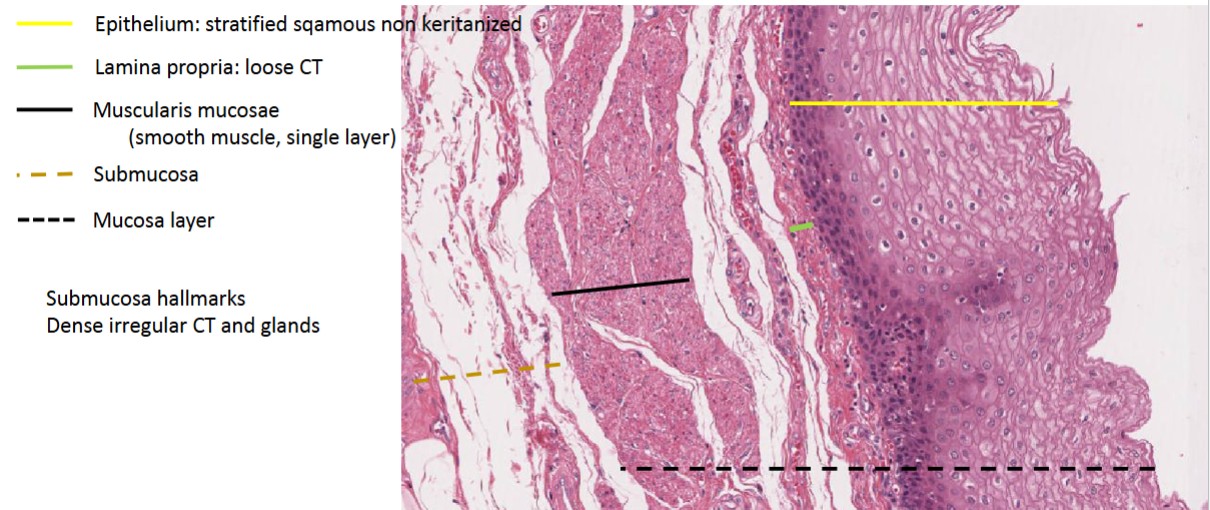
Esophageal Submucosa is made out of ?
Dense irregular connective tissue and glands
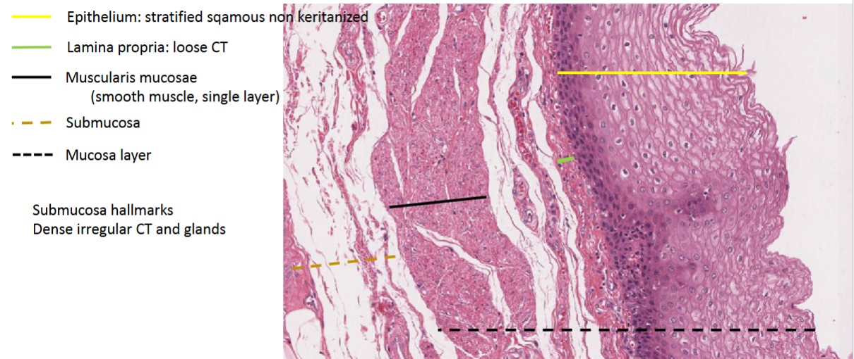
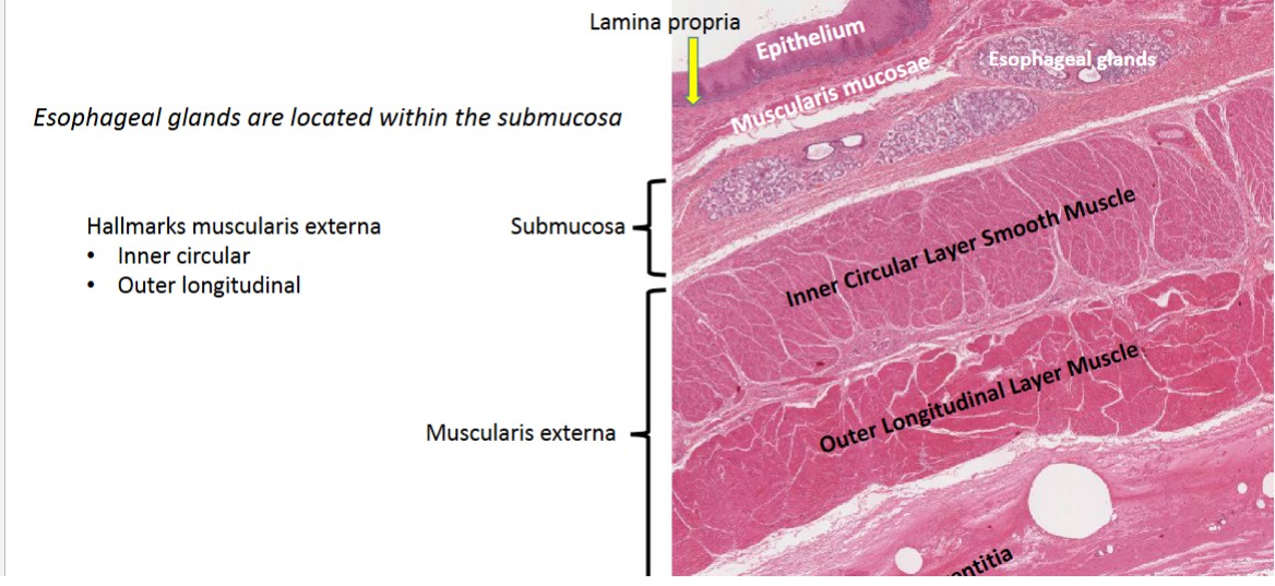
Overarching Esophageal Layers [including muscularis externa]
STAIN: AFB GO
(Acid-Fast Bacteria): Finds and differentiates acid-fast
bacteria (EX: bacteria that cause TB and leprosy)
An acid-fast bacilli (AFB) stain is typically used to see bacteria in lung tissue, lymph nodes, and skin biopsies
STAIN PAS GO
(Periodic Acid Schiff): Finds and differentiates simple
polysaccharides, neutral mucins, basement membranes, fungus
sometimes used in the renal system (kidneys)
Masson’s Trichrome stain GO
Demonstrates presence and density of
fibrosis in tissue. Differentiates CT muscle and cellular
components
Diff Quick STAIN GO
Diff Quik: Finds and differentiates H. pylori and other bacteria
non-differentially
Colloidal IRON GOOO
Finds and differentiates epithelial and CT mucins,
renal cell carcinoma, mast cell granules, calcium salts, etc.
Giemsa/Wright Stain GO
Differentiates blood cells
Gram Stain GO
Differentiates Gram positive and negative bacteria
Mucicarmine STAIN GO
Finds and differentiates epithelial acid
mucopolysaccarides and cryptococcus neoformans
Hall’s Bile stain GO
Finds and differentiates bile, hemosiderin, and
lipofuchsin
IHC stain GO
silver stain, Uses specially
labeled antibodies that bind to the proteins of interest
Alimentary Canal first HALF table
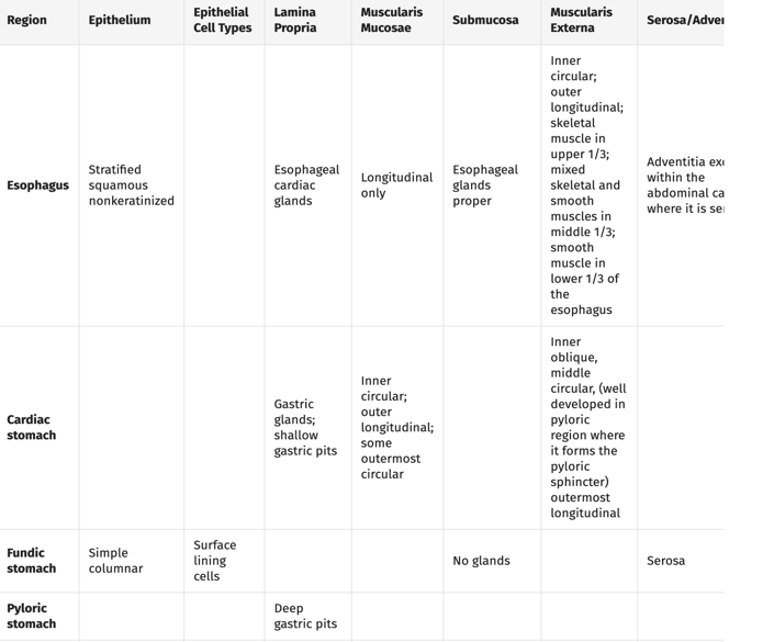
Alimentary Canal Region 2
The stomach
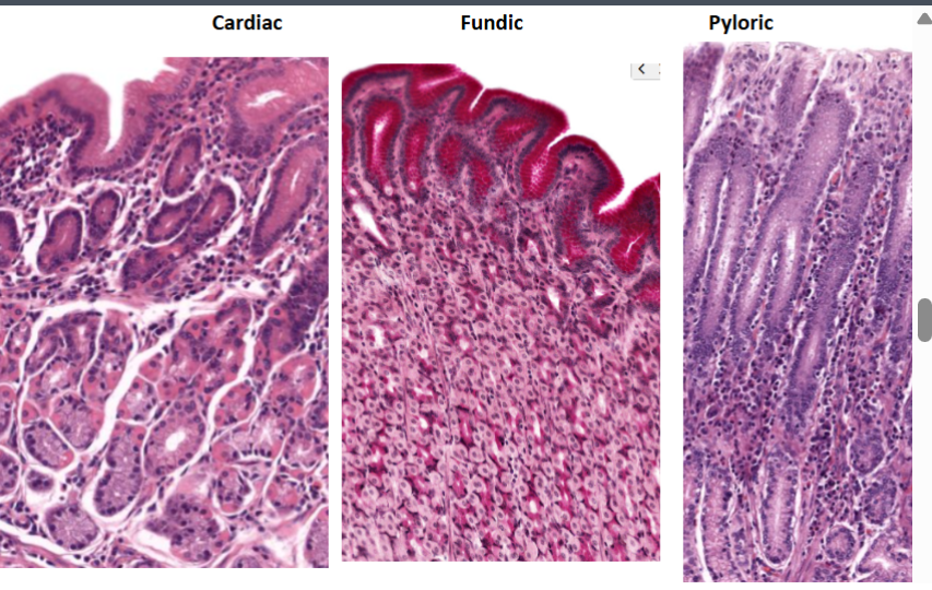
The stomach breakdown
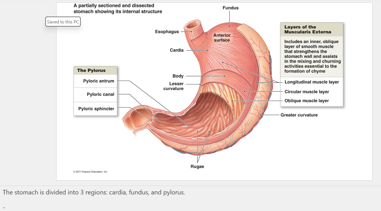
stomach layers
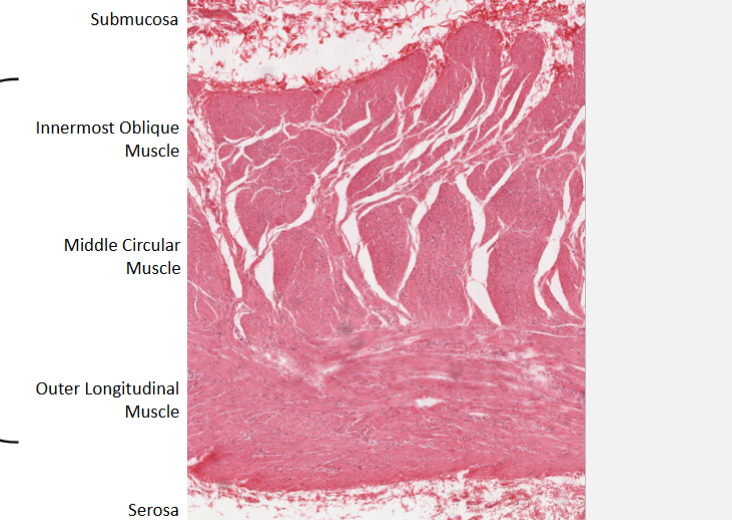
Stomach muscle layers
Junctions: Gastro esophageal
Cardiac glands, esophagus (stratified squamous keratinized)
Cardiac stomach (simple comlumnar)
Small intestine table
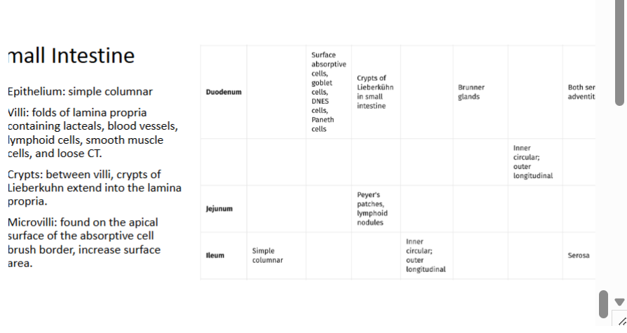
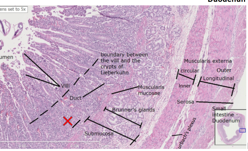
Duodenum Hallmarks
crypts (of lieberkuhn)
brunners glands (looks like bowman’s glands)
muscularis mucosae
between crypt: lacteals, and villi on top of lacteal.
paneth cells at bottom of crypt
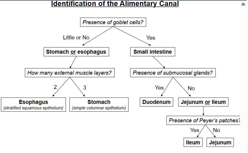
Identification of alimentary canal flow chart
Pancreas + Liver
Pancreas: dark stained, zymogen granules: centroacinar cells (stain lighter) islets of langerhans: endocrine and exocrine. Liver: looks like ground beef with big central veins and portal veins (portal got multiple holes goin on)
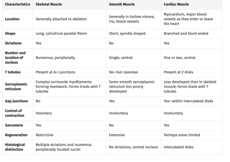
Muscle Classifications
Striated/Non- Is there contractile units present
Voluntary/Involuntary - do you have control over it
Types of muscle
Smooth, Skeletal, Cardiac
Classifications for three different types of muscle
Smooth muscle: non-striated, involuntary contraction
Skeletal Muscle: Striated, voluntary contraction
Cardiac Muscle: Striated, Involuntary contraction
Skeletal Muscle Layers
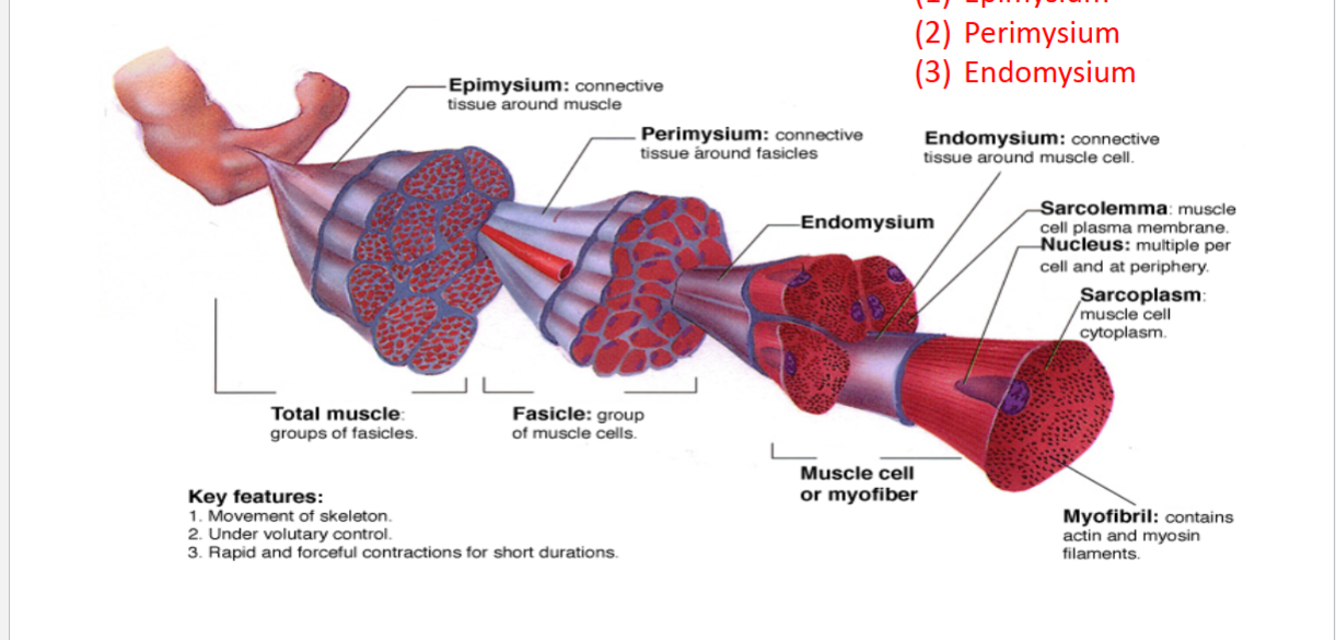
Epimysium: connective tissue surrounding muscle
Perimysium: Connective tissue surrounding Fasicles
Endomysium: Connective tissue around muscle cells
Skeletal Fasicle
Group of muscle cells
Skeletal Muscle cell (myocyte) components
Myofibrils (actin and myosin filaments)
Sarcolemma: myocyte plasma membrane
Nuclei: Multiple by periphery of cell
Sarcoplasm: myocyte cytoplasm
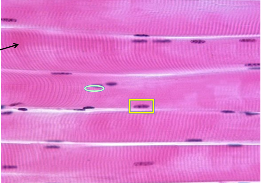
Longitudinal Smooth Muscle
“Hot dog” shape
nuclei stained dark by periphery
Longitudinal lines: are striations made by myofibrils (myosin and actin filaments aligned in sarcomere)
Endomysium (white separations
Skeletal Muscle is anchored to bone by what?
Tendons: connect the epimysium (outer layer of CT covering muscle) to the periosteum (outer layer of bone)
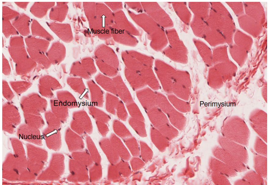
Skeletal Muscle Cross Section can see which mysium best
PERIMYSIUM
Skeletal Muscle Cross Section Hallmarks
Endomysium
Perimysium
Muscle fiber
Peripherally located nuclei
Cardiac Muscle is what kind of muscle?
heart muscle :)
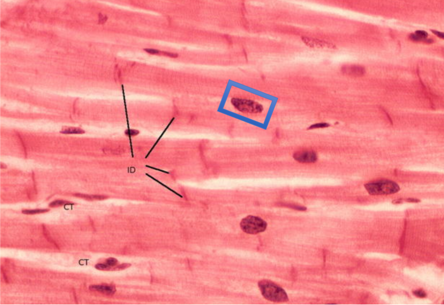
Cardiac Muscle Hallmarks
Striations, SINGLE nucleus, intercalated disks, involuntary contraction
What is the function of Intercalated disks
adhesion, and signal transduction(passing ions)
Difference in Nuclei Shape between skeletal and cardiac muscle
cardiac muscle has more circular nuclei whereas skeletal nuclei are more fried egg shape
Cardiac Muscle (longitudinal)
Hallmarks: Intercalated Discs, endomysium (CT)
Cardiac muscle makes up wall of the ??
Heart (myocardium)
Cardiac Muscle Cross Section Hallmarks
centrally located nuclei and perimysium
Smooth Muscle Hallmarks
Spindle Shaped cells (Fusiform), NON STRIATED (actin myosin mesh not sarcomere), centrally located nuclei, and involuntary contraction
Where are smooth muscle cells found?
walls of hollow organs:
stomach
intestines
urinary bladder
and
uterus
in the walls of passageways:
arteries and veins of the circulatory system
and the tracts of the respiratory, urinary, and reproductive systems.
In the eyes:
the ciliary muscles, a type of smooth muscle, dilate
and contract the iris.
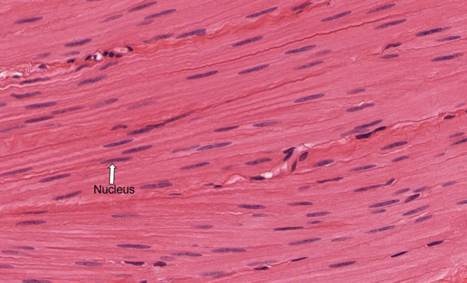
What kind of Muscle is this
Smooth muscle (longitudinally)
Hallmarks:
non-striations
nuclei
Cross section: MORE FATTY LOOKING VERSION OF CARDIAC MUSCLE (or less marbled, cells clumps closer together)
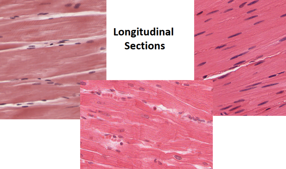
Muscle Longitudinal Sections (identify)
Skeletal, cardiac, smooth (from left to right)
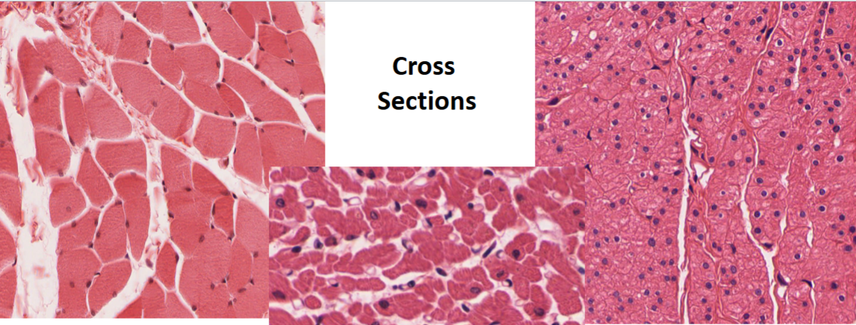
Muscle Cross Sections
Skeletal, Cardiac, smooth (left to right)
Muscle overview Table
Neurons + Types:
Multipolar, Bipolar, and Unipolar
Multipolar: one signal to multiple axons; seen in brain and spinal cord
Bipolar: Found in retina or eye, upper section (ROOF) of nasal cavity and inner ear
Unipolar: found in dorsal root ganglia of spinal nerves and sensory ganglia of cranial nerves.
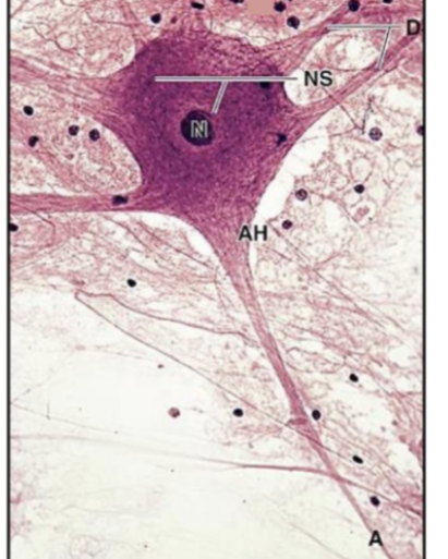
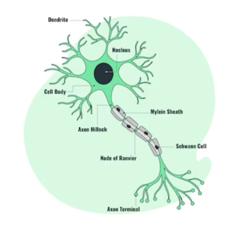
Neuron Parts:
Nucleus, cell body/soma, dendrite, Axon hillock, myelin sheath, node of ranvier, schwann cell, Axon terminal
Sympathetic Ganglion (sympathetic nervous system)
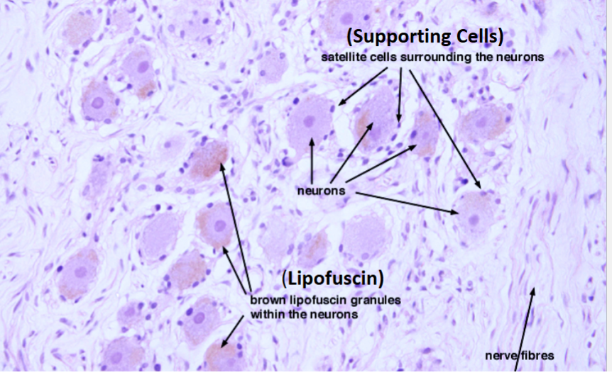
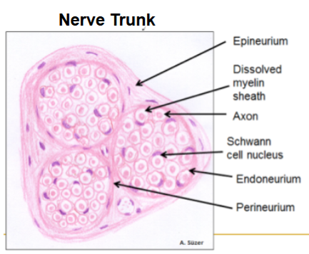
Sections of the Peripheral Nerve
the NEURIUMS
Epineurium/Perineurium
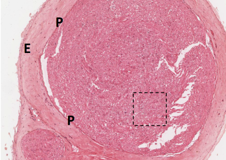
Peripheral Nerve (close)