Lecture 6: Connective tissues: Connective tissues proper and Adipose tissue
1/99
There's no tags or description
Looks like no tags are added yet.
Name | Mastery | Learn | Test | Matching | Spaced | Call with Kai | Chat |
|---|
No analytics yet
Send a link to your students to track their progress
100 Terms
Highly vascular tissue responsible for connecting other tissues and structures with abundant ECM composed of protein fibers and ground substance
What is the definition of connective tissue?
1. Organ structure
2. Mechanical strength
3. Space filling
4. Physical and metabolic support for other (more) specialized tissues
What are the functions of connective tissue? (4)
cells and extracellular matrix (ECM)
What are the two major components of connective tissue (CT)?
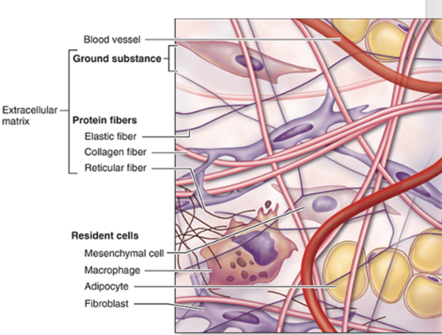
Extracellular matrix (ECM).
What is the major constituent of connective tissue?***

1. Collagen fibers
2. Elastic fibers
3. Reticular fibers.
What protein fibers are found in connective tissue ECM? (3)***

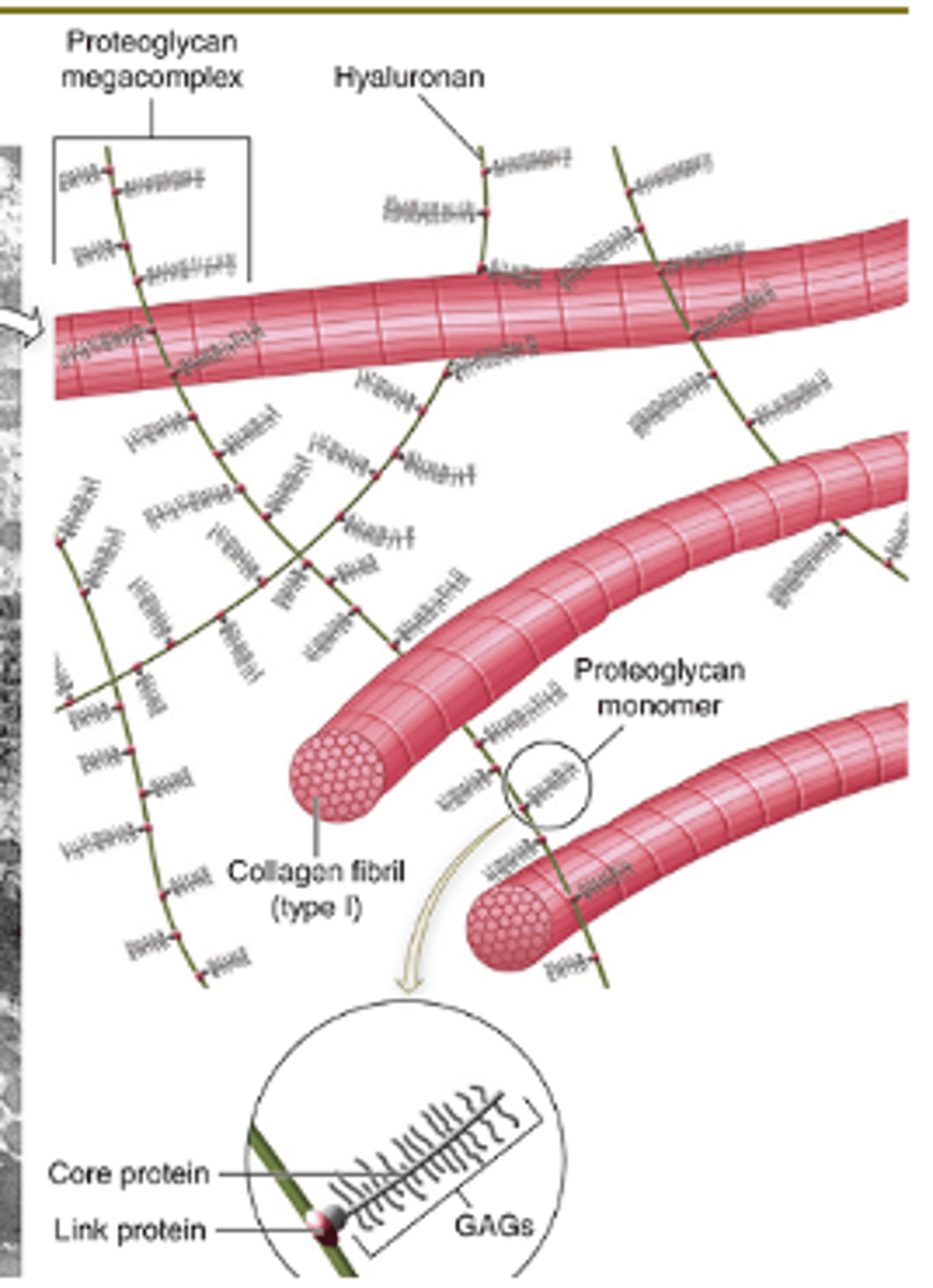
1. Proteoglycans (hydrophilic)
2. multiadhesive glycoproteins
3. glycosaminoglycans (GAG)
What makes up the amorphous ground substance of connective tissue? (3) *****

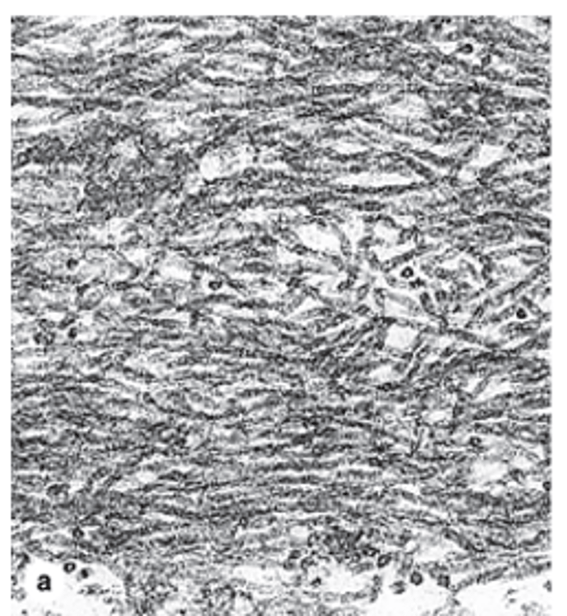
Collagen fibers; provide tensile strength (resistance to pulling, tearing, and stretching).
What fibers are formed by the collagen protein family, and what is their function?
Elastic fibers; provide elasticity (structures return to original shape after mechanical distortion).
What fibers are formed by elastin protein, and what is their function?
Provides volume, enables exchange of nutrients and metabolic wastes, lubrication, and serves as a barrier against microorganism penetration.***
What are the functions of ground substance in connective tissue? (4)
1. Fibers
2. Ground substance
What is the ECM composed of? (2)
1. Collagen
2. Reticular fibers
What are the fibers of connective tissue? (2)
Fibroblasts
Which cells mainly synthesize and secrete collagen proteins?***
30%
What percentage of the human body’s dry weight is collagen?
28 types; formed after extracellular polymerization.
How many different types of collagen fibers exist, and how are they formed?
They are extremely strong and resistant to normal shearing and tearing forces.
What mechanical property makes collagen fibers important?
•Connective tissues
•Basement membranes
•External laminae of nerve and muscle cells
Where are collagen and reticular fibers most notably localized? (3)
Type I collagen fibers
What is the most abundant type of collagen fiber?
Eosinophilic
What is the staining characteristic of Type I collagen fibers?
High resistance to TENSION!
What mechanical property do Type I collagen fibers provide?
Very thick
How thick are Type I collagen fibers compared to other types?
1. Ligaments
2. Tendons
3. Organs capsules
4. Dermis
5. Bone
6. Dentin
Where are Type I collagen fibers commonly located? (6)***
Eosinophilic
What is the staining characteristic of Type II collagen fibers?
Less abundant and less thick than Type I.
How does the abundance and thickness of Type II collagen compare to Type I?
High resistance to PRESSURE.
What mechanical property do Type II collagen fibers provide?
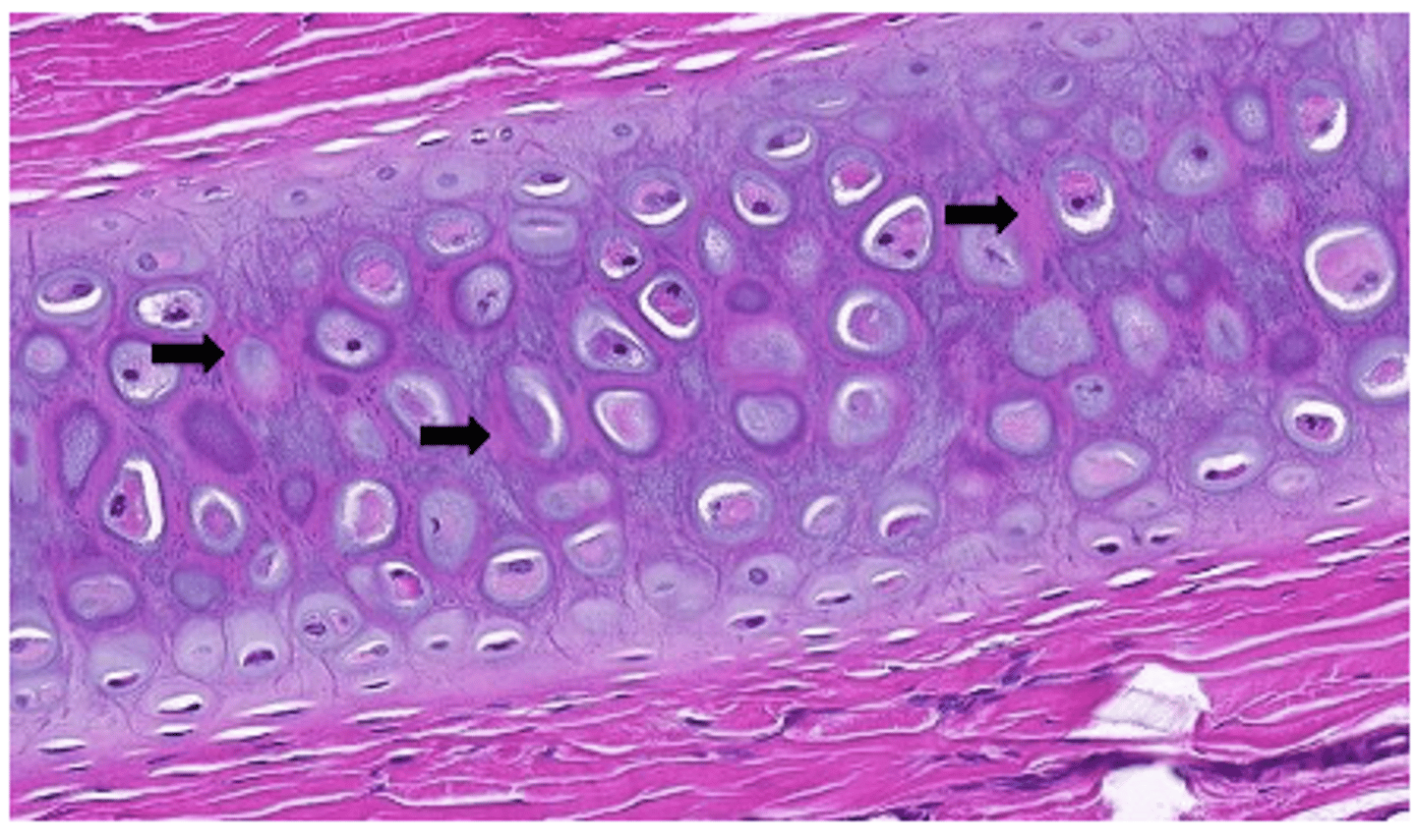
1. Cartilage
2. Vitreous body (eye)
Where are Type II collagen fibers commonly located?
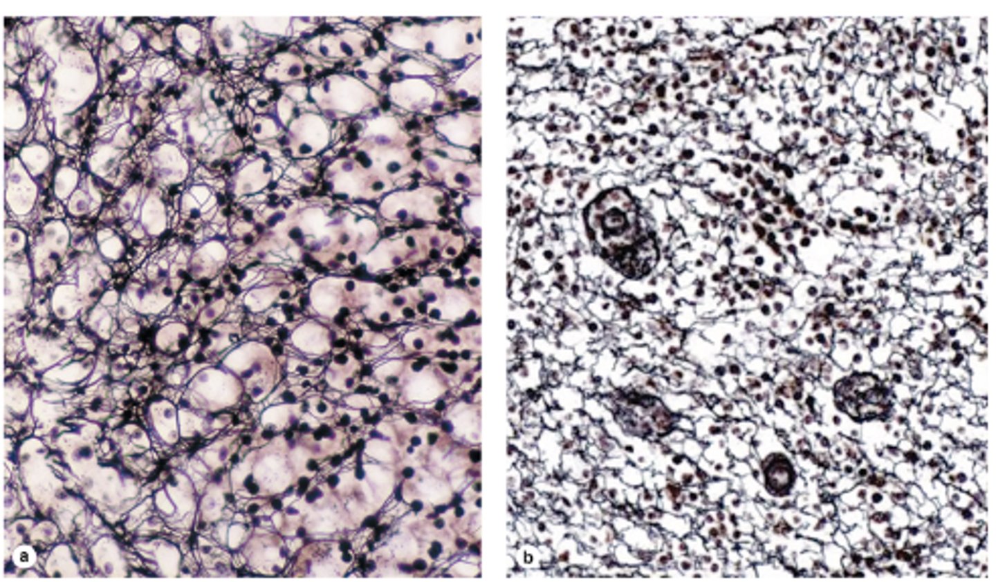
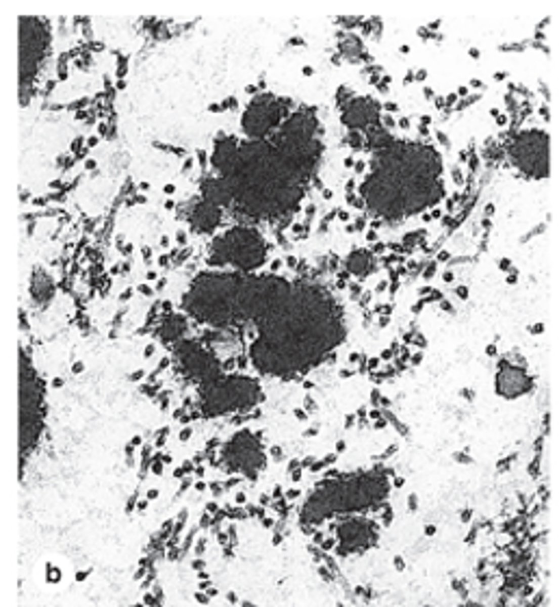
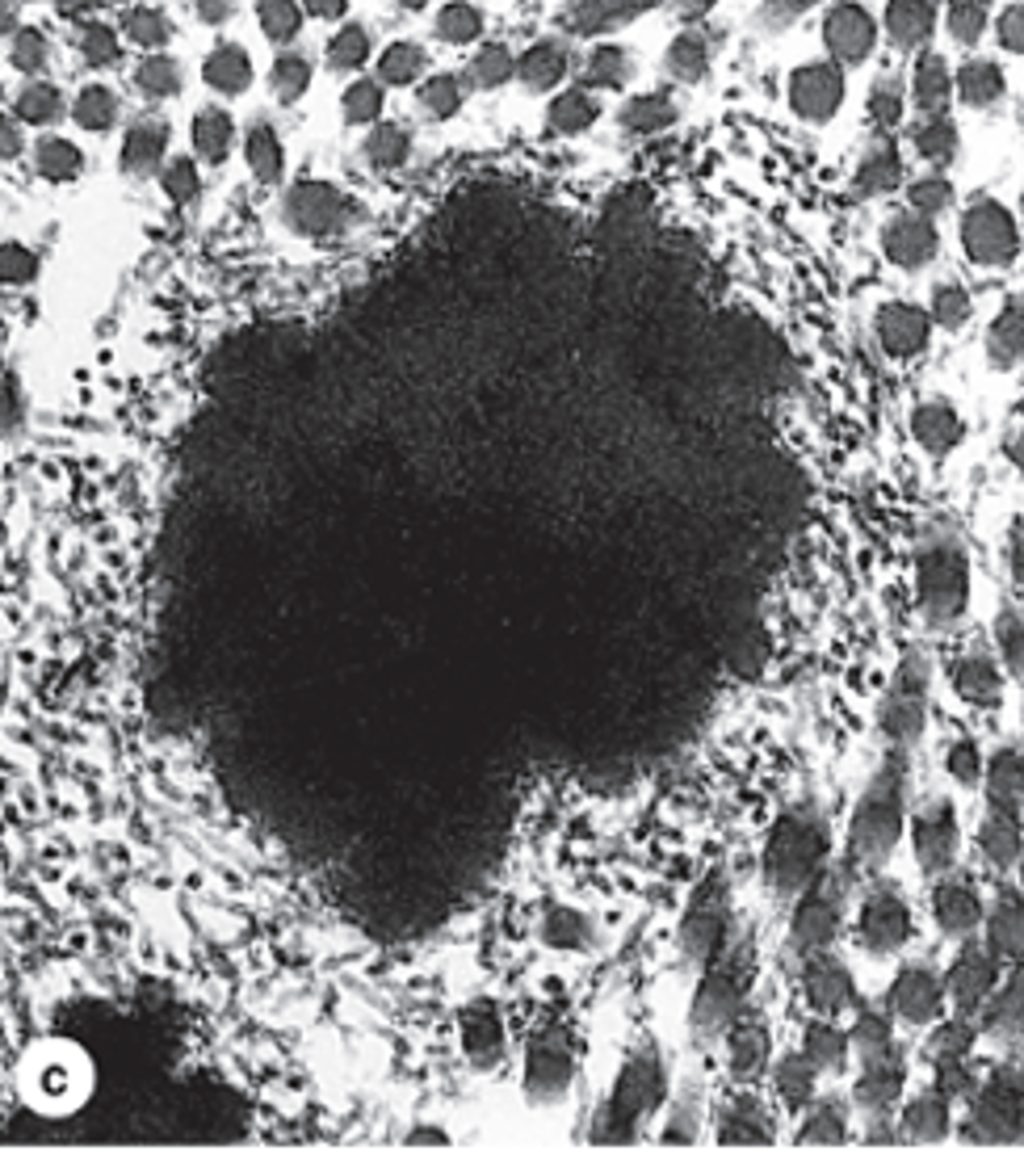
Reticular fibers.
What type of fibers do Type III collagen fibers form? ***
Not well seen.
How do Type III collagen fibers appear in routine H&E staining?
Black (argyrophilic).
How do Type III collagen fibers appear with silver impregnation preparations?
Fuchsia; PAS-positive due to ~10% carbohydrate content.
How do Type III collagen fibers appear with PAS staining?
STRUCTURE MAINTENANCE
What is the main function of Type III collagen fibers?
1. Lymphoid organs (except thymus)
2. Liver
3. Endocrine glands
4. Bone marrow
5. Delicate stroma of expandable organs
Where are Type III collagen fibers commonly located? (5) ***
fibrillin and elastin
What are elastic fibers composed of? (2) ***
They are thinner (similar to reticular fibers).
How does the thickness of elastic fibers compare to Type I collagen fibers?
FLEXIBILITY and resistance to distention.
What property do elastic fibers provide to tissues?
1. Sparse networks with collagen bundles
2. Elastic lamellae (fenestrated sheets) of blood vessels (mainly arteries
Where are elastic fibers commonly located? (2)
They stain poorly.
How do elastic fibers stain with H&E?
They stain darker than collagen.
How do elastic fibers appear with orcein and aldehyde fuchsin stains compared to collagen?
Fibroblasts and smooth muscle cells.
Which cells secrete elastic fibers? (2)
Elastin, secreted by fibroblasts and smooth muscle cells, is deposited into the fibrillin scaffold and cross-linked into larger assemblies.
How is elastin incorporated into elastic fibers?
Elastin accumulates in the electron-dense center of the fiber, while fibrillin microfibers remain visible at the surface.
Where does elastin accumulate during elastic fiber formation, and what remains visible on the surface?
1. Highly hydrated
2. transparent
3. viscous
What are the key physical properties of ground substance? (3)
1. Glycosaminoglycans (e.g., hyaluronic acid/hyaluronate***)
2. Proteoglycans (sulfated GAGs attached to a core protein)
3. Multiadhesive proteins (large molecules with branched oligosaccharide chains)
What are the main components of ground substance? ***
Allows diffusion and acts as a barrier against microorganism penetration.
What functional role does the viscosity of ground substance provide? (2)
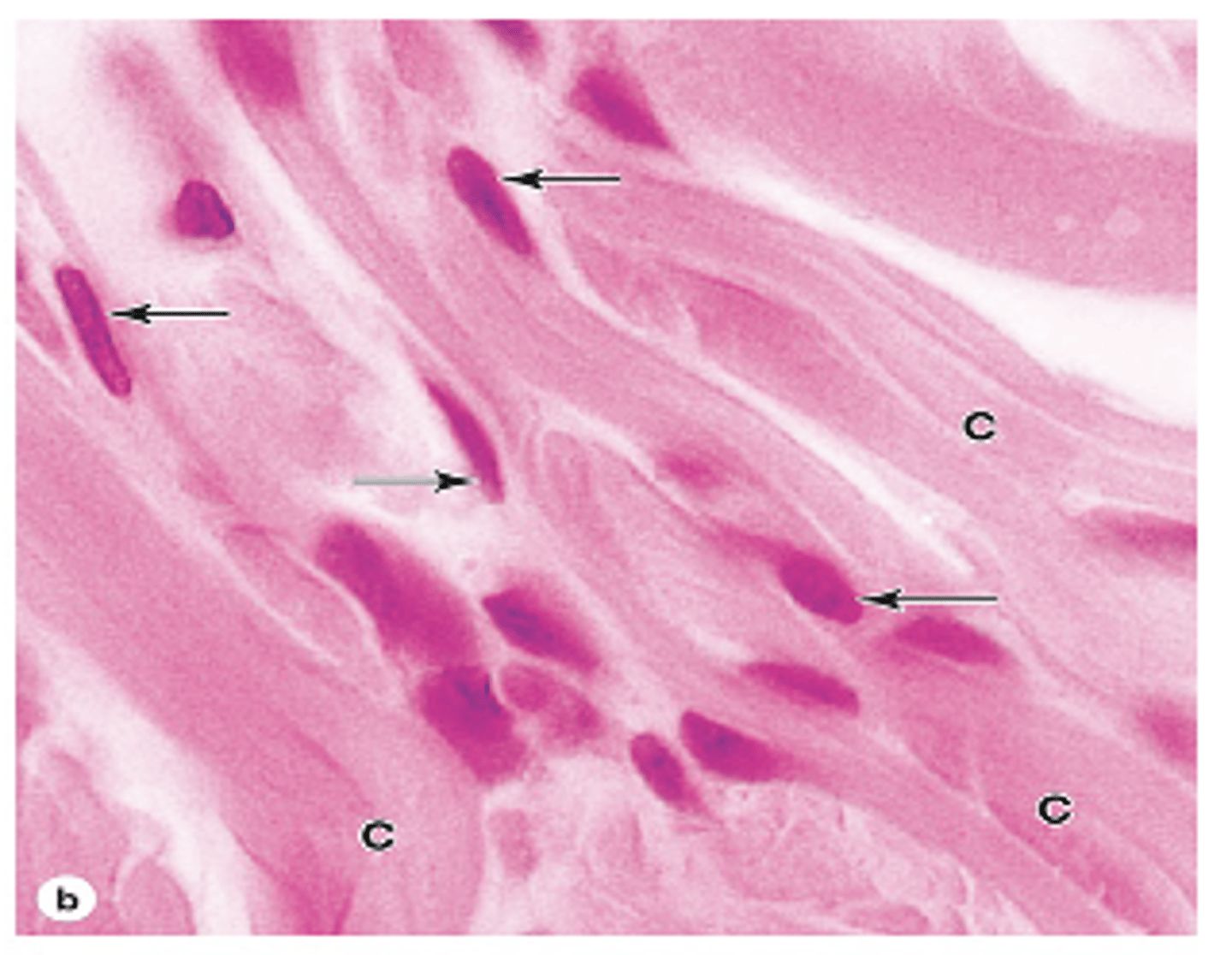
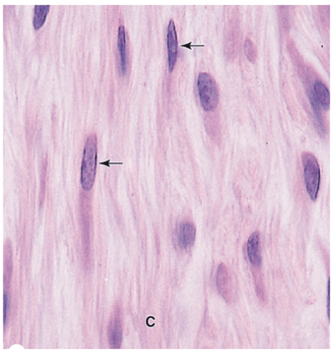
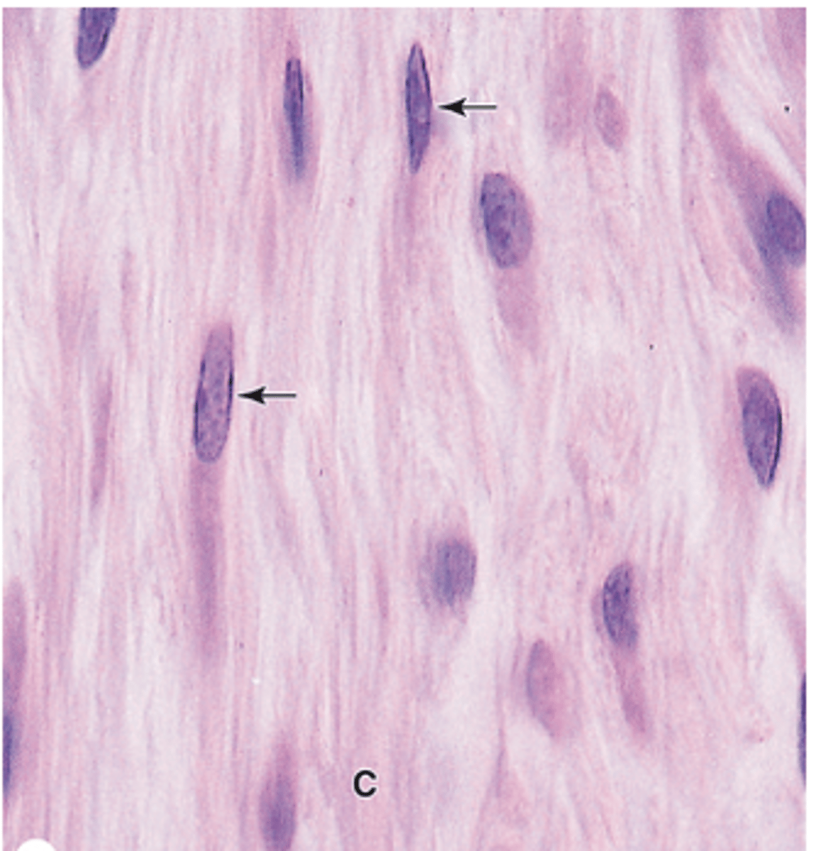
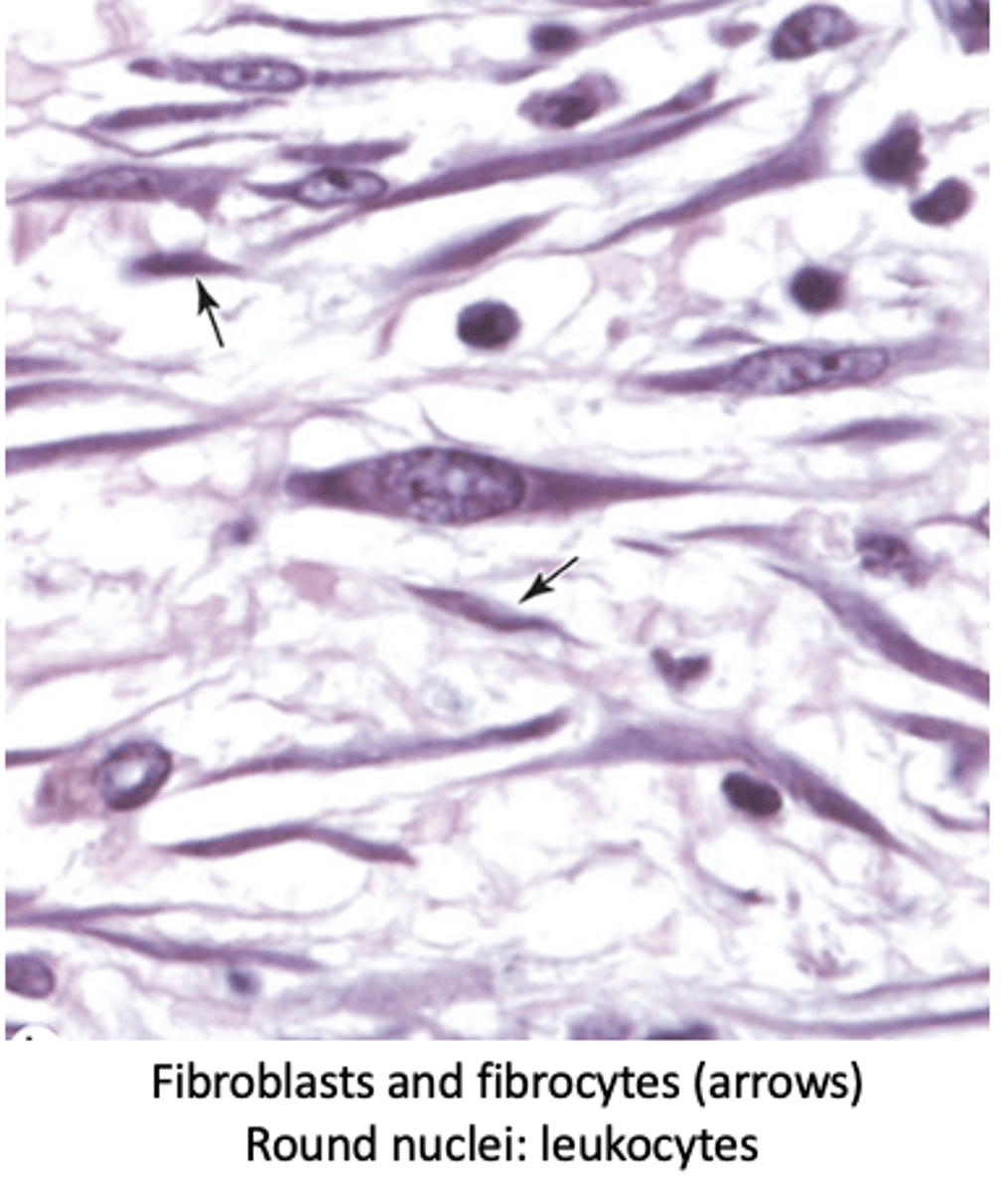
Fibroblasts
What is the most common cell in connective tissue proper?
Mesenchymal cells
From which cells do fibroblasts originate?
1. Collagen and elastin
2. Glycosaminoglycans
3. Proteoglycans
4. Multiadhesive glycoproteins
What are the main extracellular components produced and maintained by fibroblasts? (4)
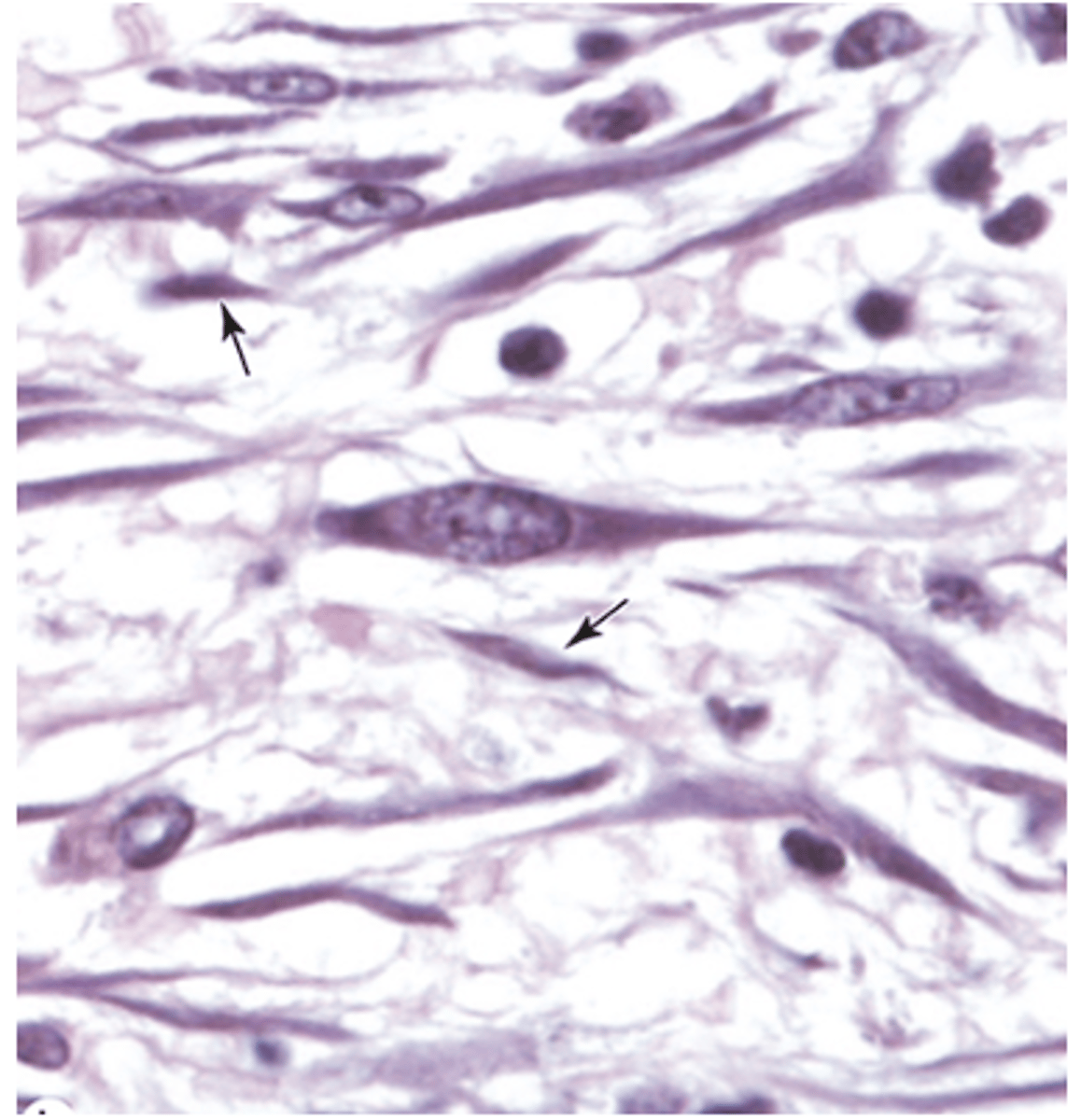
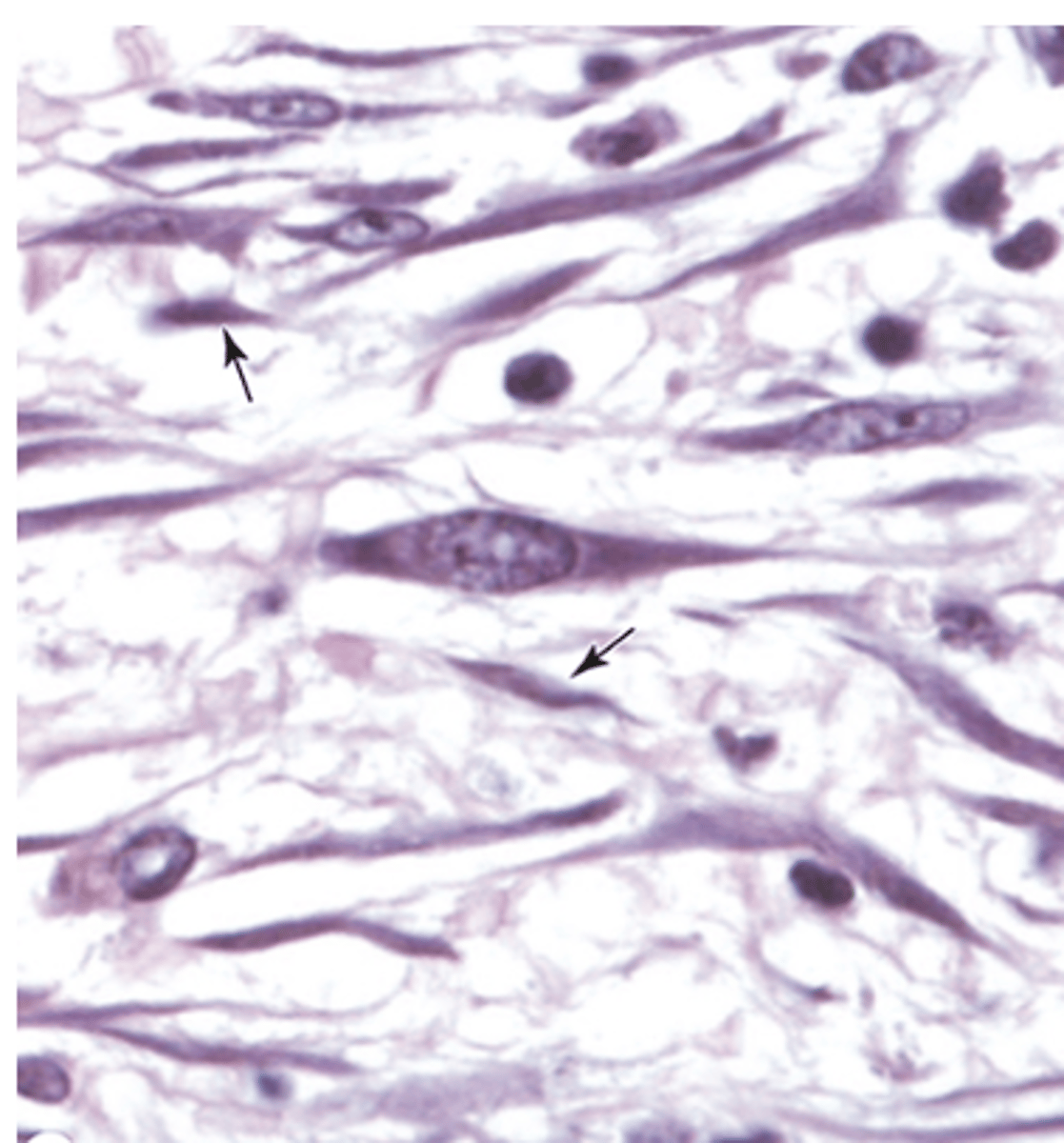
1.Active
2.Passive
3. Myofibroblasts
What are the three types of fibroblasts based on structure and function?
Spindle-shaped
What is the shape of active fibroblasts?
Intense synthetic activity
What type of activity do active fibroblasts exhibit?
•Abundant
•Eosinophilic cytoplasmic processes resembling collagen bundles
•Well-developed RER (basophilia)
•Golgi apparatus
Describe the cytoplasm of active fibroblasts.
•Large
•Ovoid
•Euchromatic (active)
•Prominent nucleolus
Describe the nucleus of active fibroblasts.

•Divide only if needed
How often do active fibroblasts divide?
Spindle-shaped
What is the shape of fibrocytes
Low synthetic activity
How active are fibrocytes in synthesis?
Smaller than fibroblasts
How does the size of fibrocytes compare to active fibroblasts?
•Less abundant
•Fewer cytoplasmic processes
•Less RER
•Cytoplasmic acidophilia
Describe the cytoplasm of fibrocytes.
•Small
•Heterochromatic (inactive)
Describe the nucleus of fibrocytes.
Involved in wound healing
What is the main function of myofibroblasts?***
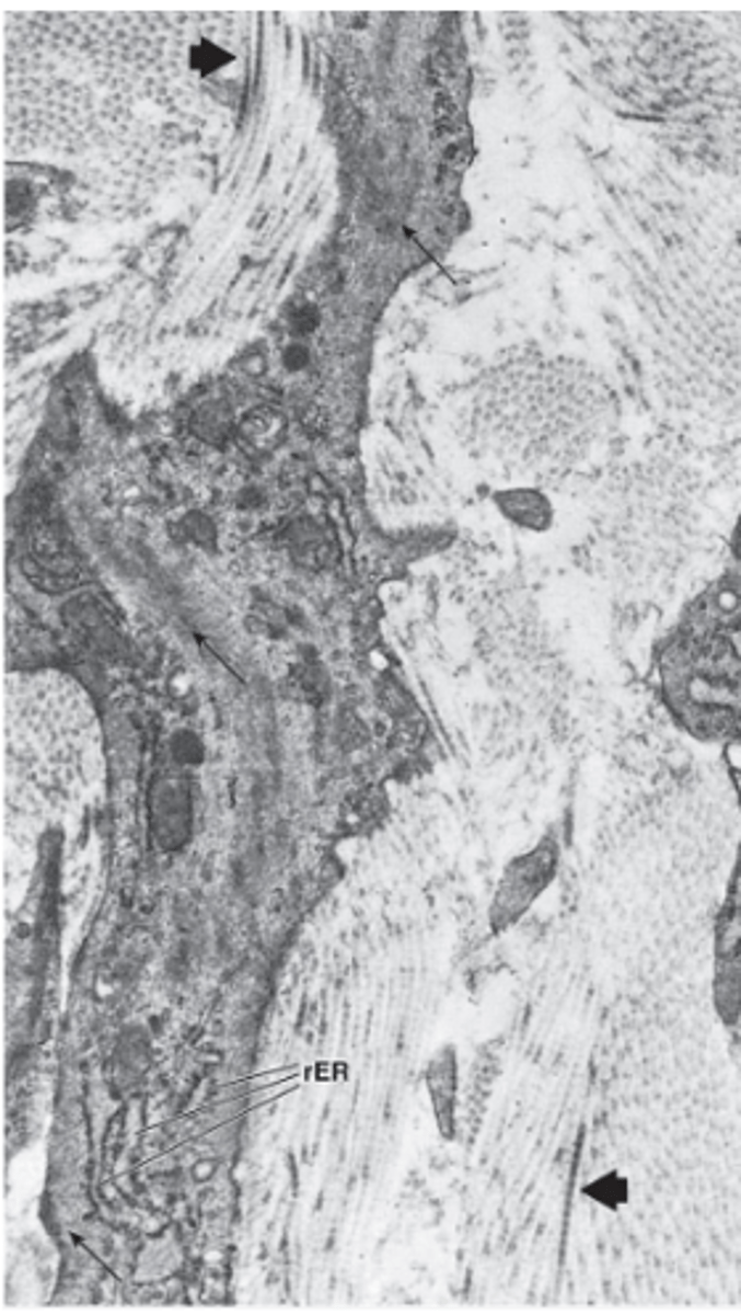
•Elongated cells
What is the shape of myofibroblasts?

•RER
•Golgi apparatus
•Abundant actin and myosin
Which organelles and cytoskeletal components are prominent in myofibroblasts? (3)

Nucleus with undulating surface (surface with a wavy, rippling)
Describe the nucleus of myofibroblasts.

Not identifiable in routine H&E preparations.
Can myofibroblasts be identified in routine H&E staining?
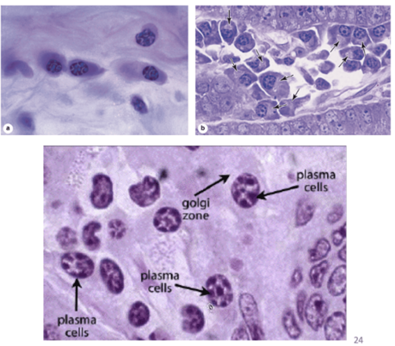
•Originate from B lymphocytes
From which cells do plasma cells originate?

•Produce antibodies
What is the main function of plasma cells?
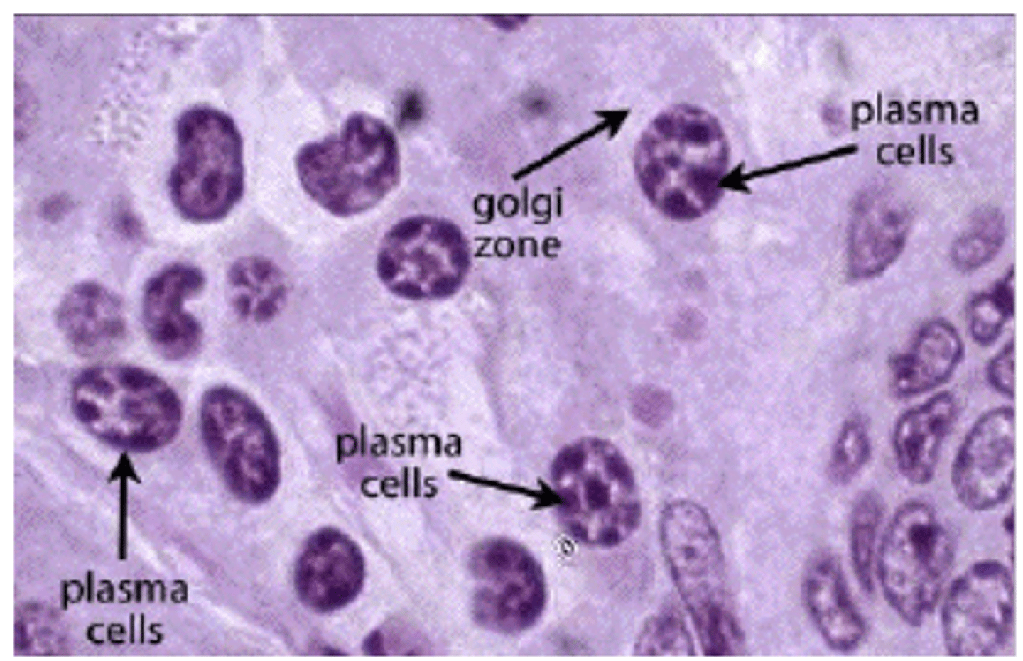
Large, ovoid cells
Describe the shape and size of plasma cells.

•Much RER (intense basophilia)
•Large Golgi apparatus
Describe the cytoplasm of plasma cells. (2)

•Eccentric (cell's nucleus is not centrally located)
•Large
•Spherical
- Clockface appearance
•Peripheral heterochromatin
•Large areas of euchromatin
Describe the nucleus of plasma cells.
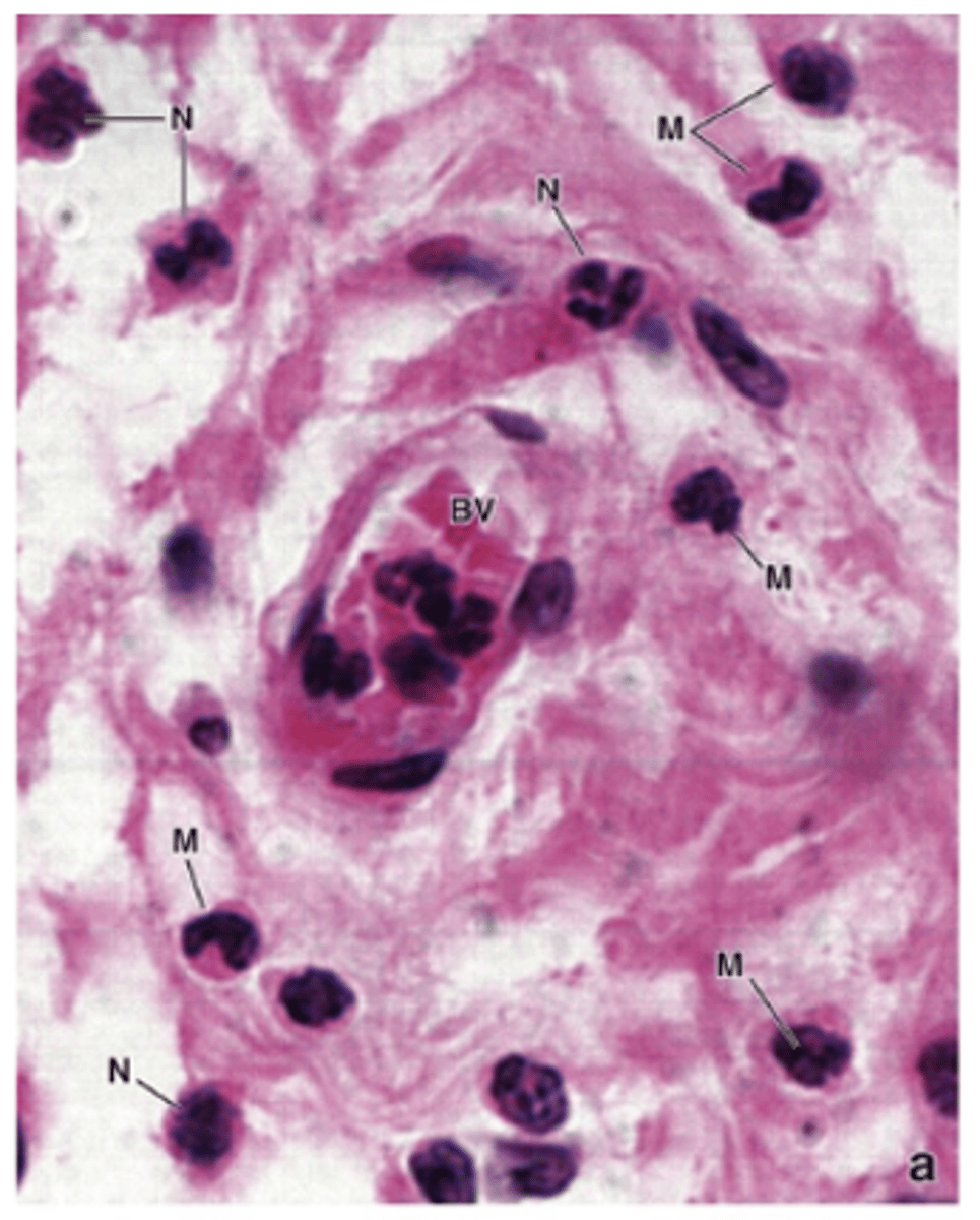
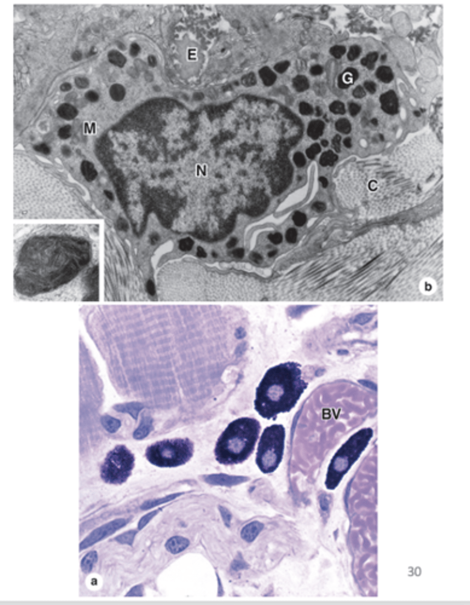
Monocytes
From which cells do macrophages originate?

Phagocytic cells
What is the main function of macrophages?

Connective tissue of most organs
Where are macrophages commonly found?

- Eccentric (cell's nucleus is not centrally located)
- Oval or kidney-shaped
Describe the nucleus of macrophages. (2)
- Well-developed Golgi complexes
- Abundant lysosomes
Describe the cytoplasm of macrophages. (2)

•Abundant in sites of inflammation
Where are macrophages particularly abundant?

Proteins, apoptotic cells, tissue debris, and other particulate material
What materials do macrophages help turnover or clear?

Irregular surface
•Variable size and shape
Describe the surface and shape of macrophages.

Cytoplasmic acidophilia
What is the cytoplasmic staining characteristic of macrophages?
Macrophage-derived, long-living cells
What is the origin of cells in the mononuclear phagocyte system (MPS)?
In the stroma of most organs
Where are Mononuclear phagocyte system (MPS) cells distributed?
1. Debris removal
2. Uptake, processing and presentation of antigens
3. Activation of lymphocytes
What are the main functions of Mononuclear phagocyte system (MPS) cells? (3)
1. Increased cell size
2. Increased protein sysnthesis
3. Increased number of GA and lysosomes
What changes occur when monocytes differentiate into macrophages? (3)
•Oval or irregular shape.
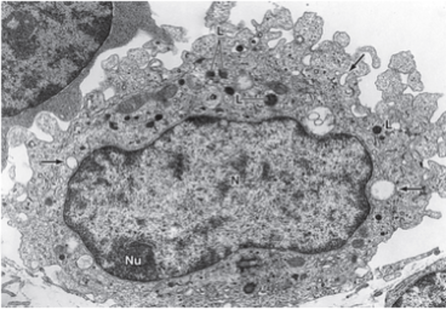
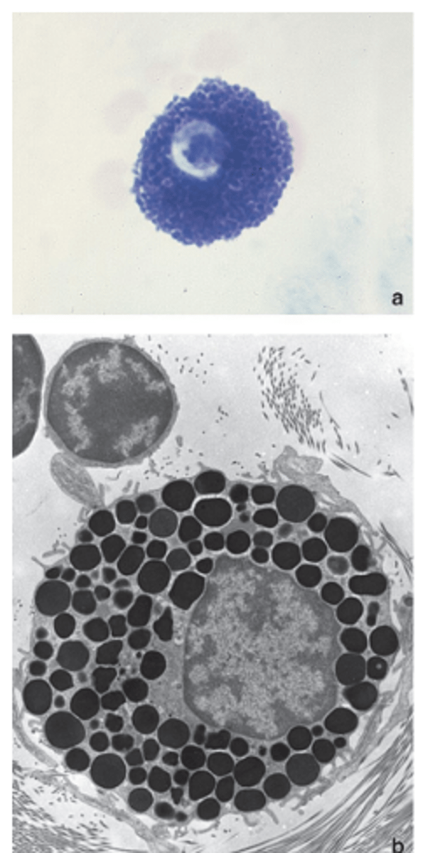
What is the shape of mast cells?

- Abundant basophilic granules
(metachromasia)
- Occasional mitochondria
- Little RER
- Golgi apparatus
Describe the cytoplasm of mast cells. (4)
Obscured by cytoplasmic granules
Describe the nucleus of mast cells.

Present in CT of many organs
Where are mast cells commonly found?

1. Perivascular mast cells: close to blood vessels (skin and mesenteries).
2 Mucosal mast cells: mucosa of digestive and respiratory systems.
What are the two types of mast cells and where are they located?
Originate from hematopoietic stem cell in bone marrow
From which cells do mast cells originate?

They circulate in the blood and enter connective tissue where they mature
How do mast cells mature?

IgE produced by plasma cells binds to mast cell receptors
What happens during the first exposure to an antigen in mast cell-mediated hypersensitivity?
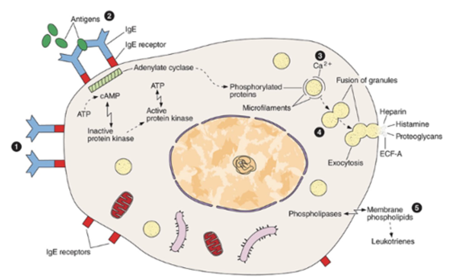
Cross-linkage of IgE receptors occurs after the antigen binds to IgE
What happens during the second exposure to the same antigen in mast cell-mediated hypersensitivity?

Activation of adenylate cyclase and protein phosphorylation
What intracellular processes are activated after IgE receptor cross-linking in mast cell-mediated hypersensitivity?

Ca²⁺ enters the cell, triggering exocytosis of granules
How does calcium contribute to mast cell activation in mast cell-mediated hypersensitivity?

Phospholipases act on cell membrane phospholipids, leading to leukotriene release
How are leukotrienes released from mast cells in mast cell-mediated hypersensitivity?

•Fat cells
What type of cells are adipocytes?

Mesenchymal cells
From which cells do adipocytes originate?

In the connective tissue of diverse organs
Where are adipocytes widely distributed?

Large
Describe the size of adipocytes.

1. Lipid (triglycerides) storage
2. Heat production
What are the main functions of adipocytes? (2)
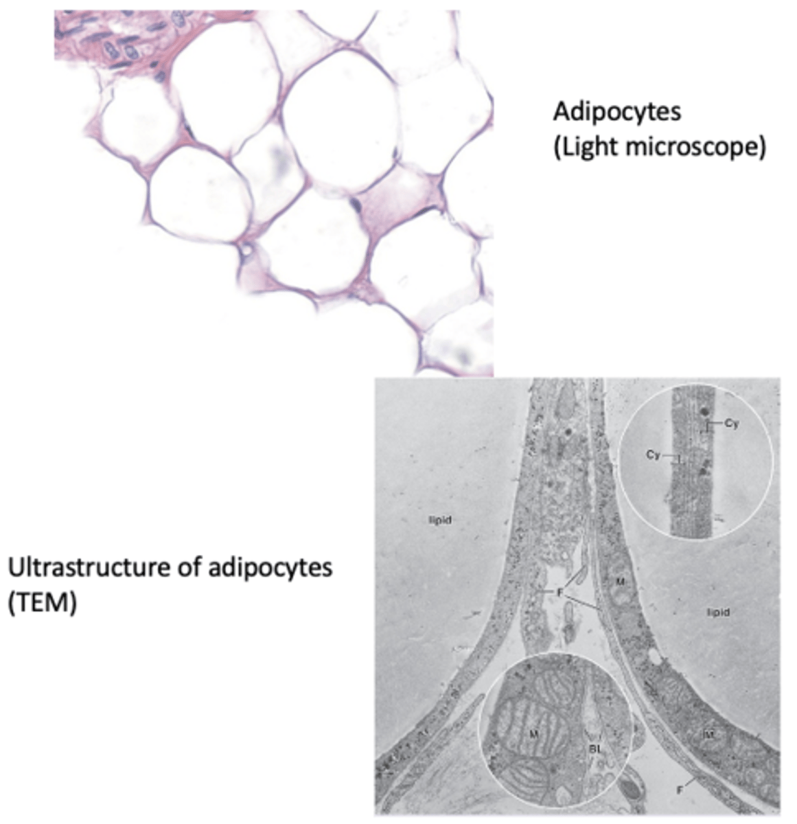
Adipose tissue
What are large aggregates of adipocytes called?

Cells that move from the blood into connective tissue, mostly during inflammation
What type of cells are wandering cells in connective tissue?
Circulating blood cells
From where do wandering cells originate?