2. tubes and lines pt 1
1/62
Earn XP
Description and Tags
specialty catheters
Name | Mastery | Learn | Test | Matching | Spaced | Call with Kai |
|---|
No analytics yet
Send a link to your students to track their progress
63 Terms
list the 2 types of specialty catheters
central lines/central venous catheters (CVCs), pulmonary artery flow directed catheters (PACs)
structurally, what is a central line
a long flexible catheter
where are CVCs inserted into (3)
through the skin and into the SVC, the cavo-atrial junction, or within the RA
role of CVCs
allow for long term infusions of meds
what type of patients require CVCs
those in critical condition + requiring continuous support with multiple IV meds
T or F: CVCs allow for large volumes of fluid boluses
true
why are CVCs a useful method for delivering medicine
many meds are irritating to peripheral blood vessels
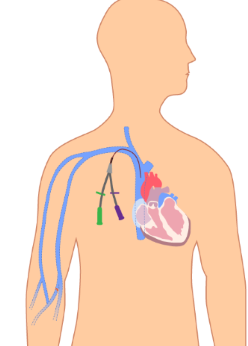
list the 3 categories of CVCs
non-tunneled catheters, tunneled catheters, peripherally inserted central catheter
purpose of non-tunneled catheters
allow for fast and reliable venous access for meds and blood draws
describe now NTCs are inserted at bedside
inserted, then secured with a dressing until placement is confirmed
once NTC placement is confirmed, how are they secured
via sutures
which vessels are NTCs inserted into
subclavian or jugular veins
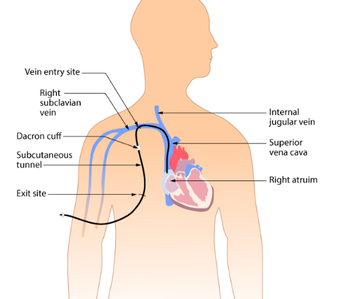
NTCs are inserted into either subclavian or jugular veins. which side of the body is preferred + why
right side; most direct path to the SVC
what are tunneled catheters used for
for patients that are well and physically able, but require ongoing venous access for outpatient therapies
list 3 common types of tunneled catheters
hickman lines, broviac lines, permacath catheters
structurally/functionally, how is a tunneled catheter different from a non tunneled catheter
tunneled also go to the jugular or subclavian veins, but first it is tunneled through subcutaneous tissue beneath the chest skin
role of the tunnel of tunneled catheters
reduces the risk of infection, provides protection against accidental dislodgement
describe how the tunnel for tunneled catheters is created
inject local anesthetic by making two small incisions; one at the vein entry site and one 2-3 inches below the clavicle. skin is separated from underlying tissue = the tunnel. catheter is positioned in the vein and some is threaded through while the rest remains outside the skin
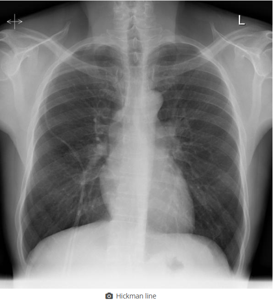
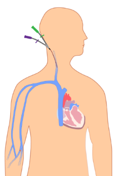
on this image, find the tunneled catheter
see picture

a port-a-cath line is a type of tunneled catheter. describe how it’s structure is unique
contains an implantable port rather than an external port. it is sutured in place beside the ribcage, and the pt will not have any part of the catheter dangling
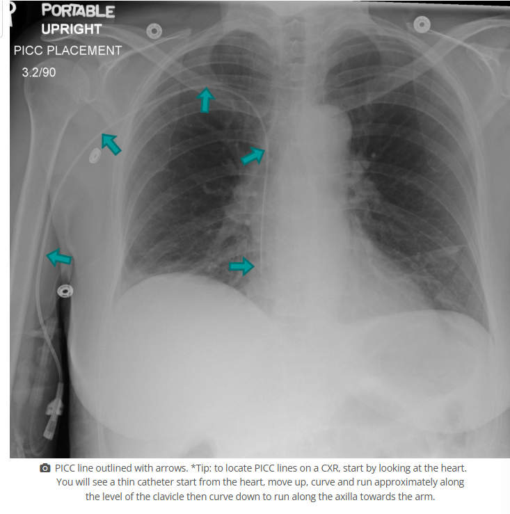
peripherally inserted central catheters (PICCs) are a type of CVC. describe the insertion site
upper arm and into the basilic vein
role of PICCs
for long or short term use for pts who are mostly well but require vascular access for treatment
benefits of PICCs
reduced infection rates compared to other CVCs, have reduced risks during insertion, and are easy for the pt to take care of
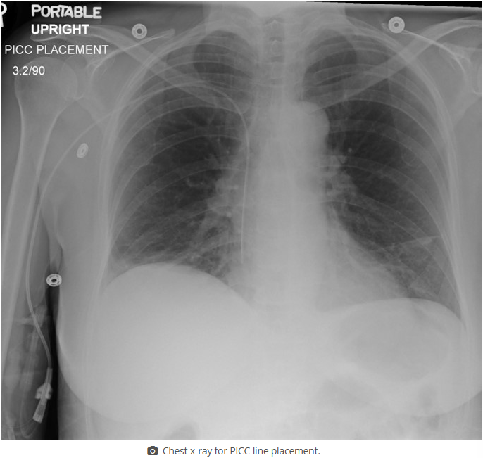
find the PICC line in this image
see image
which catheter type does this image show (be specific)
non tunneled CVC

what is the entry point shown in this image for a non-tunneled CVC
subclavian vein
what type of catheter is shown in this image (be specific)
non-tunneled CVC

what is the insertion point shown for this non-tunneled CVC
jugular vein

what catheter type is shown in this image (be specific)
tunneled CVC
what catheter type is shown in this image (be specific)
PICC (type of CVC)
how do we see CVCs on xrays
they all have a radiopaque strip
what must a tech pay attention to when doing a CXR to confirm CVC line placement
don’t dislodge the line (it’s not secure yet), have good technique for visualization, position correctly so placement confirmation is accurate
what do techs do for follow up imaging after a CVC placement
we compare with previous images to see if the line has moved/withdrawn
T or F: techs are qualified to access central lines
FALSE
techs are not qualified to access central lines, but what can we use them for
injecting contrast media, however the radiology nurse or pts ward nurse must access all connections and disconnections
tip location for CVCs vary, but roughly where should they terminate
between SVC and RA
ideally, the tip of the CVC should be in the same vertical plane as which structure
SVC
what is the result if the catheter terminates before the SVC (ie in the subclavian or brachiocephalic veins)
higher risk of infection, thrombosis
role of catheters that terminate in the SVC
fluid injections
for SVC placements of catheters, what will this look like on a radiograph
tip ends 2cm above the level of the carina
for CVC placements in the SVC, what risks are there if the tip extends below the level of the carina
cardiac tamponade
role of the catheters that terminate at the cavo-atrial junction
long term use, infusion of meds that might be irritating (ie chemo)
for catheters that terminate at the cavo-atrial junction, what will the tip look like on a radiograph
ends approx 2 vertebral bodies below the level of the carina
T or F: typically we don’t want catheters to terminate in the atrium
true
why would catheters migrate to the RA over time
due to patient position over time
list 3 risks associated with atrial CVC placement
cardiac tamponade, tissue erosion, perforation
after CVC placement, which factor (other than time) could contribute to the migration of the catheter tip into the RA
lines are placed when the pt is supine or trendelenburg, so it might move once the pt is placed upright
in which scenario would we want the catheter to end in the RA
for hemodialysis
undiagnosed CVC malpositioning leads to ___ and ___
morbidity and mortality
give some examples of what issues result from undiagnosed CVC malpositioning (6)
vessel erosion, perforation, venous thrombis formation, subsequent migration, catheter dysfunction, cranial infusion rather than central circulation
most frequently malpositioned CVCs are within which vessel
jugular vein
most frequently malpositioned CVCs are within the jugular vein. describe this
tip points up the vein rather than down towards the heart
where does the PICC line terminate in this image
RA
pulmonary artery flow directed catheters (PACs) are the second type of specialty catheter in this course. what is their role
measure cardiac output and BP within the heart
what type of patients need PACs
ones that require intensive monitoring (ie following open heart surgery, and pulmonary hypertension)
what does the info we get from PACs help us with
diagnosing heart failure, elevated stress on heart function, and monitoring oxygen saturation between both sides of the heart
T or F: PACs aid in continuous temp monitoring
true
T or F: PACs can deliver fluids + meds
true
brand name of PACs
Swan-Ganz catheter
which vessels are PACs inserted into (3)
subclavian, jugular, or femoral veins
where does the tip of the PAC end
RA
what do we do with the PAC once it’s in the RA
using a balloon tip, we direct it to the left or right pulmonary artery
what does a PAC look like radiographically
seen making a large U turn within the heart shadow, tip rests in the pulmonary trunk below the carina or is to the right/left of the carina in one of the pulmonary arteries