Exam 1 -- Bony Thorax (Sternum and Ribs)
 Call Kai
 Learn
Learn Practice Test
 Spaced Repetition
 Match
Match Flashcards
 Knowt Play
1/56

There's no tags or description
Looks like no tags are added yet.
Name | Mastery | Learn | Test | Matching | Spaced | Call with Kai | Chat |
|---|
No analytics yet
Send a link to your students to track their progress
57 Terms
bony thorax
made up of: sternum, thoracic vertebrae, lungs, 12 pairs of ribs
sternoclavical joint (SC joint)

manubrium

sternal angle
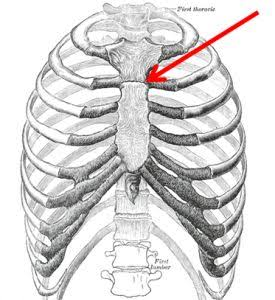
body (corpus, gladiolus) of sternum

sternum

xiphoid process
level of T9-T10

topographic landmarks
jugular notch (T2-T3)
sternal angle (T4-T5)
Xiphoid tip (T9-T10)
end of rib cage (L2-L3)
sternal rib articulations
ribs 1 through 7 articulate with sternum
ribs 8 through 10 articulate/merge with rib 7
rib

head of rib
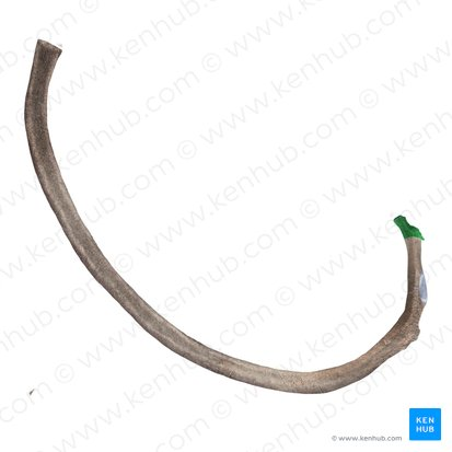
neck of rib

tubercle of rib

body (shaft) of rib

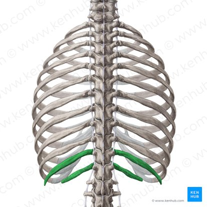
true ribs
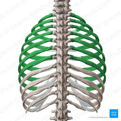
false ribs

floating ribs
costal cartilage

angle of rib
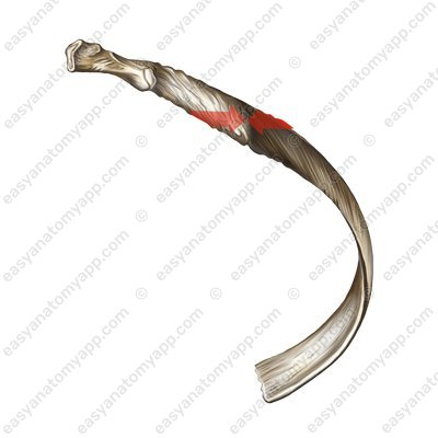
vertebral end (posterior)
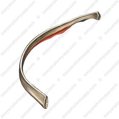
costal groove of rib
for blood vessels and nerves
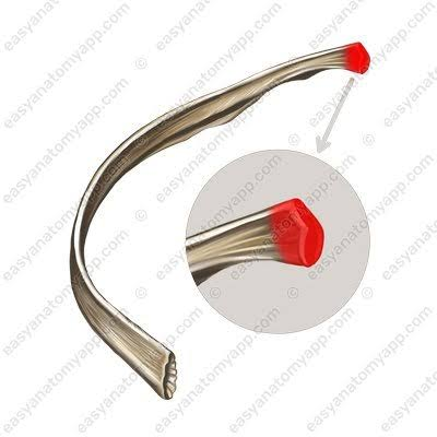
sternal end of rib (anterior)

posterior bony thorax articulations
synovial type with plane movement
costotransverse joint
costovertebral joint
costotransverse joint
plane (gliding) motion
diarthrodial
between rib and transverse process

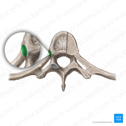
costovertebral joint
plane (gliding) motion
diarthrodial
between rib and vertebral body
oblique sternum considerations
RAO
degree of obliquity
large, barrel chested thorax = 15 degrees
thin chested thorax = 20 degrees
technical considerations for the sternum
breathing technique (2 to 3 seconds)
80 to 85 kV - digital
short but safe SID (never use SID less than 40 inches or 100 cm)
positioning considerations for AP ribs above diaphragm
erect if possible
inspiration
kV (80-85 digital)
CR 3 to 4 inches (8 to 10 cm) below jugular notch
positioning for AP ribs below diaphragm
recumbent
expiration
80-85 kV digital
CR midway between xiphoid process and lower ribs
overall positioning considerations for ribs
area of interest closest to IR (AP or PA)
axillary ribs: rotate spine away from area of interest to elongate
marking site of injury
LPO ribs
elongates left posterior and axillary rib
RPO ribs
elongates right posterior and axillary ribs
sternum routine
RAO, lateral
sternoclavicular joint routine
PA, anterior oblique
RAO sternum
15 to 20 degree RAO
CR to center of sternum (1 inch or 2.5 cm to left of midline and midway between jugular notch and xiphoid process)
orthostatic breathing technique — 3 seconds
trauma alternative: 15 to 20 degree cross table — angle
AEC is not recommended
SID > 40 inches
evaluation criteria for RAO sternum
entire sternum visualized
correct rotation
optimal exposure factors
lateral sternum positioning
CR to center of sternum
SID — 60 to 72 inches (150 to 180cm) recommended
suspended on inspiration
AEC is not recommended
lateral sternum evaluation criteria
entire sternum visualized
no rotation
optimal exposure factors
PA sternoclavicular joint positioning
true PA
CR perpendicular to T2-T3 (3 inches, or 7 cm, distal to vertebra prominens)
suspend on expiration
evaluation criteria for PA SC joints
medial portion of clavicles and SC joints visualized
no rotation
optimal exposure factors
anterior oblique positioning for SC joints
10 to 15 degree rotation
CR to level of T2-T3
1 to 2 inches towards upside
right SC joint
RAO rotation
left SC joint
LAO rotation or a shallow RAO rotation
evaluation criteria for anterior oblique SC joints
manubrium and medial clavicle visible
SC joint open and shifted away from spine
optimal exposure factors
Rib routine
basic
AP or PA projection (area of injury closest to IR)
unilateral or bilateral study (follow department protocol)
axillary portion of ribs — 45 degree anterior or posterior oblique position (rotate spine away from side of interest)
option
chest study if pulmonary injury is suspected
evaluation criteria for AP rubs above diaphragm
first to ninth posterior ribs visualized above diaphragm
no motion
no rotation
optimal exposure factors
evaluation criteria for AP ribs below diaphragm
eighth through twelfth ribs visualized
no motion
no rotation
optimal exposure factors
positioning considerations for posterior or anterior oblique upper ribs
above diaphragm
45 degree oblique
CR to T7
positioning considerations for posterior oblique below diaphragm
can be done recumbent or erect
45 degree oblique
CR midway between xiphoid process and iliac crest
evaluation criteria for oblique ribs above diaphragm
axillary portion of ribs appear elongated
no motion
optimal exposure factors
rib fracture
commonly caused by trauma or underlying pathology; may cause injury to adjacent lung or cardiovascular structures (pneumothorax, pulmonary or cardiac contusion)
fractures of first rib are often associated with injury to the underlying arteries or veins, whereas fractures to the lower ribs may be associated with injury to the spleen, liver, or kidney
flail chest
occurs when ribs are fracture in two or more places on multiple adjacent ribs, creating a segment of ribs that are unattached to the bony thorax.
this type of injury can lead to instability of the chest wall and create paradoxical chest movement during breathing
is the result of severe trauma, such as blunt trauma, and is associated with underlying pulmonary injury
if injury is suspected, perform rib studies erect if the patient’s condition permits for best visualization
sternum fractures
typically caused by blunt trauma, are associated with underlying cardiac injuries
pectus carinatum (pigeon breast)
this defect is characterized by anterior protrusion of the lower sternum and xiphoid process; it is usually a benign condition but could lead to cardiopulmonary complications in rare cases
pectus excavatum
also referred to as funnel chest, this deformity is characterized by a depressed sternum; this condition rarely interferes with respiration but often is corrected surgically for cosmetic reasons
metastases
primary malignant neoplasms spread to distant sites via blood and lymphatics; ribs are common sites for lesions, which may be characterized and visualized on the image as follows:
osteolytic, osteoblastic, or combination osteolytic and osteoblastic
osteomyelitis
localized or generalized infection of bone and marrow can be associated with postoperative complications of open-heart surgery, which requires the sternum to be split; the most common cause of this is a bacterial infection