OT543: Kinesiology Exam 2C
 Call Kai
 Learn
Learn Practice Test
 Spaced Repetition
 Match
Match Flashcards
 Knowt Play
1/102

There's no tags or description
Looks like no tags are added yet.
Name | Mastery | Learn | Test | Matching | Spaced | Call with Kai | Chat |
|---|
No analytics yet
Send a link to your students to track their progress
103 Terms
What are the Components of the Wrist?
"Functional Unit" of:
-Distal Radius & Ulna
-8 Carpal Bones
-Proximal Metacarpal Bones
-Soft Tissue Structures
What is the Purpose of the Wrist "Unit"?
-Position the hand (for grasp/dexterity)
-Increases the mechanical advantage of the hand muscles (ex Tenodesis Action)
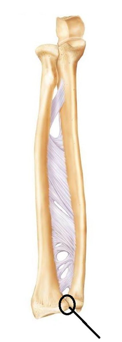
Distal Radioulnar Joint
(Pivot Joint)
between head of ulna & ulnar notch of radius
-loose, surrounding capsule allows for pronation/supination
-stability via ligaments & interosseous membrane
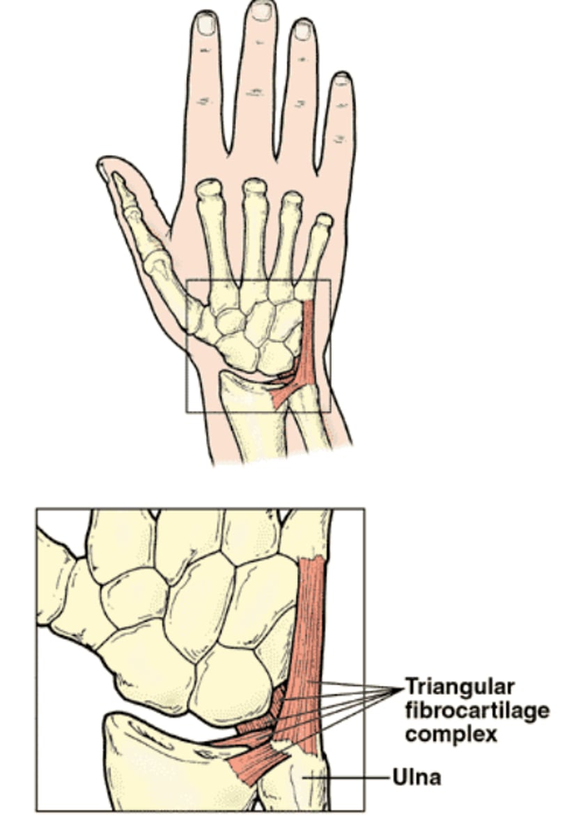
Triangular Fibrocartilage Complex (TFCC)
Complex structure comprised of radioulnar disc & fibrotic attachments that provide primary support for the distal radioulnar joint
-located on the medial aspect of the wrist
---cushion between the ulna & carpal bones
-disruption can impact the ulna/ulnar side
-functionally considered an extension of the radioulnar joint & the distal radius (articular surface)
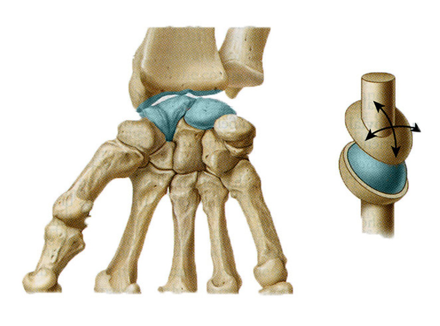
Radiocarpal Joint
(Condyloid Joint)
Where the distal radius articulates with the proximal row of carpal bones
-50% of wrist motion occurs here
-Consists of:
---Radius
---Radioulnar Disc
---Scaphoid
---Lunate
---Triquetrum
-Motions: Flexion/Extension & Radial/Ulnar Deviation
-Ligaments: Ulnar & Radial Collateral (anterior & posterior)
Midcarpal Joint
(Plane/Gliding Joint)
(Functional unit rather than anatomical)
Between the proximal & distal carpal rows
-Motions: ALL motion; but mainly Extension & Radial Deviation
-Ligaments for stability including the Transverse Carpal Ligament
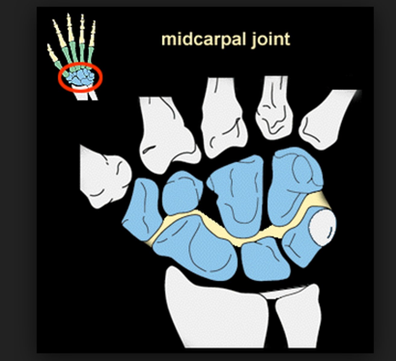
What do the Midcarpal & CMC Joints Form?
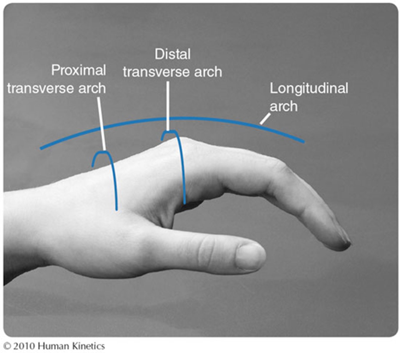
The arches of the hand
What Carpal Bones are in the Proximal Row?
-Scaphoid
-Lunate
-Triquetrum
-Pisiform
What Carpal Bones are in the Distal Row?
-Trapezium
-Trapezoid
-Capitate
-Hamate
Carpal Movement in Wrist Flexion
Proximal carpal row glides POSTERIORLY
Carpal Movement in Wrist Extension
Proximal carpal row glides ANTERIORLY
Carpal Movement in Radial Deviation
Proximal carpal row glides ULNARLY
Carpal Movement in Ulnar Deviation
Proximal carpal row glides RADIALLY
Which Carpal Row has the Most Movement in Flexion/Extension?
Proximal Row
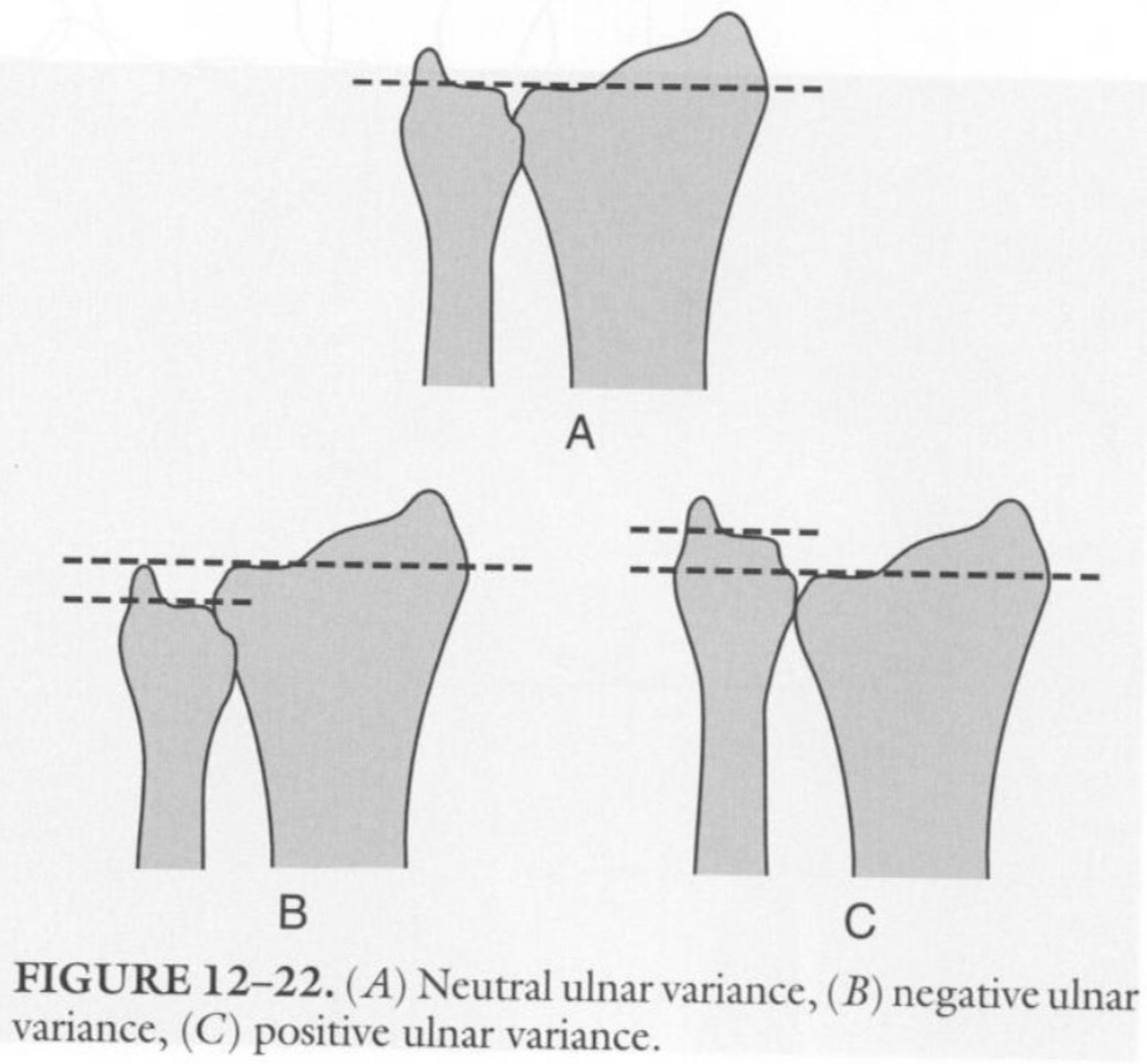
Ulnar Positive Variance
The ulna is TOO LONG
-possible TFCC Impingement
---causes pain when loading wrist & during ulnar deviation
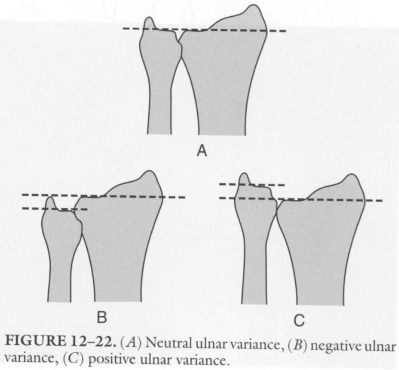
Ulnar Negative Variance
The ulna is TOO SHORT
-Kienbock's Disease
---when the lunate collapses & dies (avascular necrosis) from taking ALL the pressure
What are the 2 Volar Carpal Ligaments?
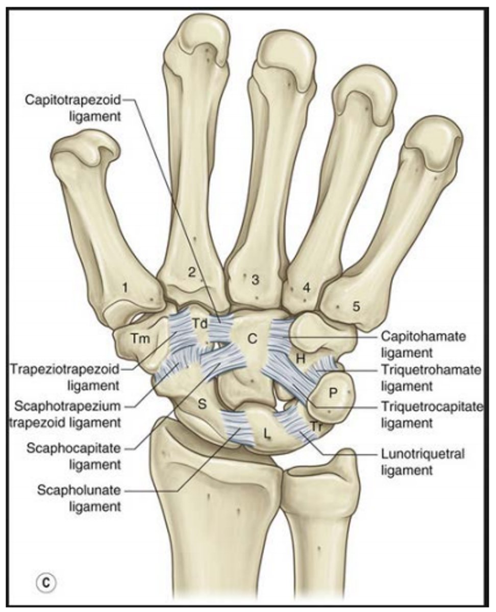
1) Scapholunate Interosseous Ligament
2) Luntoriquetral Interosseous Ligament
Scapholunate Interosseous Ligament
Responsible for wrist stability, as it stabilizes the scaphoid
-hooks the scaphoid to the lunate
-Scaphoid Instability: common wrist issue
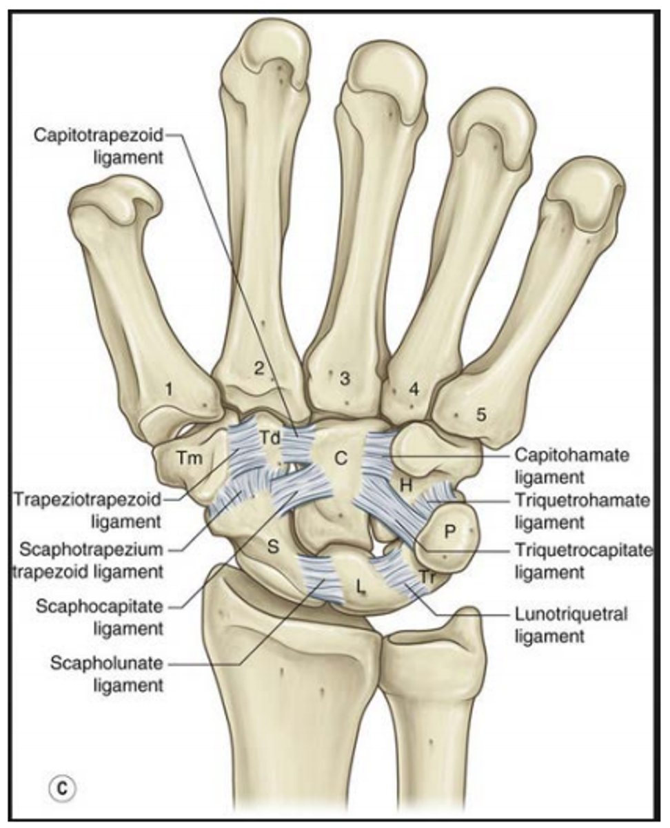
Luntoriquetral Interosseous Ligament
Stability between the lunate & triquetrum
-Avascular: can contribute to lunate instability
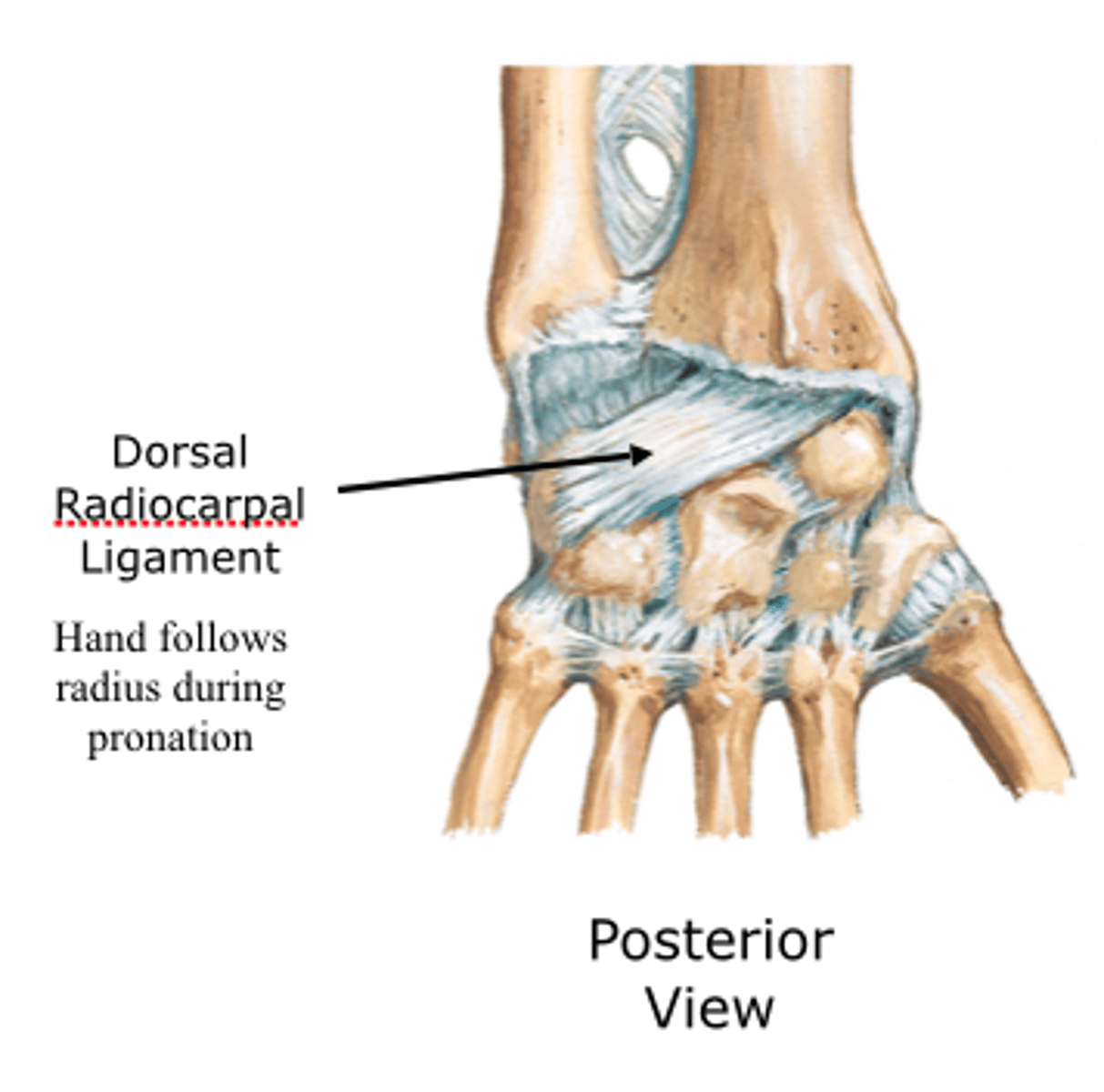
Dorsal Radiocarpal Ligament
Connects radius to posterior scaphoid & lunate
-contributes to wrist stability- especially of the scaphoid during wrist ROM
-TAUT in wrist flexion
---keeps carpal bones in place
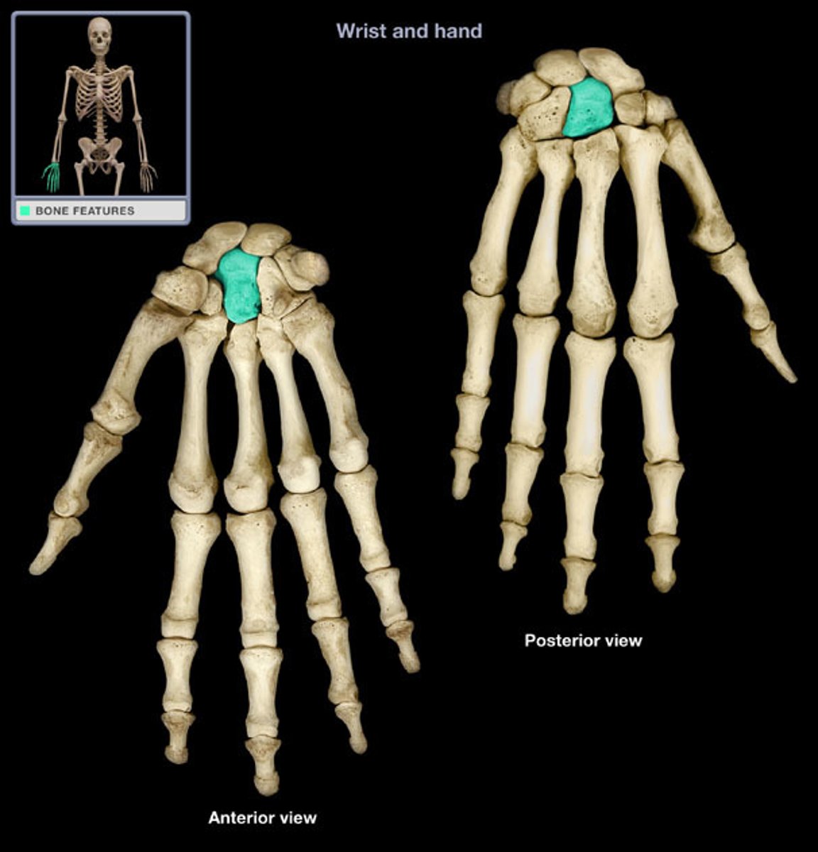
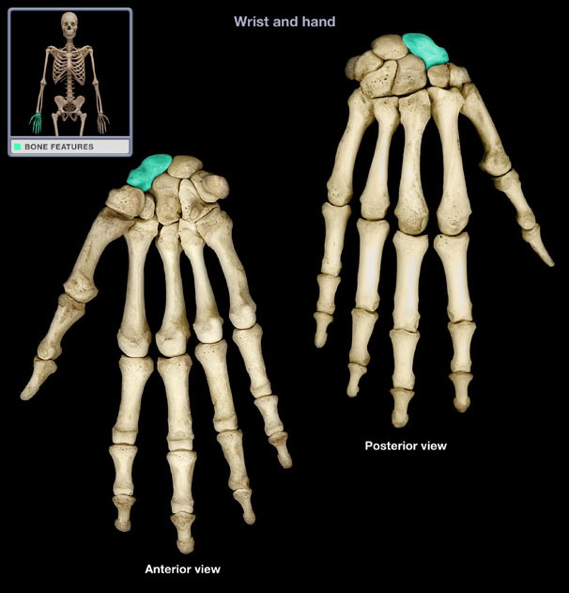
Capitate Bone
Considered the CENTER of wrist motion ("KEYSTONE")
-LARGEST carpal bone
Lunate Bone
Moves the LEAST in flexion/extension
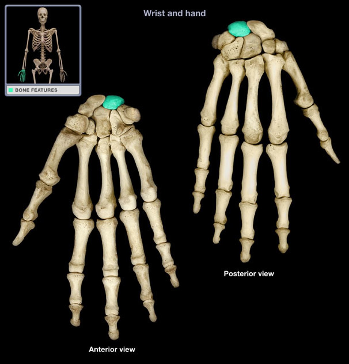
Scaphoid Bone
Links the proximal & distal carpal rows
-important for stability
-MOST MOTION of carpal bones during wrist flexion/extension
**Source of COMMON wrist injuries
Functional Movement of the Wrist
(FULL Flexion to Extension)
Movement initiated at the distal carpal row & metacarpals by the wrist extensors
-DISTAL carpals glide ON the PROXIMAL carpals
At Neutral: Close-Packed Position of the Scaphoid & Capitate
(Complete Wrist Extension)
Proximal carpals move as ONE unit on the radius & TFCC
-Ligaments taut in FULL extension = Full Close-Packed Position
Carpal Joints in Radial & Ulnar Deviation
Reciprocal motion of the proximal row in RD & UD
-MOST ROM = Neutral Position
-Proximal Row: simultaneous flexion of the proximal carpals & extension of the distal carpals during RD also occurs
-Scaphoid Flexion: related to ligament laxity
-Full Radial Deviation: radiocarpal & midcarpal joints are close packed
-Extreme Flexion or Extension: little radial/ulnar deviation available
What are the Most Important Motions for Function?
-Wrist Extension
-Ulnar Deviation
Wrist ROM for Most ADLs (functional requirements)
-Flexion: 54°
-Extension: 60°
-Ulnar Deviation: 40°
-Radial Deviation: 17°
What is the Primary Roles of the Wrist Muscles?
-Provide a stable base for the hand
-Position hand for optimal length-tension relationship in the long finger muscles
Wrist Flexor Work Capacity
MORE than TWICE than that of extensors
-but the wrist can position the extensors to have a mechanical advantage
Are Flexors or Extensors Stronger?
Flexors
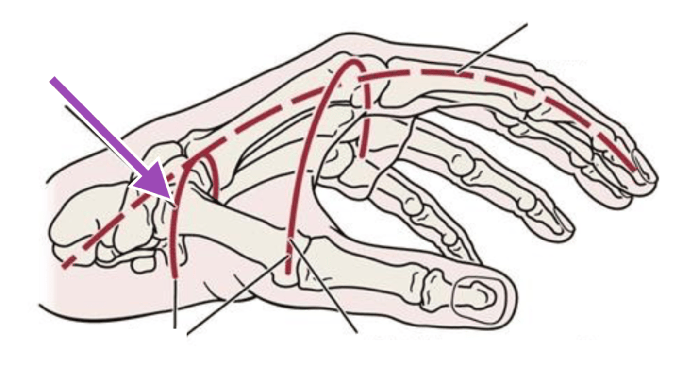
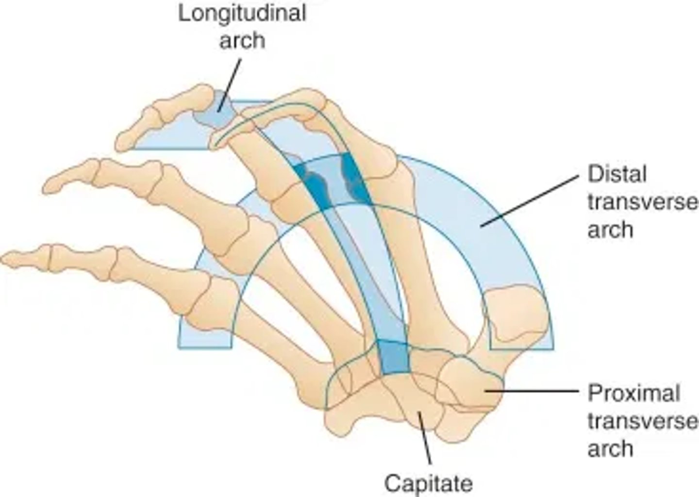
Proximal Transverse Arch
(aka. Carpal Arch)
Formed by the distal row of carpal bones
-arch right above wrist on the palm side
-forms the "Carpal Tunnel"
Contains:
-Median n.
-9 Extrinsic Flexor Tendons
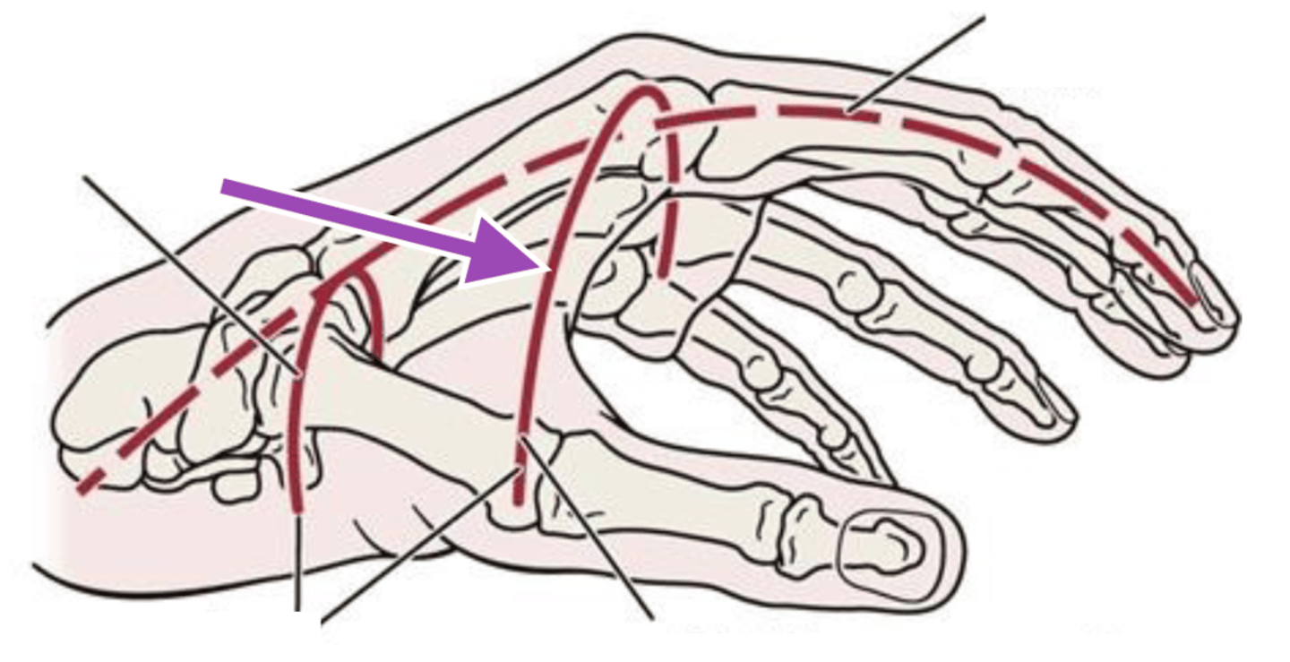
Distal Transverse Arch
Found at the level of the MCP joints
Longitudinal Arch
Begins at the wrist & runs the length of the metacarpals & phalanges for each digit
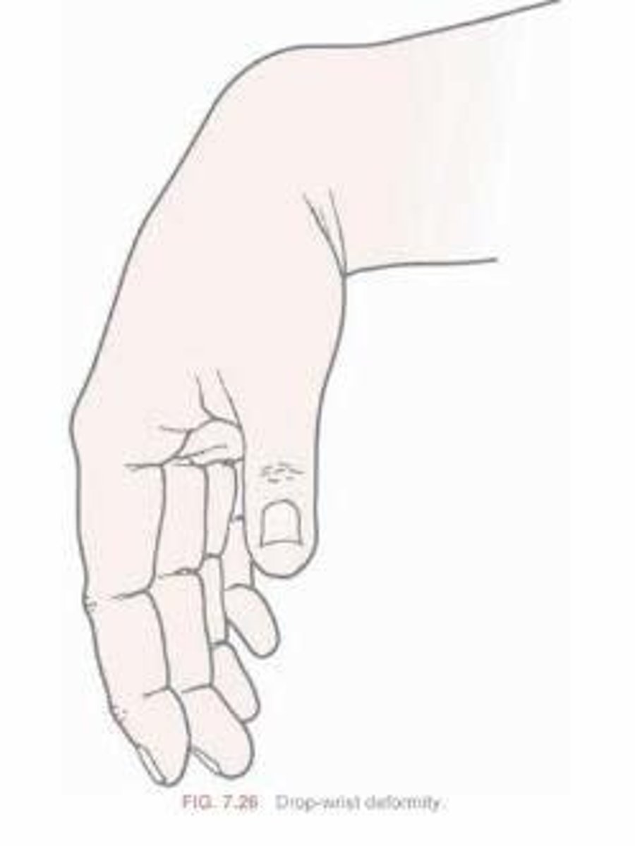
Drop Wrist
Radial n. Palsy
-CAN'T extend wrist
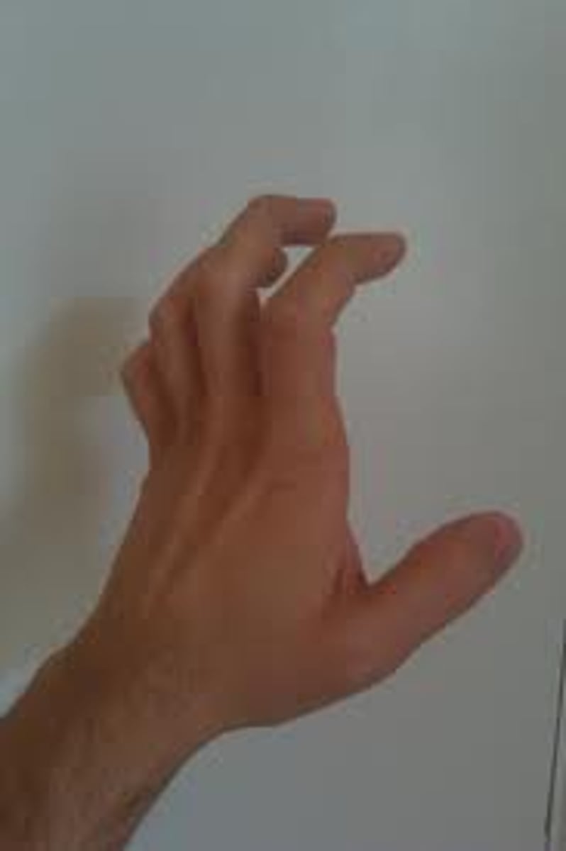
Claw Hand
Ulnar n. Injury
-CAN'T do the intrinsic plus position
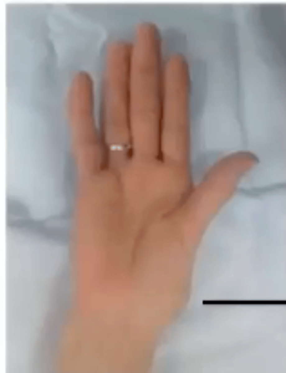
Ape Hand
Median n. Injury
-wasting of the thenar eminence
-flat hand/palm (lack of opposition)
---less prominent arches
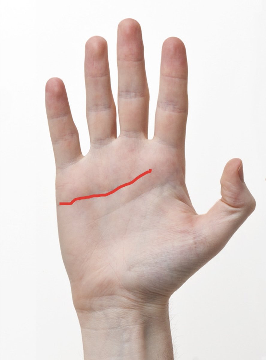
Distal Palmar Crease
Transverse crease that start at the medial side & goes between index & middle fingers
**MP Flexion**
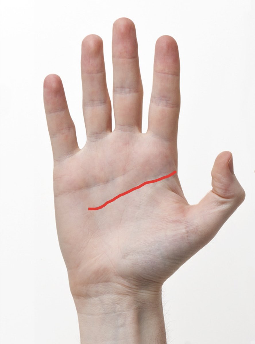
Proximal Palmar Crease
Transverse crease commencing on the lateral side of the palm, extending medially & fading out on the hypothenar eminence
**Middle crease**
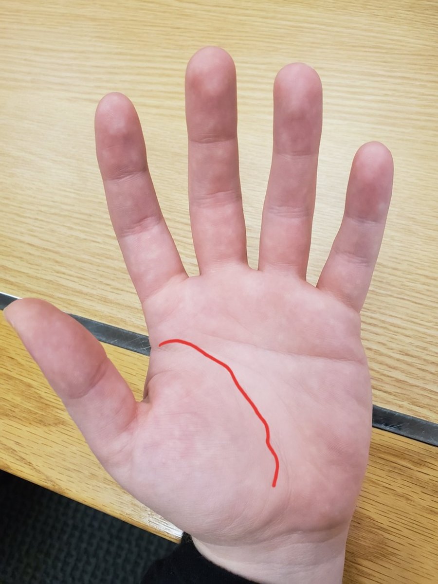
Thenar Crease
Crease around the thenar eminence
Thumb Motion: opposition, CMC, & palmar abduction
CMC Joints of the Fingers
Between the carpals & metacarpals
-Index & Middle Finger = STABILITY
-Ring & Small Finger = MOBILITY
Order of Most -> Least Mobile CMC Joints
5, 4, 2, 3
Thumb CMC Joint
(Saddle Joint)
Trapezium meets the metacarpal
-EXTREMELY MOBILE
Motions:
-Flexion/Extension
-Radial & Palmar Abduction/Adduction
-Rotation
-Opposition
-Circumduction
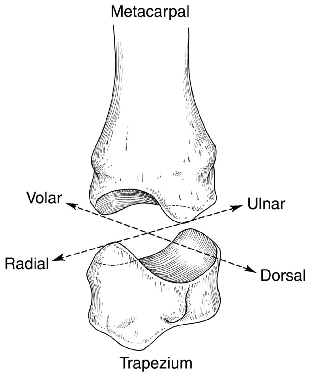
When are the Thumb CMC Ligaments Taut?
Abduction/Adduction
MP Joints of Fingers & Thumb
(Diarthrodial (biaxial)/Condyloid) = MOBILITY
-Radial Side = LESS ROM
-Ulnar Side = MORE ROM
MP Joint Collateral Ligaments
On BOTH sides
-Taut in flexion which limits abduction/adduction
MP Joint Capsule
-Taut in Flexion
-Lax in Extension
Ulnar Collateral Ligament Injury of Thumb
(Skier's Thumb)
Forced hyperextension and/or hyperabduction stress to the thumb MP joint
-occurs 10x MORE often than radial collateral injuries
-results in thumb instability
Radial Collateral Ligament Injury of Thumb
Disruption results in pain during ADDucted activities (ex. opening jar)
What Type of Joints are the PIPs or Thumb IP?
Synovial Hinge Joints
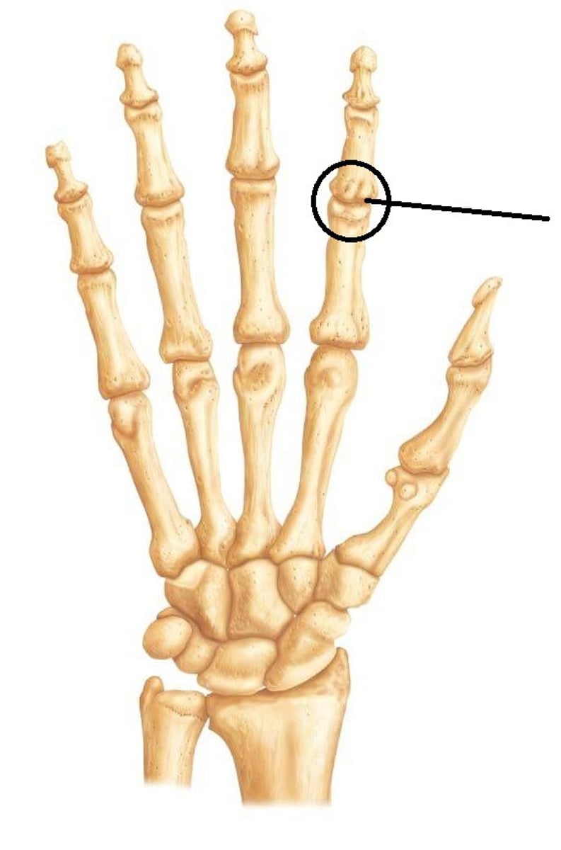
PIP/IP Collateral Ligaments
Taut in flexion OR extension
-important for STABILITY
-RADIAL = MORE likely to be injured
What is the Strongest PIP Collateral Ligament?
RADIAL Collateral Ligament of INDEX FINGER
-used for key pinch
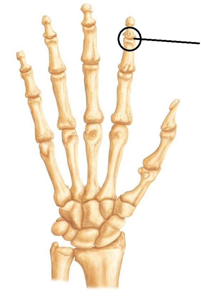
What Type of Joints are the DIPs?
Hinge Joints
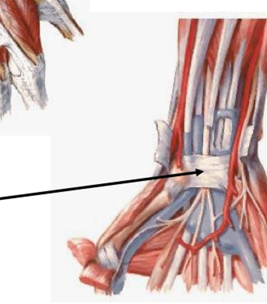
What is the Function of the Fascia at the Wrist?
Prevents bowstringing & INCREASES the mechanical advantage
Deep Transverse Ligament
Part of the flexor retinaculum
-incised during carpal tunnel release
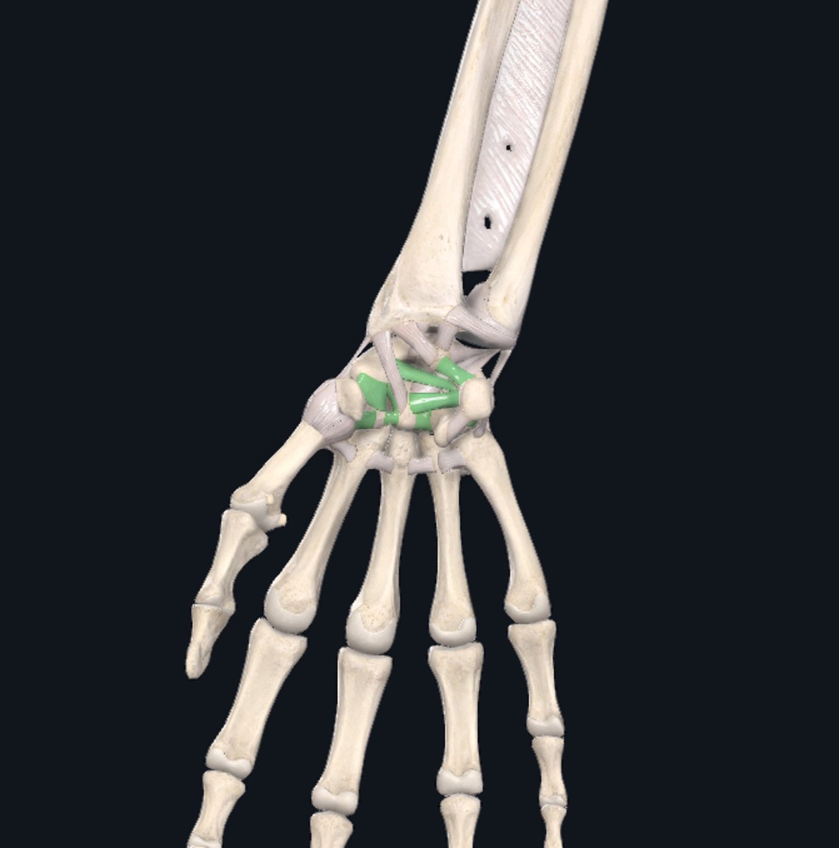
Intercarpal Ligaments
Connect the carpal bones
-contribute to the carpal tunnel
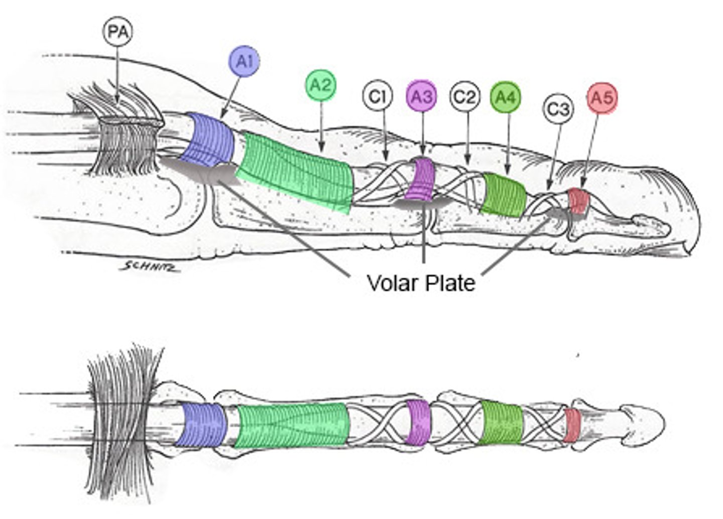
"Pulley" System of the Digits
Fibrous Digital Sheath
-Annular Ligaments:
---5 per digit
---3 at each thumb
-Cruciate Ligaments:
---4 per digit
---2 at each thumb
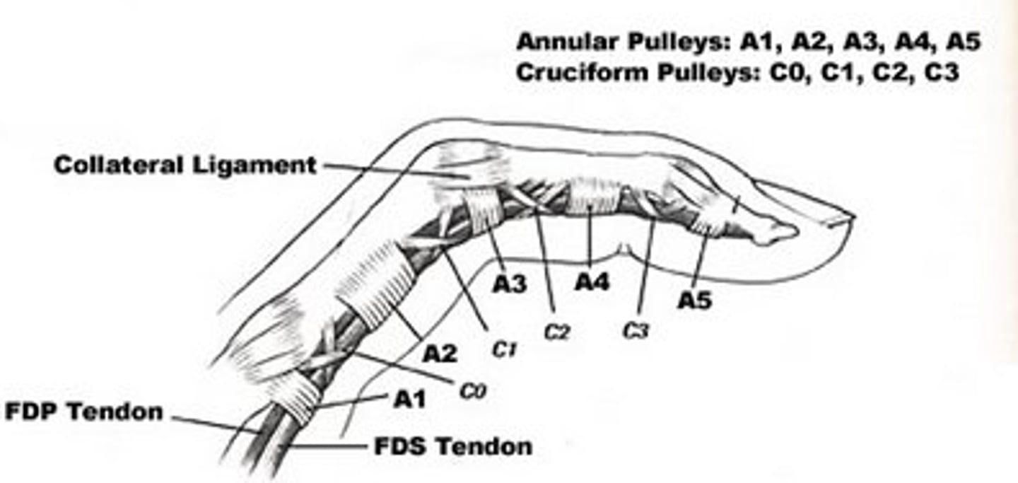
A1 Pulley
Band of tissue holding flexor tendon closely to finger bones, near palm
- responsible for trigger finger
Optimal Splinting Positioning to Prevent Deformity
Wrist Extension: 15-20°
MP Flexion: 70-80°
IP/DIP/PIP Extension: 0° (full extension)
Thumb: always leave FREE unless it needs to be included then:
---Palmar Abduction
Flexor Tendon Zones
5 Different Zones:
Zone 1: Distal to Flexor Digitorum Superficialis insertion
Zone 2: A1 Pulley to Flexor Digitorum Superficialis insertion
Zone 3: Distal end of the Carpal Tunnel to A1 Pulley
Zone 4: Within the Carpal Tunnel
Zone 5: Proximal to the Carpal Tunnel
3 Thumb Zones:
-Zone 1: Distal to IP joint
-Zone 2: Between the MP & IP joints
-Zone 3: Thenar Eminence
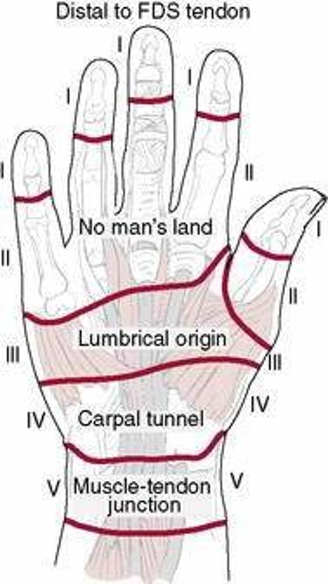
What is Significant About Flexor Zone 2?
("No Man's Land")
-HIGH risk for tendon adhesions
What is Significant About Flexor Zone 5?
("Spaghetti Wrist")
-severe, multi-structural lacerations of the palm side of the wrist
Extensor Tendon Zones
8 Different Zones:
-Zone 1: DIP joint
-Zone 2: Middle Phalanx
-Zone 3: PIP joint
-Zone 4: Proximal Phalanx
-Zone 5: MP joint
-Zone 6: Metacarpal
-Zone 7: Dorsal Retinaculum
-Zone 8: Distal Forearm
5 Thumb Zones:
-Zone 1: IP joint
-Zone 2: Proximal Phalanx
-Zone 3: MP joint
-Zone 4: Metacarpal
-Zone 5: CMC joint/Radial Styloid
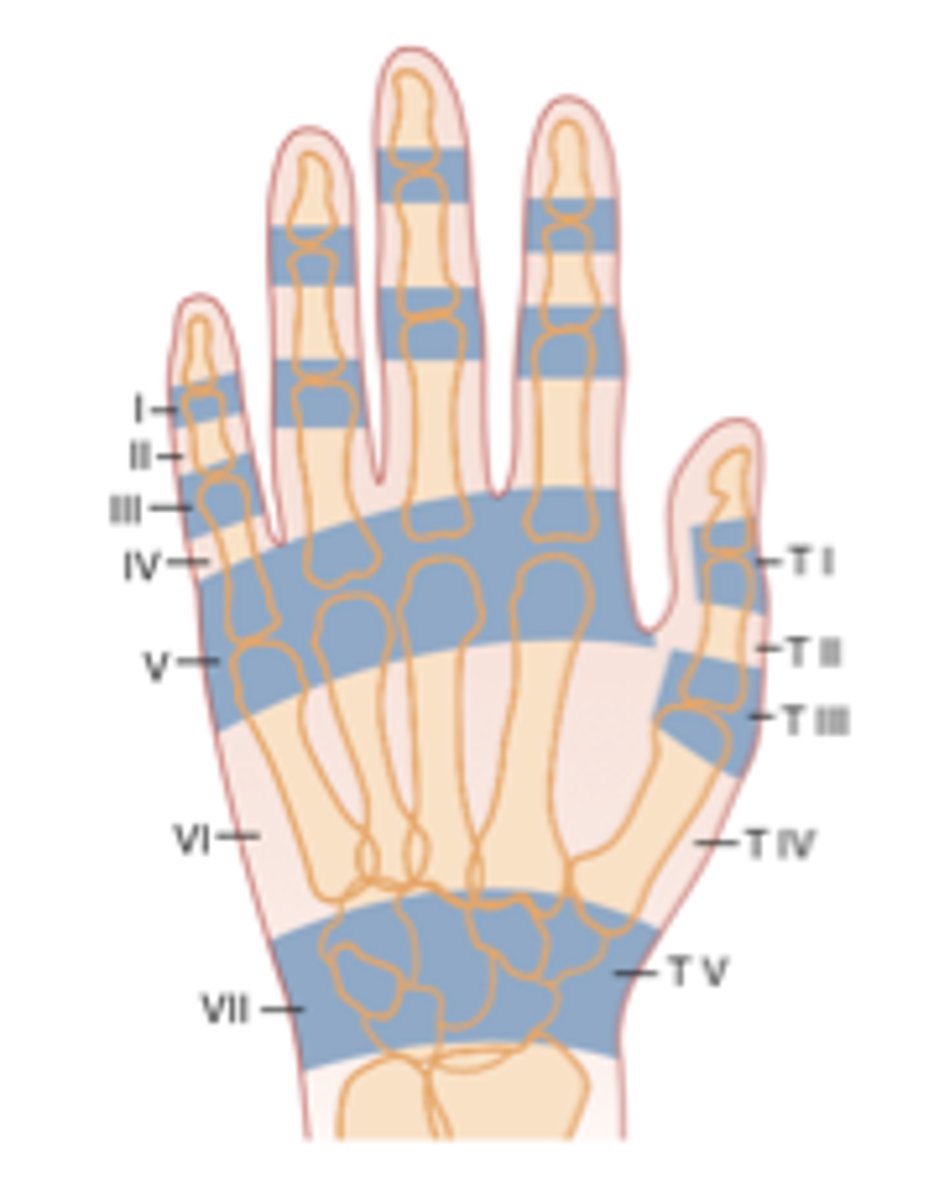
What Tendon is Injured the Most in Extensor Zone 1?
Extensor Digitorum
Extensor Expansion
Specialized connective tissue structure by which the extensor tendons insert onto the phalanges
AKA:
-Extensor Aponeurosis
-Extensor Hood
-Extensor Mechanism
-Dorsal Hood
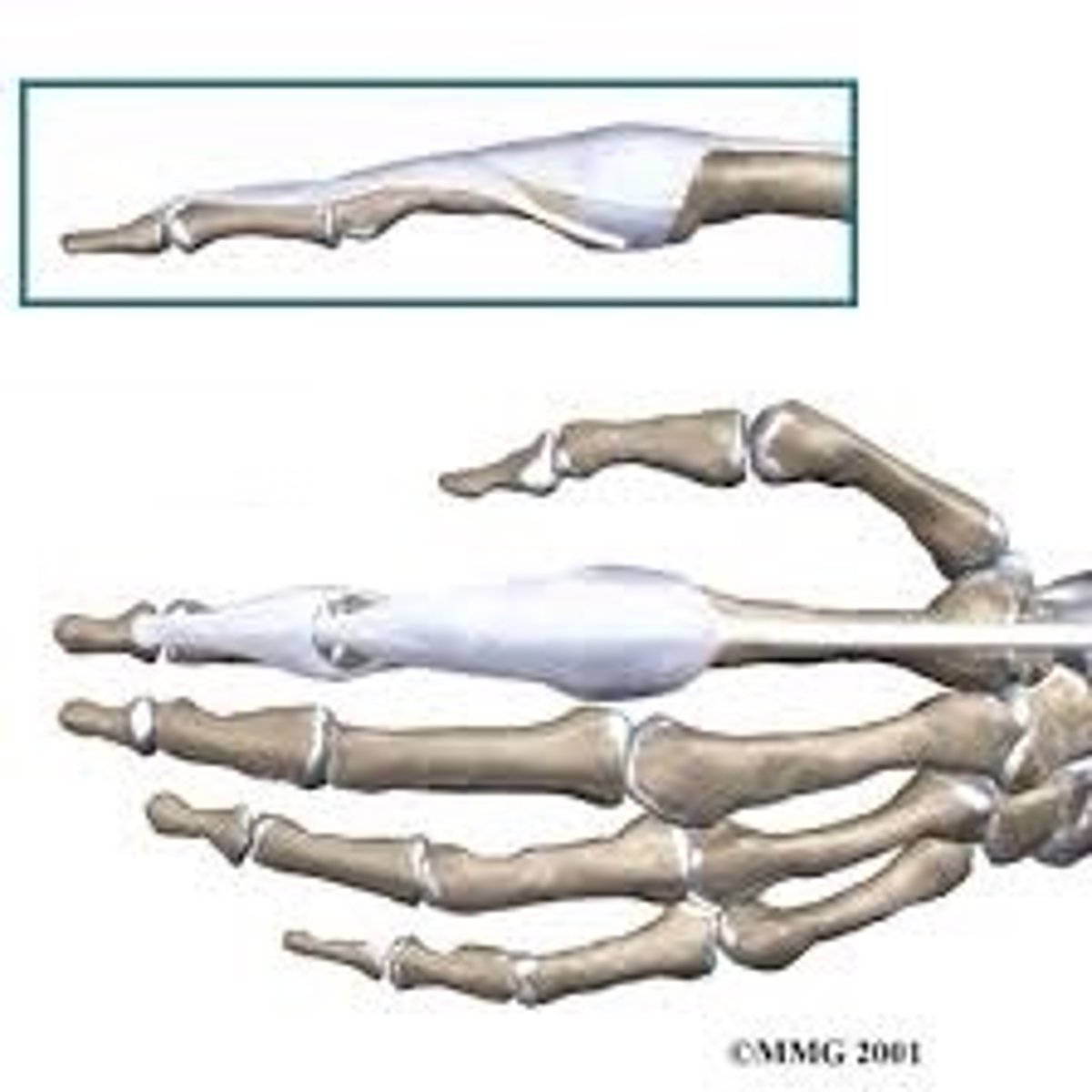
Central Band/Central Slip
Inserts into the base of the MIDDLE phalanx
-is an extension of the Extensor Digitorum & Interossei
-EXTEND the PIP joints
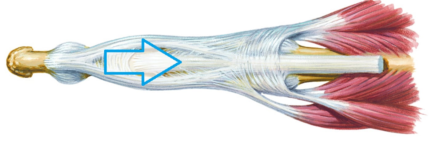
Lateral Bands
Inserts into the base of the DISTAL phalanx
-is an extension of the Extensor Digitorum, lumbricals & Interossei
-EXTEND the DIP joints
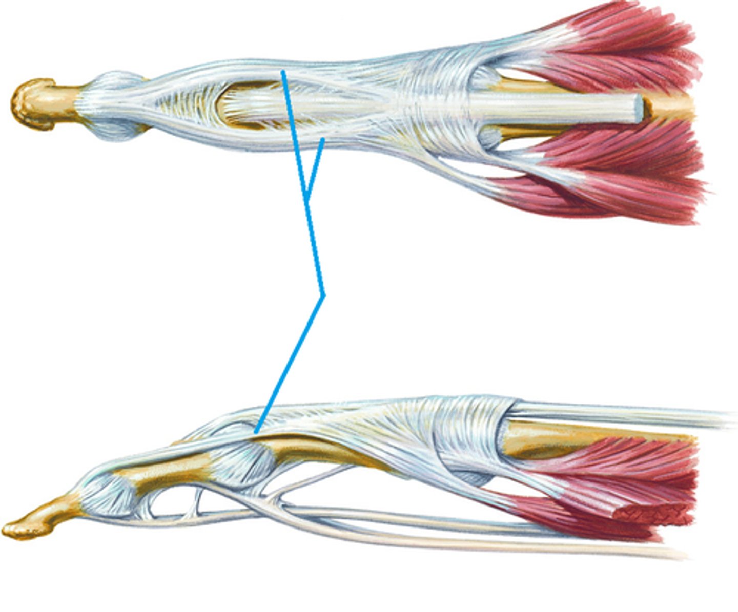
Retinacular "Link" Ligaments
They pass ANTERIOR to the PIP joint axis & POSTERIOR to the DIP joint axis
-they "link" the PIP & DIP joint motions
---if the DIP is flexed, it becomes taut & the PIP tends to flex
------if the PIP is extended, it becomes taut & the DIP is extended
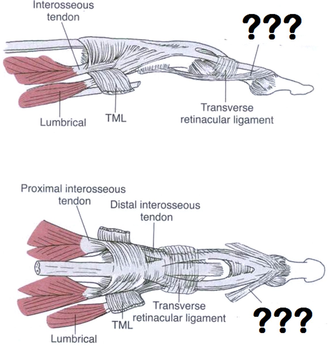
What is the Main Function of the Extensor Mechanism?
EXTENDS PIP & DIP joints
Extensor Mechanism Summary
-When the PIP extends, the DIP extends
-DIP cannot actively extend with PIP flexed
- When the DIP actively flexes - the PIP actively flexes
-Simultaneous MP, PIP, & DIP flexion = use ALL available extensor mechanism slack
Swan Neck Deformity
PIP extended & DIP flexed
-lateral bands slide POSTERIOR
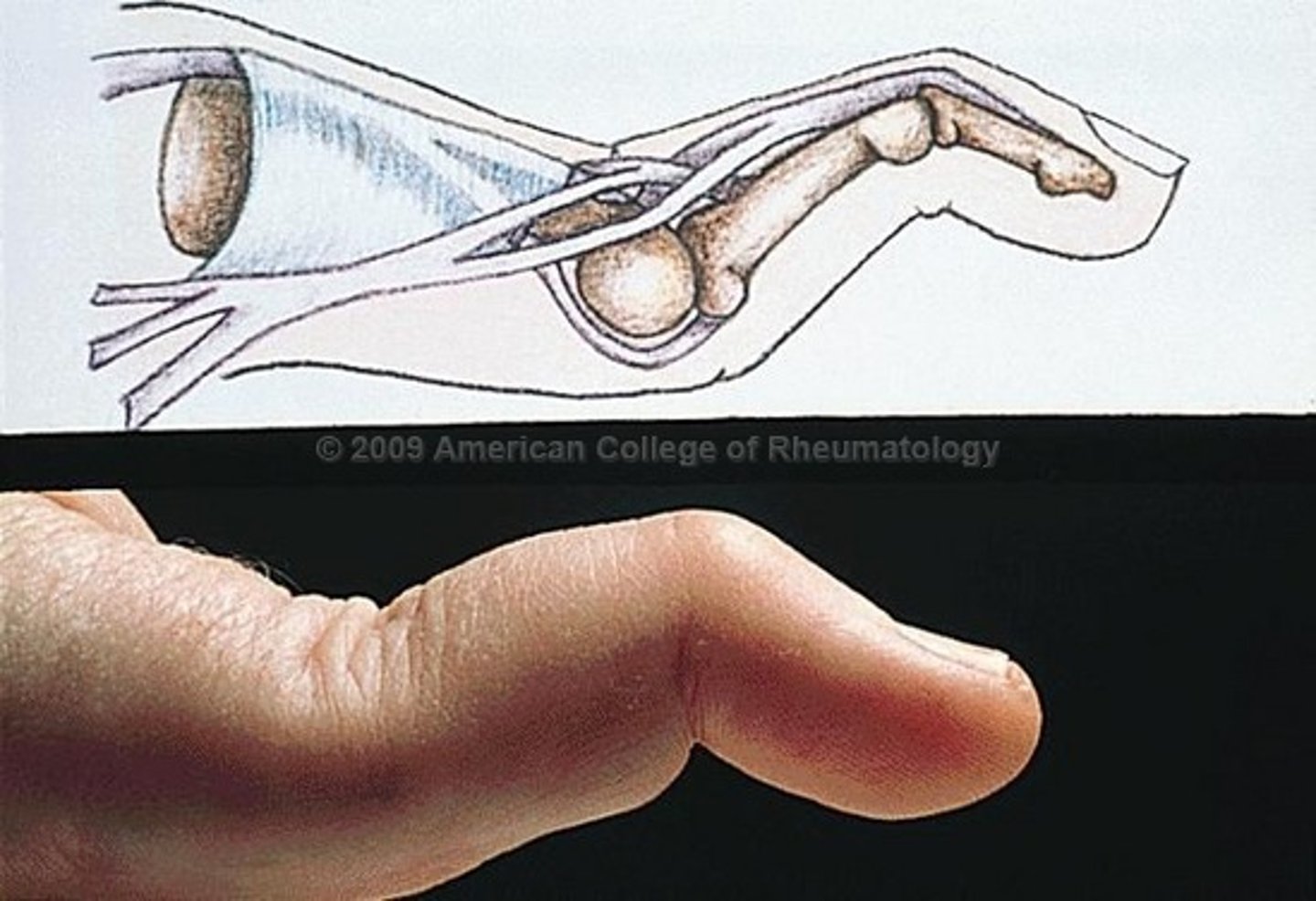
Boutonniere Deformity
PIP flexed & DIP extended
-lateral bands get too short/tight & slide ANTERIOR
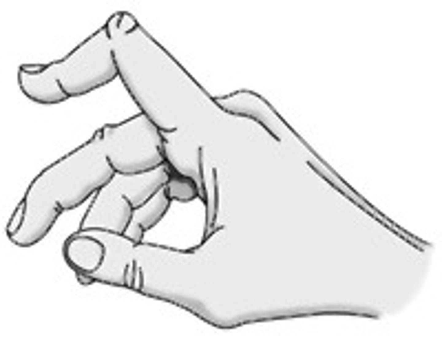
Boxer's Finger
Fracture of the 5th Metacarpal
-common when striking something with a closed fist
-can be treated with a closed reduction or surgically
CMC Osteoarthritis
Arthritis of the CMC joint
-more common in women
Symptoms:
-thumb pain at CMC joint
-positive grind test
-achy wrist
Trigger Finger
Pathologic thickening of the flexor tendon sheath
-Most Common at the A1 pulley , FDS
---Thumb FPL
-Most Common Digits:
---Thumb
---Ring Finger
---Middle Finger
SLAC Wrist
Scapholunate Advanced Collapse
-form of scapholunate instability
DISI Wrist
Dorsal Intercalated Segment Instability
-form of scapholunate instability (tear of the ligament)
-Lunate tilts posteriorly
-Dorsiflexion Instability
VISI Wrist
Volar Intercalated Segment Instability
-volar rotation of the scaphoid & lunate w/ extension of the triquetrum w/ lunotriquetral instability (tear of the ligament)
----Lunate tilts anteriorly
What is the Functional Position of the Hand & Wrist?
-Forearm in Midposition
-10° of Ulnar Deviation
-20° of Wrist Extension
-MPs are Slightly Flexed & Abducted
-Thumb is Abducted & in Slight Opposition
-IP/DIPs are Flexed
Prehension
Grasping or taking hold of an object between any 2 surfaces
-power grip OR precision handling
Power Grip
General term to describe ALL gripping
-OR full grip which includes the thumb
Includes:
-Cylindrical Grip
-Spherical Grip
-Hook Grip
-Lateral Prehension
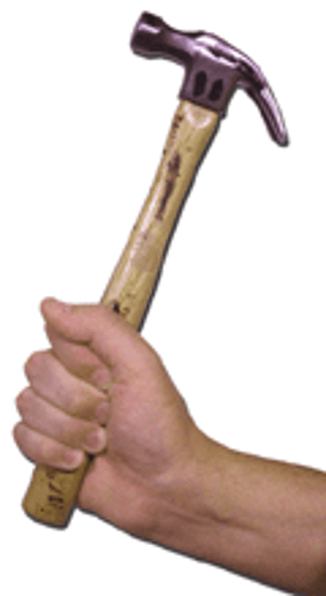
Cylindrical Grip
ALL fingers flexed around the object
-Thumb & fingers in opposite direction
-Ex: holding a cup
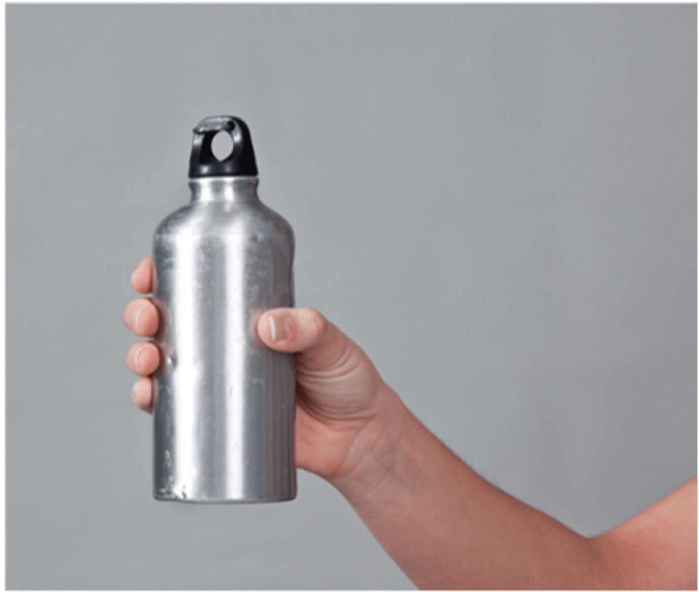
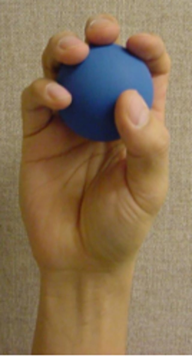
Spherical Grip
All fingers & thumb adducted around an object
-Fingers spread apart
-Palm is often not involved
-Ex: holding a ball
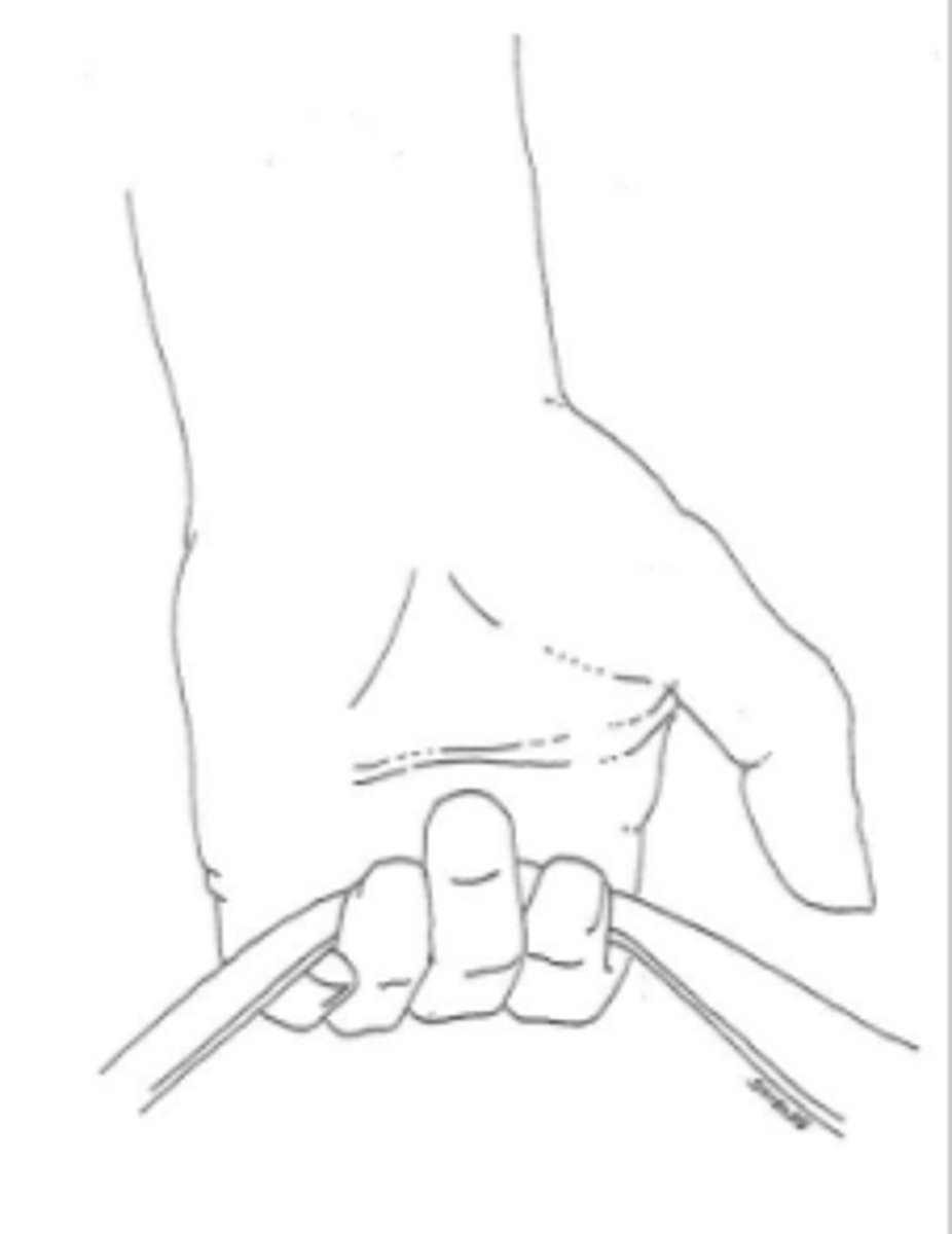
Hook Grip
Thumb NOT included
-looks like intrinsic minus position
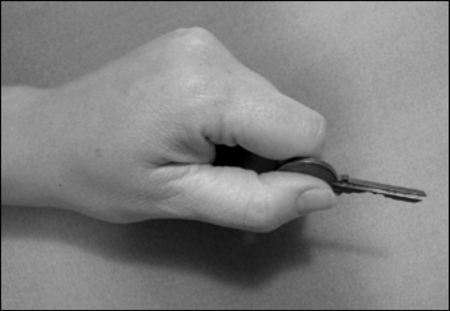
Lateral Prehension
Thumb pad to lateral surface of index finger
-"KEY PINCH"
What is the Optimal Wrist Position for Power Grip?
-20° of Extension
-5° of Ulnar Deviation
**In this position the Index & Middle Fingers each absorb approximately 32% of the force
When is Grip the Least Powerful?
40° of WRIST FLEXION
What are the 3 Types of Precision Handling?
-Pad to Pad
-Tip to Tip
-Pad to Side
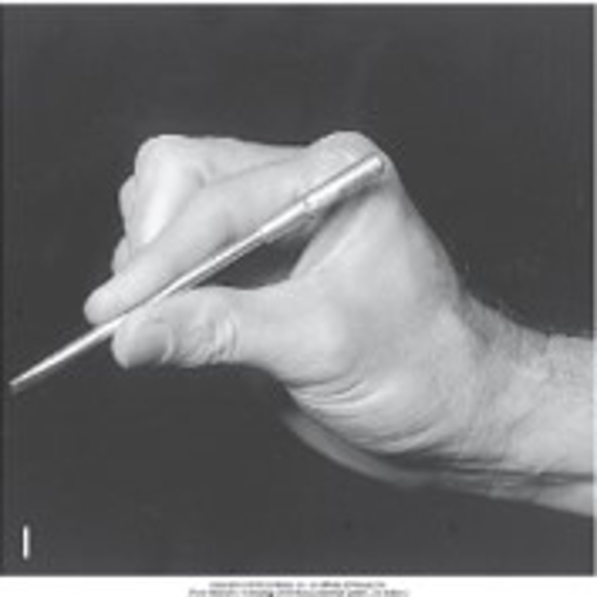
Pad to Pad Prehension
Opposition of pad of thumb to pad of finger
-Tripod Pinch
-Palmar Pinch
Tip to Tip Prehension
Tip of thumb against tip of index finger
-Tip Pinch
-Pincer Pinch
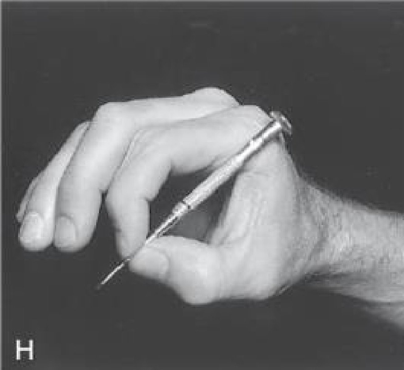
Pad to Side Prehension
Tip of thumb to side of index finger
-LEAST precise type
-Key Grip
-Key Pinch
-Lateral Pinch
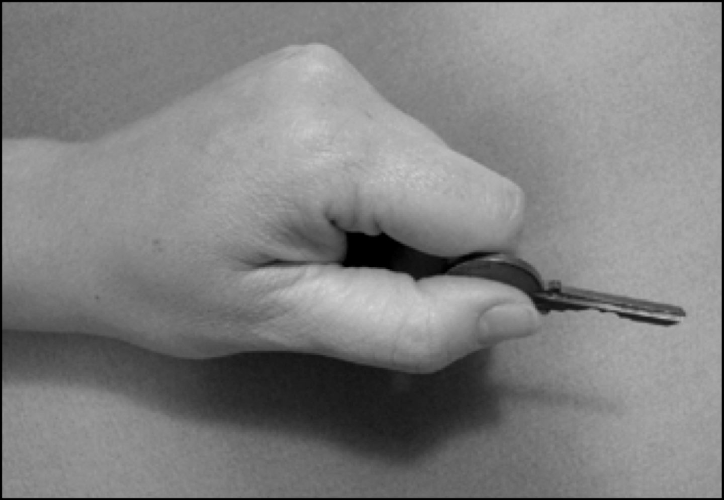
What is the Best Way to Measure Edema for the Entire Wrist/Hand?
Volumeter
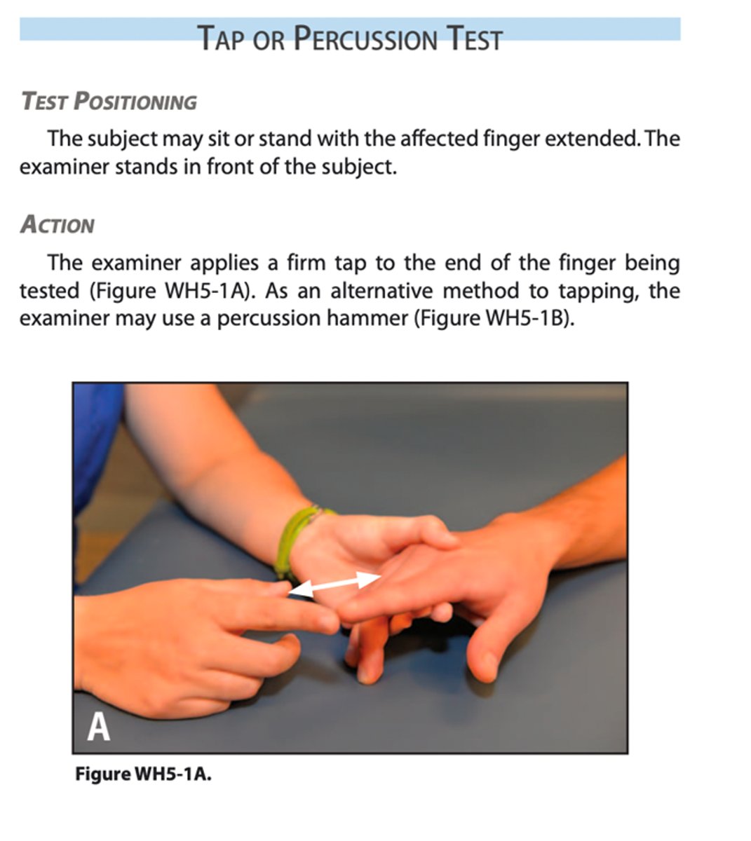
Tap or Percussion Test
Patient sitting/standing with forearm resting on table & affected finger extended
--examiner taps end of finger
Positive Test = Fracture
--pain at the injury site
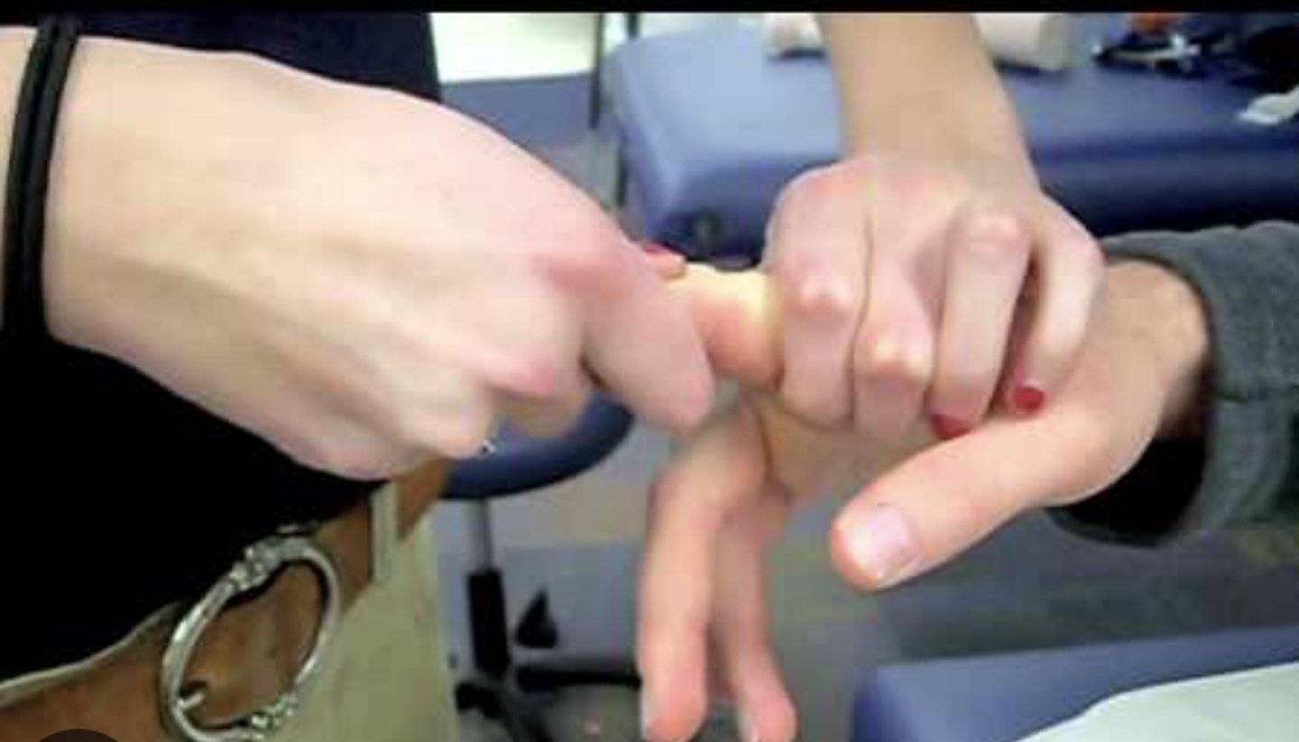
Compression Test
Patient sitting/standing with forearm resting on table & affected finger extended
--examiner holds distal phalanx & applies compression DOWN
Positive Test = Fracture
--pain at the injury site
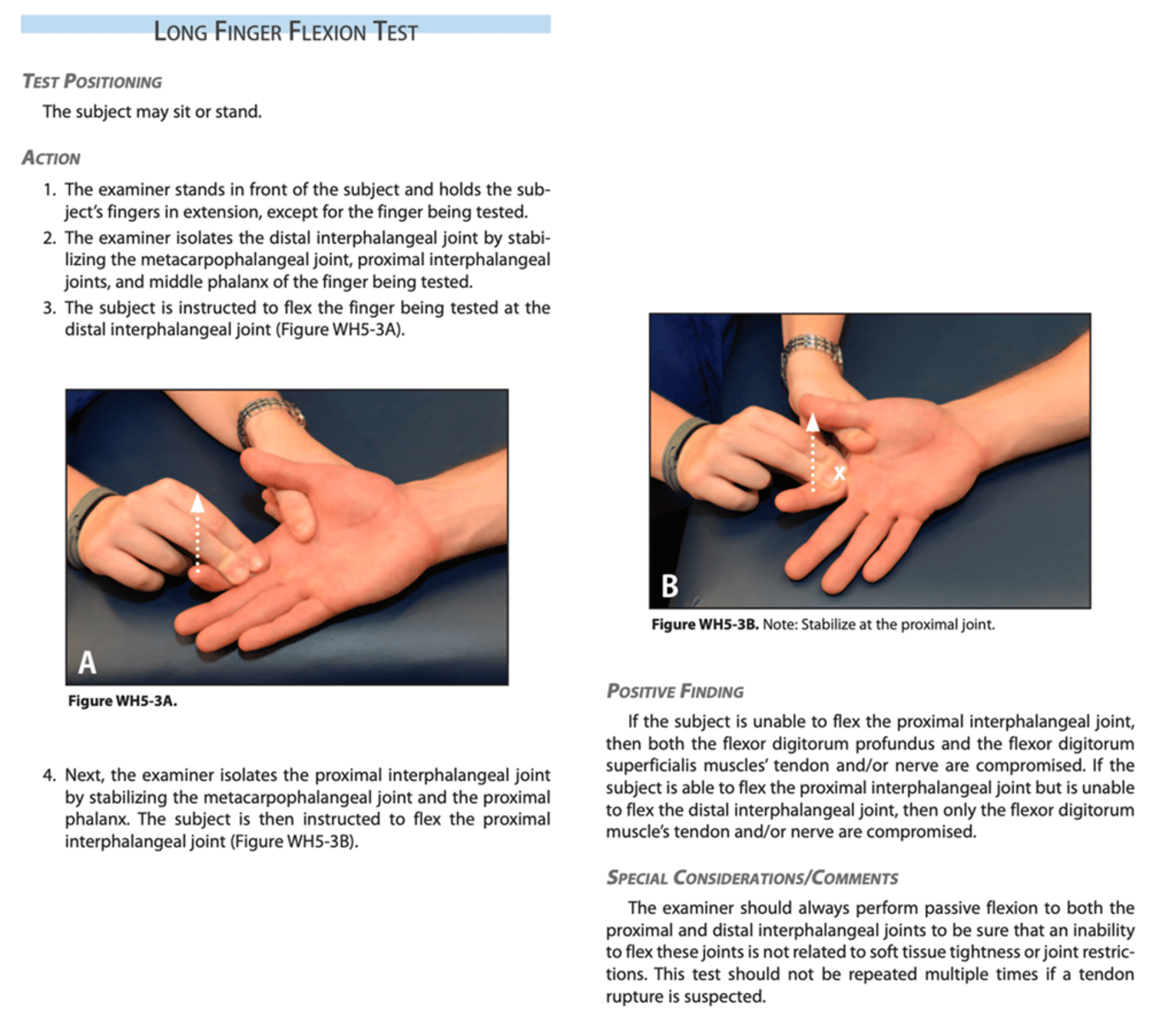
Long Finger Flexion Test
Patient sits with forearm supinated & resting on table
--examiner stabilizes all NONTEST finger & has the person flex both PIP & DIP
Positive Test = Flexor Digitorum Superficialis/Flexor Digitorum Profundus and or Nerve Compromised
--unable to flex the PIP/DIP joints
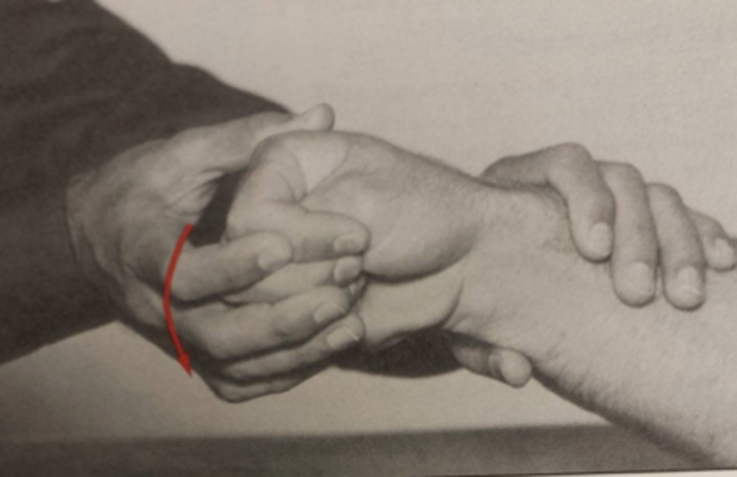
Finkelstein Test
Patient sits/stands & forms a first around thumb with forearm in neutral position
--examiner pushes down on hand while supporting forearm
Positive Test = DeQuervain's
--pain on the radial side of the wrist
Phalen Test
Patient sits/stands with elbow extended & forearm pronated
--examiner pushes hand DOWN into flexion & holds for 1 minute
Positive Test = Carpal Tunnel Syndrome
--numbness/tingling on the median n. distribution of the fingers
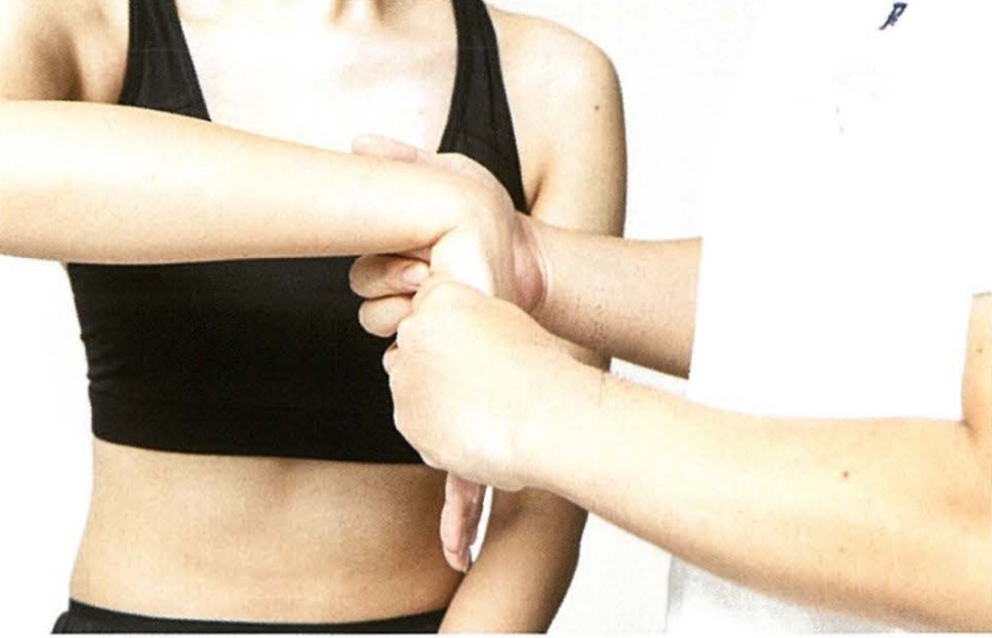
Reverse Phalen Test
Patient sits/stands with elbow extended & forearm pronated
--examiner pushes hand UP into extension & holds for 1 minute
Positive Test = Carpal Tunnel Syndrome
--numbness/tingling on the median n. distribution of the fingers

Tinel's Sign
Patient sits with forearm supinated & resting on table
--examiner taps over the carpal tunnel
Positive Test = Carpal Tunnel Syndrome
--tingling, paresthesia, or pain in the area of the median n. distribution of the fingers

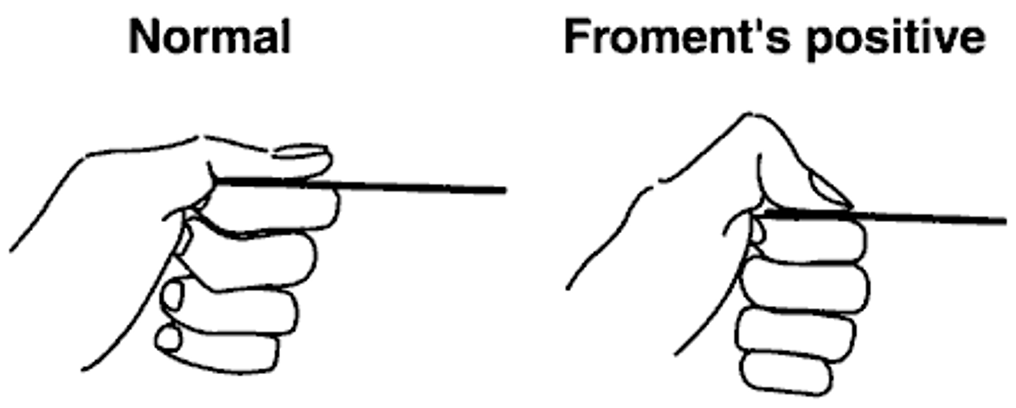
Froment's Sign
Patient sits/stands & holds a piece of paper between their thumb & index finger
--examiner tries to pull paper out
Positive Test = Ulnar n. Compromise
--flexion of the distal IP joint of the patient's thumb