Epithelial and Connective Tissues
 Call Kai
 Learn
Learn Practice Test
 Spaced Repetition
 Match
Match Flashcards
 Knowt Play
1/33

There's no tags or description
Looks like no tags are added yet.
Name | Mastery | Learn | Test | Matching | Spaced | Call with Kai | Chat |
|---|
No analytics yet
Send a link to your students to track their progress
34 Terms
Cutaneous Membrane
Skin, Epithelial Tissue (epidermis), Connective Tissue (dermis), Dry membrane
Mucous Membranes
Lines all body cavities open to the environment, Epithelial tissue (stratified sqamous or simple columnar) , Connective Tissue (areolar lamina propria), moist membrane
Serous Membranes
In closed body cavities, made of epithelial tissue (simple squamous), connecti
Serous membranes are what
moist membranes of the closed ventral body cavity
ventral body cavity
a large fluid-filled space located on the front (anterior) side of the human body that houses and protects major internal organs.
Skins Properties
Sensation: monitors temperature, touch, and pressure via nerve endings
Thermoregulation: optimal body temp
Excretion: excretes salts, water wastes (via glands)
Physical Barrier: defense against pathogens + foreign substances
bound together by tight junctions, acid mantle (lower pH)(thin film on the surface of the skin that protects against bacteria)
Barrier to water and light:
Keratinized cells prevent gain/loss of water
Melanin of cells prevent UV damage from sun
Why is sun exposure necessary
Vitamin D Synthesis by cholesterol
What are the two main regions of the skin
Epidermis, Dermis
Epidermis
most outermost layer, made up of stratified squamous epithelial tissue, keratinized, made up of tightly joined together cells by tight junctions and desmosomes
Dermis
Connective tissue layer, made up of:
collagen, elastic and reticular fibers
muscle (arrector pili - a tiny smooth muscle attached to a hair follicle that pulls the hair upright when it contracts)
blood and nerve supply
glands opening onto surface
Hypodermis
subcutaneous layer, fat layer under dermis:
composed of adipose and areolar connective tissues
anchors and insulates
fat is not uniformly distributed (low levels on scalp, high levels in abdomen)
not technically a skin layer
Stratum Corneum
first layer of epidermis, made up of 20+ layers of dead keratinocytes tightly joined by desmosomes
flattened cells with protein packed lipid envelope
water resistant lipid covering
barrier to water, not water proof
Stratum Granulosum
second layer of skin, made up of 1-5 layers of flat cells, water resistant
Stratum Spinosum
3rd layer of epidermis, thickest layer, 8-10 layers of cells filled with pre-keratin filaments
cells flatten as they approach the top of the spinosum layer
dendritic cells present (immune cells)
Stratum basale (germinativum)
fourth layer of epidermis, Basement layer, miotic (produces keratinocytes)
includes stem cells, tactile cells, melanocytes
epidermal ridges extends into the dermis
What is the stratum lucidum?
located in thick skin under the stratum corneum, pale featureless cells packed with eleidin (clear intracellular protein) and keratin
Colleen Likes Good Soup Bowls (CLGSB)
Corneum, Lucidum, Granulosum, Spinosum, Basale
Keratinocytes
most numerous in epidermis, 30-45 day lifespan, anchored together by desmosomes and tight junctions
produced in basale layer and pushed upward
as keratinocytes migrate up the skin layer, they become more resilient - accumulates melanin to protect against UV damage
packed with keratin filaments to provide resistance and protection
coats surface with lipids to resist water
undergoes apoptosis when it reaches the top of the epidermis
Stem Cells
divides to generate keratinocytes
confined to the basal layer (stratum basale), in direct contact with cell tissue beneath
Melanocytes
Located in basal layer (basement layer)
branches protrude between keratinocytes
Produces melanin
releases melanin from surface in granules, melanin pigments picked up by keratinocytes and accumulate on outward facing edge on cell (sunny side up)
Dermis
the connective tissue that is deep to the epidermis, the connective tissue layer is made of collagen, elastic and reticular fibers, has extenside blood and nerve supply (which provides O2 and nutrients and processes stimuli)
involved in immune defense which includes:
macrophages
white blood cells
mast cells
Fibroblasts
produces semifluid matrix, deposits collagen
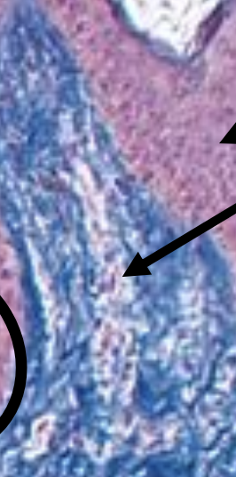
Papillary Layer
Loose connective tissue - Areolar (collagen and elastic fibers) - Good for immune defense - Many small blood vessels
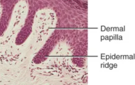
Dermal Papillae
extends to epidermis, includes Capillaries, nerve endings, and touch receptors

Epidermal Ridge
downward projections of epidermis
How are friction ridges made
dermal papillae + epidermal ridges
Messiner’s corpuscles
Specialized mechanoreceptors located near the epidermal : dermal border (mainly in hairless skin- fingertips, palms)
detect light touch
encapsulated nerve ending, not a nerve
surrounded by flattened Schwann cells (protects nerve ending)

reticular layer
extends from the papillary layer to the subcutaneous layer
made of Dense irregular connective tissue - Thicker collagen fibers for strength and stability - Fewer cells than in papillary layer - Elastic fibers for stretch and recoil - Adipose tissue
cleavage lines
made of Collagen fibers and elastic fibers form parallel bundles
Perpendicular cuts take longer to heal and are pulled apart by elastic fibers
Cuts parallel heal more quickly with less scaring
collagen fibers
provide strength
elastic fibers
provide structure/stretch and recoil (think of a rubber band)
Strech Marks
generated by tears in dermis
Stretch exceeds elastic limits
• Wrinkles/creases- don’t go away

Pacinian corpuscles
deep pressure mechanoreceptors in the reticular layer of the dermis
Pacinian corpuscles are deep pressure mechanoreceptors
• Detect pressure and vibrations
• Generally oval shaped
• Looks like slices of onion
Myelinated nerve enters corpuscle and loses myelination
Interstitum
a fluid filled space in between other cellular structures and tissues that drains into the lymph nodes and is supported by a complex network of thick collagen bundles.