How the brain works quiz 2
 Call Kai
 Learn
Learn Practice Test
 Spaced Repetition
 Match
Match Flashcards
 Knowt Play
1/73

There's no tags or description
Looks like no tags are added yet.
Name | Mastery | Learn | Test | Matching | Spaced | Call with Kai | Chat |
|---|
No analytics yet
Send a link to your students to track their progress
74 Terms
Luigii Galvani
Discovered animal electrcity by an electric spark hit from a metal surgical tool attached to nerve in frog leg
Gustav Fritsch and Eduard Hitzig
Used a dog brain to stimulate brain movement, mid/front of brain: leg movement, back of brain; no effect due to muscle contraction via motor nerves (motor cortex)
Walter Hess
Used cat brain to stimulate different behaviors: rage, fear, sniff, pant, eat; hypothalamus regulates homeostasis and coordinates behaviors needed for survival
Epilepsy
Sudden uncontrollable increase in brain activity in the cerebral cortex; activity spreads uncontrolled to the rest of the brain
Wilder Penfield
Used technique to map the function of the human brain; patient is fully conscious during procedure, patients describe what they are experiencing after each brain region
Motor homunculus
Motor cortex (pre-central gyrus), stimulation = body movement; contralateral response: right brain: left body, left brain: right body, motor control represnted in larger brain area (face, fingers)
Sensory homunculus
Sensory cortex (post-central gyrus), stimulation = body sensation (touch, tickle, itch, pain); contralateral lresponse, highly sensitive parts of body represnted by larger brain area
Auditory cortex
Temporal lobe; stimulation = non recognizeable sounds
Visual cortex
Occipital lobe; stimulation = flashes of light, no distinct images because of organized activation of dorsal and ventral streams
Temporal lobe
Stimulation = experimental and interpretive response; reliving past experiences with very vivid memory flashbacks or sudden change in interpretation of current experiences
Deja vu
New seems familiar
Jamais vu
Familiar becomes strange
Transcranial magnetic stimulation (TMS)
Non-invasive way to induce electrical stimulation of the brain using strong magnetic fields; high: increase brain activity (treatment for depression, does not cause epilepsy), low: decrease brain activity (treatment for schizophrenia)
Deep brain stimulation
Invasive technique to implant electrodes semi-permanently into particular brain regions (treats depression, parkinson’s disease, epilepsy)
Electrical stimulation and recording
Stimulation: stimulate location electrically to observe behavioral effects; recording: observe electrical events that correlate to behavior
Electrial recording
Stimulating the brain electrically through electrodes can induce motor behavior or sensory effects; different behavioral or cognitive brain states should correlate with different patterns of electrical activity
Electroencephalography (EEG)
Developed by Hans Berger; 20-60 electrodes are placed on scalp and recordings for 20-30 minutes, must pass skull and skin; signals are detected by produced synchronous activity in brain regions
Brain waves
Electrical signals produced by synchronous activity in large networks of the brain are read by the EEG as waves with varying frequency and amplitude, including delta, theta, alpha, beta, and gamma waves
Frequency
number of peaks per second (Hertz or Hz)
Amplitude
height of the peaks, indication of how strong the signal is

Complex brain waves
Sum of combination of frequencies and amplitudes

Alpha frequency range
High frequency fast wave activity (8-13 Hz)

Beta frequency range
HIgh frequency fast wave activity (13-30 Hz)

Theta frequency range
Low frequency slow wave activity (4-7 Hz)

Delta frequency range
Low frequency slow wave activity (1-3 Hz)

EEG Waves correlate with behavioral states
Beta (alert), Alpha (relaxed), Theta (drowsy), Delta (deep sleep)
REM sleep: dreaming stage
Stage where no muscle tone, no movement (except eyes), inhibitory signals from brainstem block muscle activity, hallucinating brain in a paralyzed body
Rapid eye movement (REM)
State where eye movement controlled by cranial nerve III; body muscles controlled through the spinal cord and spinal nerves, 70-95% of dreams occur; The “paradoxical state”: EEG resembles awake state, yet you are asleep and dreaming
Slow-wave sleep
No thoughts or normal everyday thoughts (10% claim dreams) EEG: low frequency, high amplitude, Decreased muscle tone, but still able to move; Sleep walking appears to occur
Parasomnias: groups of sleep disorders
Non - REM sleep parasomnias: Sleep terror, Sleep walking, Confusional arousals; REM sleep parasomnias: Nightmare disorder, REM sleep behavior disorder, Recurrent isolated sleep paralysis
REM sleep behavior disorder
In REM sleep behavior disorder, the paralysis that normally occurs during REM sleep is incomplete or absent
People can “act out” dreams, particularly those that are vivid, intense, and violent
Sleep paralysis
Flip side of REM sleep behavior disorder
Muscle paralysis (or atonia, lack of muscle tone) occurs either before sleep begins, or persists after sleep ends

Basal (ventral) forebrain:
Promotes slow wave sleep (deep sleep) in the hypothalamus, lesion in this region can produce insomnia

Part of the hypothalamus
Coordinates other sleep centers, regulating orderly stages of sleep
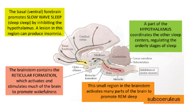
Brainstem
Contains reticular formation, activates and stimulates the brain to promote wakefulness
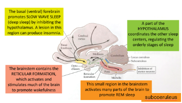
Subcoeruleus (small region in the brainstem)
Activates parts of the brain to promote REM sleep
Sleep is important for
Conservation of metabolic energy, consolidation of memory in long-term storage, clearance of toxins from the brain
Using EEG to determine brain functions
Exposes subject to some kind of sensory stimulus, read brain waves occurring in a subject
Changes are called evoked potentials (EPs) or event-related potentials
(“potential” refers to change in voltage=electricity)
One, called the P300, is seen when the subject sees something familiar, but unexpected in that context
The EEG technique advantages
Non-Invasive, can test living, talking human subjects
Inexpensive compared to other methods
Very fast, excellent temporal (time) resolution
The EEG technique disadvantages
Not very sensitive as signal is very small
Can not detect activity in all parts of the brain
Only the cortex because it is close to the surface
Cannot localize a given brain wave pattern to which part of the brain is active
Poor spatial resolution
EEG techniques also measure
Blood flow
Brain oxygen levels OR
Brain glucose levels
Positron Emission Tomography (PET)
Inject a radioactive form of glucose into the bloodstream (fluorodeoxyglucose)
The fluorine isotope decays and releases a positron (the antimatter version of an electron)
Distribution of this positron emission can be imaged by a computer; provides measure of glucose utilization in the brain
Functional Magnetic Resonance Imaging (fMRI)
Does not require any injections or radioactivity
Uses signals given off naturally by iron atoms in the blood’s hemoglobin to measure use of oxygen by the brain
Change in magnetization of oxygen-rich vs. oxygen-poor blood
Best resolution, least invasive
Function of fMRI
Areas that “light up” are not the only active regions
Entire brain is active all of the time
Blue or red areas are those whose blood flow changes when given a task
3 hypothesized components to love
The drive (lust, libido)
Attraction (romantic love, being in love, infatuation)
Attachment (pair bonding, long term stable relationship)
Headaches
pain or discomfort located in the head
Primary: occurs independently
Secondary: byproduct of another condition
Localized nociceptive event
Migraines
a distinct, chronic neurological disorder
Head pain is only one feature, can be absent in some patients
Systemic brain disorder
Migraines pt 2.
Can have a genetic component
Can consist of moderate to severe headaches
Typically unilateral
Nausea
Heighted (hypersensitivity) response to light and sound, etc.
Last hours to days, recurrent
May have an “aura”
Visual, sensory, or neurological warning signs before the onset of the migraine

fMRI can show
Cortical Spreading Depression in migraines - a wave of electrophysiology hyperactivity followed by a wave of inhibition
Implicated in migraine auras
Can be seen as spreading of a wave of vasoconstriction following vasodilation and prolonged sustained vasoconstrictions
Reductionist approach advantages
Can understand actual mechanisms
Critical to fixing problems
Use animal models
Utilize broad range of techniques
Reductionist approach disadvantages
Difficult to study humans
Can be difficult “to see the forest for the trees”
Not clear how individual components work together to result in a whole complex
The light microscope
One or more convex lenses in a tube
Allows magnification of up to 1000-fold
Leeuwenhoek built a microscope and applied microscopy to biology
Robert Hooke and identification of cellular structure
Also constructed a microscope
Viewed cell structure in a thin slice of pork
Thought he saw cells in a monastery
Cell theory of Schleiden and Schwann
All living matter is made of cells and products of cells
Cell is the most elementary unit that can contain life
All cells come from other cells
Cell is surrounded by plasma membrane that separates it from the environment
All cells have similar chemical composition with organelles that perform specific function
How can we see brain cells in the microscope?
First: must fix the tissue
Brain is soft, must be hardened for cutting or freezing
Prevent tissue from rotting
Second: must section the tissue
Make thin slices; millionths of a meter
Transparent and hard to see
Third: must stain these sections
Different stains stick to different organelles and reveal overall structure, cell types, and even individual molecules

Nucleus
Contains DNA (genes); transcription of DNA into messenger RNA

Ribosomes
transcribe messenger RNA into proteins (translates gene code to protein code) located on the rough endoplasmic reticulum
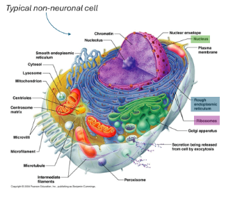
Organelle
Each is specialized for a particular task
The Nissl Stain
Stain reveals cell bodies organized into SIX layers in the cerebral cortex
Stains rough endoplasmic reticulum
Different stains give different patterns
Weigert or Weil Stain
Labels the fatty insulation found on many nerve fibers called myelin - white matter

Stains for gray matter
Nissl stain: cell bodies (darker) fiber tracts (lighter)

Stains for white matter
Weil stain: cell bodies (lighter), fiber tracts (darker)
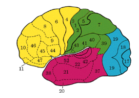
Bordmann areas defined by Nissl stain
Defined 50 brain area with different “cytoarchitecture” revealed with Nissl stain
Saw layers and clusters of neurons called nuclei (in the CNS) or GANGLIA (in the PNS)

Camillo Golgi
Created the Golgi stain “the black reaction”, filled a few % of all neurons revealing their true shape
Labels a random subset of neurons, but outlines them completely (fibers AND cell bodies)

Dendrites
Shorter, thicker/tapering, many of them, more branches to the cell body
Recieve and integrate nerve impulses

Axons
Longer, thinner/untapered, only one per neuron, fewer branches on the cell body
Transmit nerve impulses to terminals
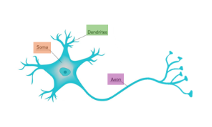
Cell body
Gene expression and protein production

Synpases
Release neurotransmitters to activate electrical and chemical changes in connected neurons

Brainbow
Genes for jellyfish and coral proteins that glow under various light wavelengths that can be inserted into the mouse genome to make fluorescent neurons
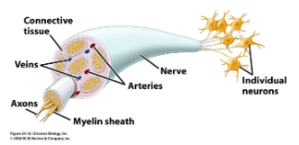
Nervous system to neuronal function
Acquire information from the external (and internal) environment
Sensory signals must enter the CNS
Produce behavior based on information
Motor signals must leave CNC to contact muscles
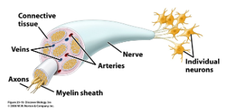
Nervous system to neuronal function pt.2
Input and output is performed through axons called “nerves” in the PNS and “tracts” white matter in the CNS
Bundle of axons arising from neurons in ganglia (clusters of neurons in the PNS) or within the CNS

Sensory neuron
Detect specific stimuli in the environment
Carry that information into the CNS via. Axons
Cell bodies lie in the periphery (PNS)
Detector can be a specialized sensory organ (tongue, nose, eye) or a free nerve ending (skin, internal organs)

Dorsal root ganglia (DRG)
Contains cell bodies of somatosensory neurons (touch, tickle, itch, pain, etc.)
Neurons have a fiber that goes to the periphery and one that provides input to the cord

Motor neurons
Sends motor signals to muscles that regulate their contraction
Spinal motor neurons have cell bodies in ventral cord
Sends axons out the ventral roots to the skeletal muscles

Interneurons
Contained in 97% of the CNS
Cell bodies and axons/dendrites lie entirely within the CNS
Perform analysis of sensory signals and initiate behavioral decision making