(A&P 1) Lab 1: Body Organization and Terminology
 Call Kai
 Learn
Learn Practice Test
 Spaced Repetition
 Match
Match Flashcards
 Knowt Play
1/52

There's no tags or description
Looks like no tags are added yet.
Name | Mastery | Learn | Test | Matching | Spaced | Call with Kai | Chat |
|---|
No analytics yet
Send a link to your students to track their progress
53 Terms
Anatomy
studies the structure of body parts and their relationship to one another
Physiology
how the body parts work (function) and carry out their activities
Homeostasis
Relatively stable internal conditions
Loss of Homeostasis
Loss of homeostasis leads to disease, failure to keep internal environmental balance.
Negative Feedback

Positive Feedback

Anatomical Position
Body is erect
feet slightly apart palms facing forward
thumbs point away from body
Palms facing forward

Anatomical Regions
(Practice Exercise on iPad)
Sagittal Plane
Divides the body in left and right parts
Produces sagittal section (on midline)

Frontal (Coronal) Plane
Divides body vertically into anterior and posterior
Produces frontal/coronal section

Transverse (Horizontal) Plane
Divides body into superior and inferior parts
Produces a cross section

Directional Position
Right and left referring to the body being observed not in terms of you the observer.
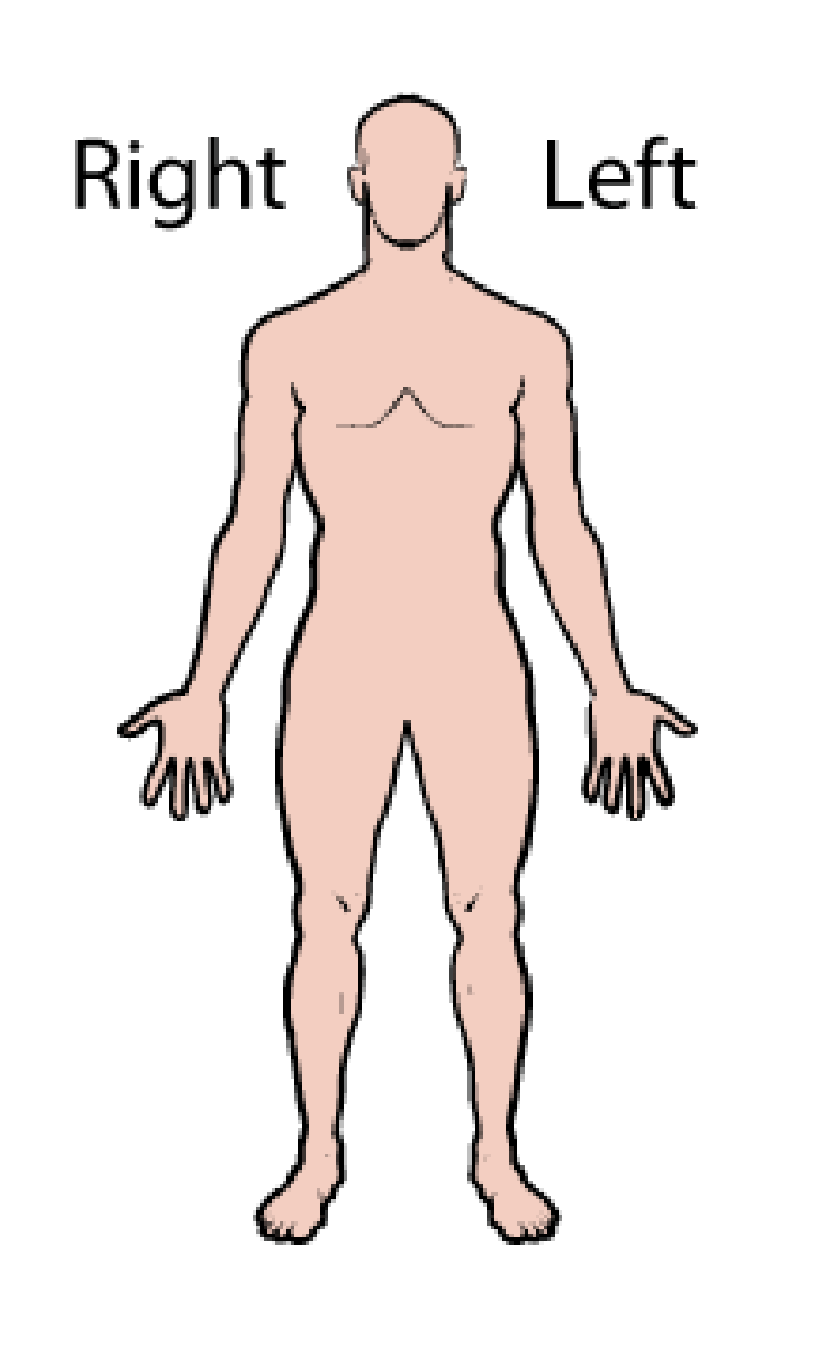
Superior

Inferior (caudal)

Anterior (ventral)

Posterior (dorsal)

Medial

Lateral

Intermediate

Proximal

Distal

Superficial (external)

Deep (internal)

Ipsilateral
On the same side of the body

Contralateral
On opposite sides of the body

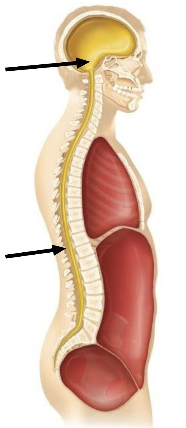
Dorsal Body Cavity (Back)
Has two subdivisions that consists of (1) Cranial Cavity and (2) Vertebral Cavity
(1) Encases the Brain
(2) Encases the Spinal Cord
Ventral Body Cavities
Houses internal organs, two subdivisions divided by the diaphragm
(1) Thoracic cavity
(2) abdominopelvic
Thoracic Cavity contains…
vital organs of respiration and circulation including the
heart
lungs
trachea
esophagus
Abdominal Cavity contains…
Contains major organs for digestion, filtration, and hormone production, including the
Stomach
Pancreas
Gallbladder
Liver
Small intestine
Large intestine
appendix
Kidneys
Adrenal Glands
Spleen
Pelvic Cavity contains…
Urinary bladder
Distal ureters
Proximal urethra
Sigmoid colon
Rectum
Ovaries
Fallopian tubes
Uterus
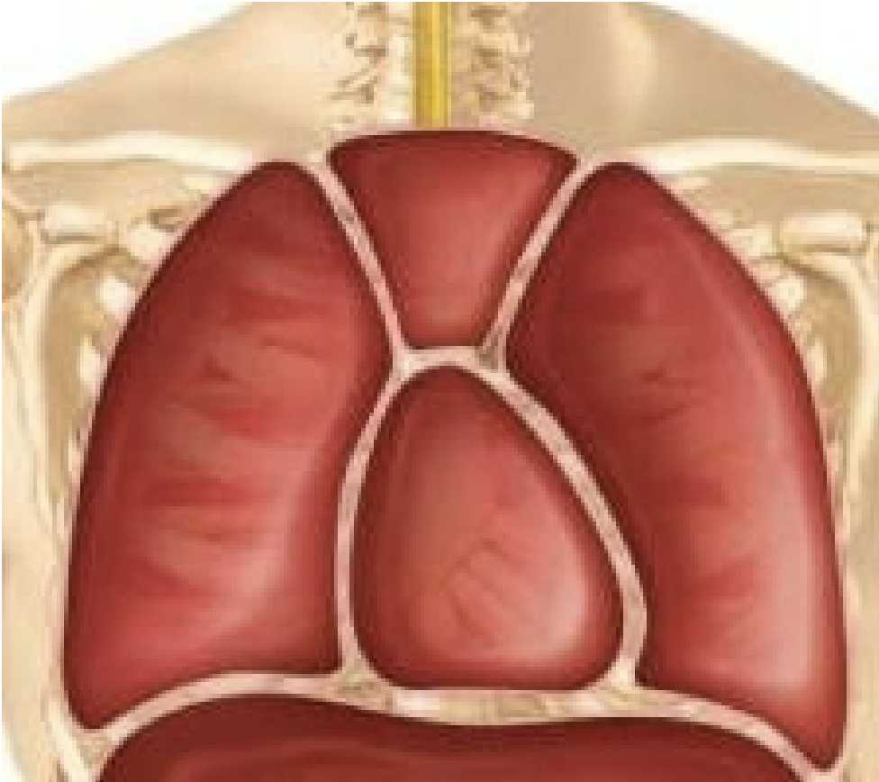
Thoracic Cavity Subdivisions
Pleural Cavities: house each lung
Mediastinum: contains pericardial cavity
Pericardial: encloses the heart
Abdominopelvic Cavity Subdivisions
Abdominal Cavity: stomach, intestines, spleen, and
liver
Pelvic Cavity: urinary bladder, reproductive
organs, and rectum

Parietal
The membrane lining the body cavity

Visceral
Membrance covering the organ
Lung
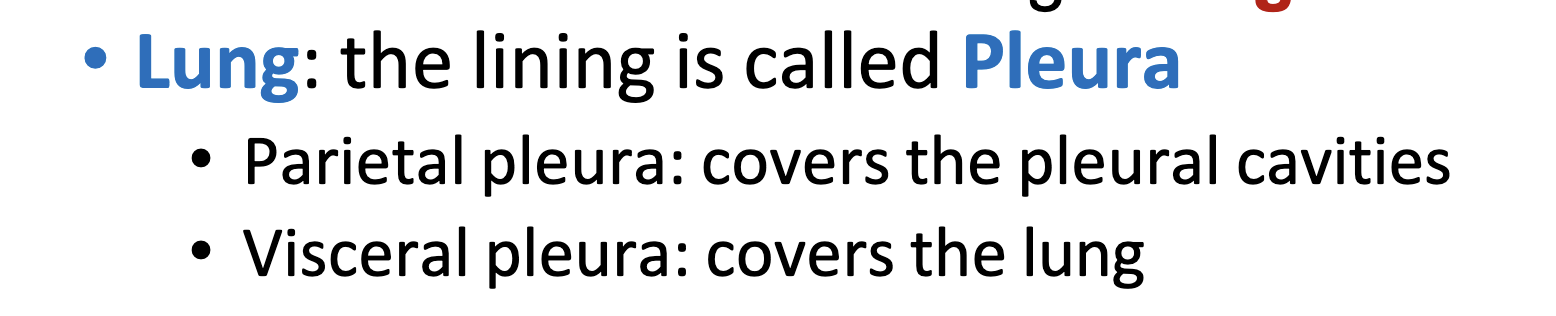
Heart

Abdomen

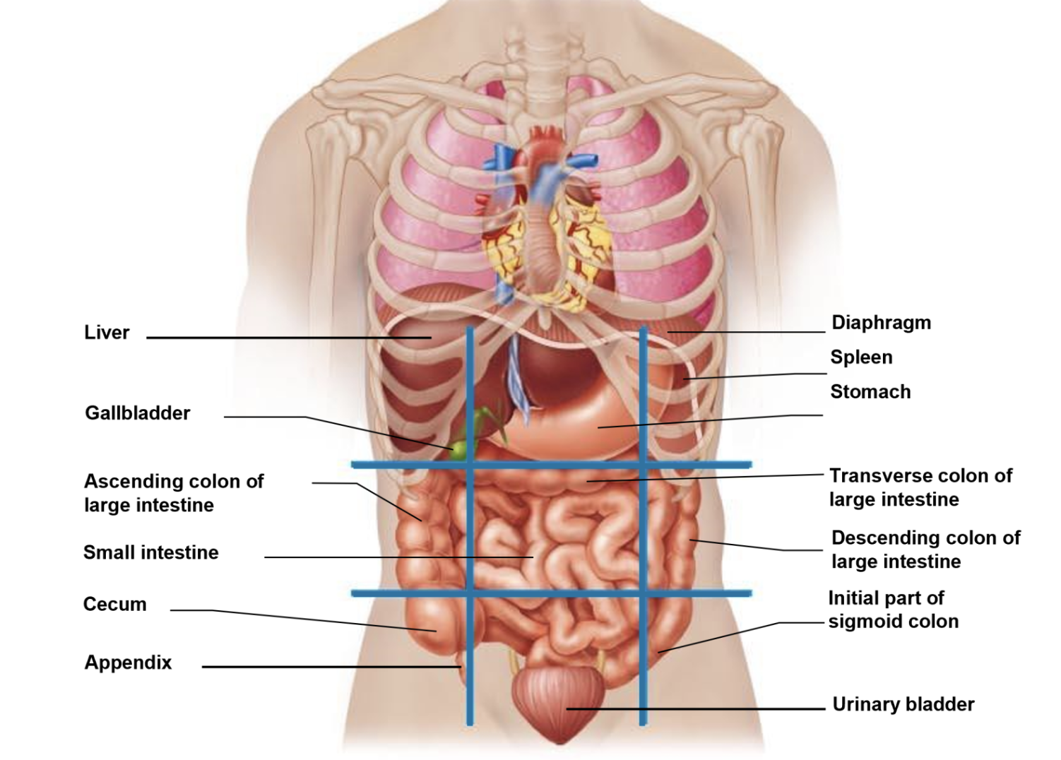
Abdominopelvic Quadrants
Exercise on iPad

Which organs are located in each quadrant?
Right Upper Quadrant (RUQ)
Liver: The large right lobe dominates this space.
Gallbladder: Tucked underneath the liver.
Duodenum: The initial segment of the small intestine.
Right kidney and adrenal gland: Located posteriorly.
Hepatic flexure: The curve where the ascending colon becomes the transverse colon.
Pancreas: Specifically the head of the pancreas.
Left Upper Quadrant (LUQ)
Stomach: Sits mostly in this upper left region.
Spleen: Located posteriorly under the left rib cage.
Left liver lobe: The smaller, left portion of the liver.
Pancreas: Specifically the body and tail of the pancreas.
Left kidney and adrenal gland: Located posteriorly.
Splenic flexure: The curve where the transverse colon becomes the descending colon.
Right Lower Quadrant (RLQ)
Cecum: The pouch at the beginning of the large intestine.
Appendix: Projecting off the cecum.
Ascending colon: The lower segment traveling upward.
Right ovary and fallopian tube: In females.
Right ureter: Channeling urine down to the pelvis.
Right spermatic cord: In males.
Left Lower Quadrant (LLQ)
Descending colon: The lower segment traveling downward.
Sigmoid colon: The S-shaped terminal portion of the colon.
Left ovary and fallopian tube: In females.
Left ureter: Channeling urine down to the pelvis.
Left spermatic cord: In males.
Abdominopelvic Regions
Exercise on iPad

What organs are located in each region?
Right Hypochondriac Region: Located in the upper right section, sheltered by the lower ribs.
Liver: Majority of the right lobe.
Gallbladder: Entirely nestled underneath the liver.
Right Kidney: The upper portion.
Epigastric Region: Located in the upper middle section, superior to the belly button.
Stomach: Majority of the stomach body.
Liver: Portions of both left and right lobes.
Pancreas: Head, body, and tail stretch across this midline space.
Duodenum: The C-loop of the small intestine.
Adrenal glands: Sitting atop both kidneys.
Left Hypochondriac Region: Located in the upper left section, sheltered by the lower ribs.
Spleen: Nestled deep beneath the left rib cage.
Stomach: The upper fundus portion.
Left Kidney: The upper portion.
Pancreas: The tip of the tail.
Right Lumbar Region: Located in the middle right section, adjacent to the lumbar spine.
Ascending colon: Traveling upward along the posterior wall.
Right Kidney: Lower pole and lower ureter.
Small intestine: Portions of the jejunum.
Umbilical Region: Located in the center section, surrounding the navel.
Small intestine: The majority of the jejunum and ileum.
Transverse colon: Stretches across the center.
Abdominal aorta and inferior vena cava: Major core blood vessels.
Left Lumbar Region: Located in the middle left section, adjacent to the lumbar spine.
Descending colon: Traveling downward.
Left Kidney: Lower pole and lower ureter.
Small intestine: Portions of the jejunum.
Right Iliac (Inguinal) Region: Located in the lower right section, near the groin and hip bone.
Cecum: The pouch where the large intestine begins.
Appendix: Blind pouch extending off the cecum.
Right Ovary and Fallopian Tube: In females.
Hypogastric (Pubic) Region: Located in the lower middle section, inferior to the belly button.
Urinary bladder: Rises into this zone when filled.
Uterus: In females, resting posterior to the bladder.
Sigmoid colon and Rectum: Terminal segments of the large intestine.
Prostate: In males, sitting beneath the bladder.
Left Iliac (Inguinal) Region: Located in the lower left section, near the groin and hip bone.
Descending/Sigmoid junction: Transition point of the large intestine.
Left Ovary and Fallopian Tube: In females.
Integumentary System (Skin)
Forms external body covering, protects from injury. Synthesis of Vitamin D, has cutaneous receptors and sweat and oil glands
Skeletal System (Bones)
Protects and supports body organs. Framework for muscles. Blood cell formation. Mineral storage
Muscular system
Allows manipulation of the environment, locomotion and facial expression. Maintains posture and
produces heat
Nervous System
Control system of the body, responds to internal and external changes
Endocrine System
Glands secrete hormones that regulate body processes
Cardiovascular System
Blood vessels transport blood, which carries O2, CO2, nutrients, waste, etc. The heart pumps blood
Lymphatic System (Immunity)
Picks up fluid leaked from blood vessels and returns it to blood. Houses WBCs involved in immunity
Respiratory System
Keeps blood supplied with O2 and removes CO2
Digestive System
Breaks down food, absorbs nutrients and eliminates feces
Urinary System
Eliminates nitrogenous wastes from the body. Regulates H20, electrolyte and acid-base balance of the blood
Male+Female Reproductive System
Production of offspring
PRACTICE LABELING EXERCISES ON IPAD NOW
:3