Unit 4: Sea Urchins early dev
1/41
There's no tags or description
Looks like no tags are added yet.
Name | Mastery | Learn | Test | Matching | Spaced | Call with Kai |
|---|
No analytics yet
Send a link to your students to track their progress
42 Terms
Define cleavage and what does cleavage result in?
Cleavage is defined as a series of mitotic divisions, by which the cytoplasm divides into many smaller, nuclear cells (blastomeres) and leads to the formation of a blastula.
What controls cleavage?
Cleavage is controlled by protein and mRNA stored within the oocyte.
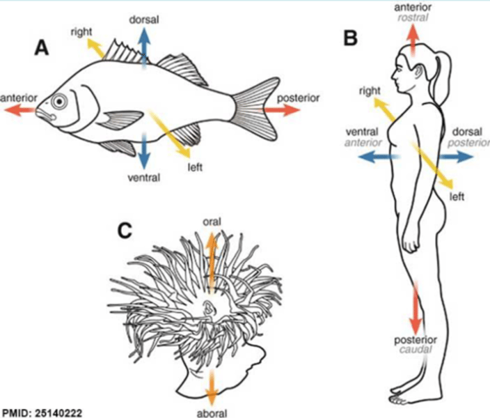
How are the axes defined?
Anterior-Posterior
Dorsal-Ventral
Left-Right
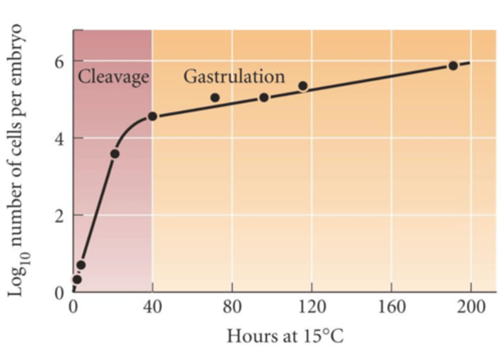
During which stage does the number of cell increase drastically and when does it slow down? (vertebrate)
During the cleavage stage the number increases exponentially and during gastrulation the increase slows down.
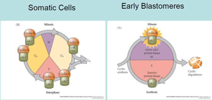
How does the cell cycle in early blastomeres differ from that of somatic cells?
- Normal cells: have the M, G1, G0, S and G2 phases.
- Early blastomeres: only have M and S phases. There is a constant shift between them and is cyclin B mediated. Cyclin B is synthesized by oocyte mRNA.
Which cyclic processes is cleavage based on?
- Karyokinesis
- Cytokinesis
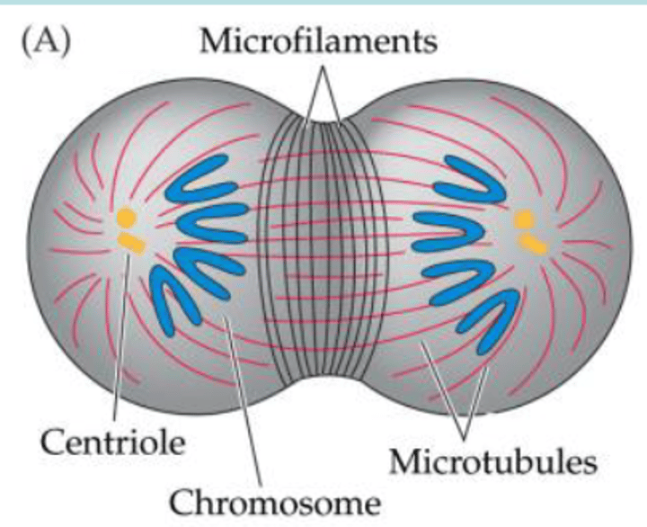
What is the role of MT and microfilaments in cell division?
- MT draw chromosomes to the centrioles.
- Cytokinesis happens by the cytoplasm being pinched off by contraction of microfilaments.
Which parameters determine cleavage patterning?
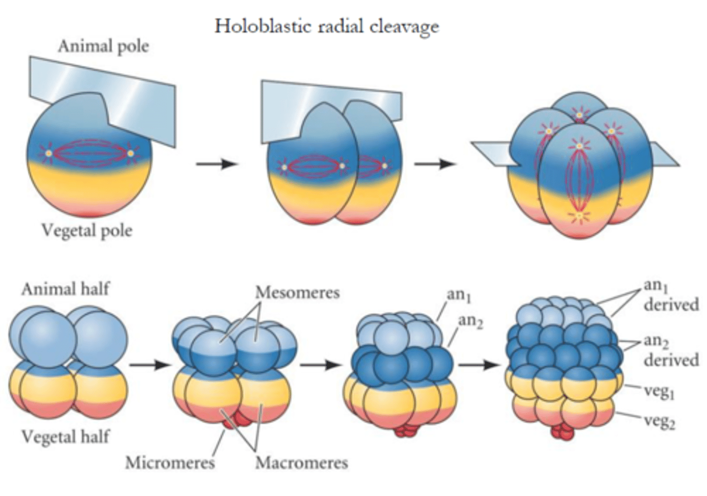
1) The amount and distribution of the yolk in the cytoplasm. (yolk rich pole: vegetal pole, opposite pole with less yolk: animal pole). In the zygote, the nucleus is frequently displaced towards animal pole.
2) Factors within the egg cytoplasm that influence the angle of the mitotic spindle and the timing of its formation.
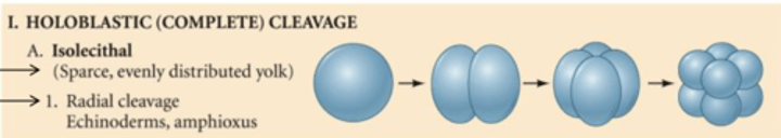
Which cleavage pattern is found in sea urchins?
Sea urchins have a holoblastic cleavage pattern which is isolecithal (sparse, evenly distributed yolk) and radial (cleavage plane perpendicular to each other).
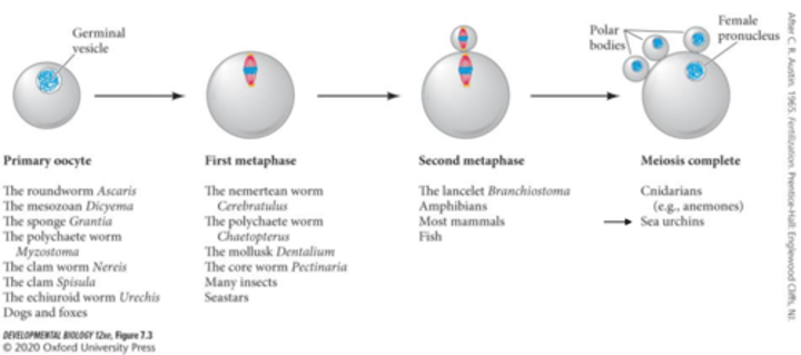
When is the sea urchin oocyte ready for fertilization?
After complete meiosis.
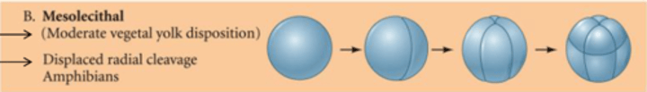
In which organisms is mesolecithal (moderate vegetal yolk distribution) yolk patterning found?
In amphibians. The moderate vegetal yolk disposition leads to displaced radial cleavage.
Define gastrulation
Coordinated cell movements that reorganizes the blastula, leading to the formation of 3 germ layers.
Which coordinated cell movements are observed during gastrulation?
1) Invagination
2) Involution
3) Ingression
4) Delamination
5) Epiboly
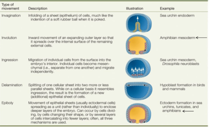
Invagination
Infolding of a region of cells into the embryo
Involution
Inturning or inward movement of an expanding outer layer, so that it spreads over the internal surface.
Ingression
Migration of individual cells from the surface layer into the interior of the embryo. The cells become mesenchymal and migrate independently
Delamination
The splitting of one cellular sheet into two more or less parallel sheets
Epiboly
Movement of epithelial sheets that spread as a unit to enclose the deeper layers of the embryo.
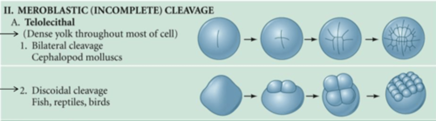
Which organisms have the meroblastic (incomplete) cleavage pattern and what characterizes this cleavage pattern?
Molluscs (bilateral) and fish, birds and reptiles (discoidal). Meroblastic cleavage is characterized by a telolecithal yolk distribution (dense yolk throughout most of the cell).
What happens to the synchronicity of the cleavage in sea urchins?
Cleavage ceases to be synchronous after the blastomere reaches a certain size.
Morula
Sea urchin embryo between the 32- and 60-cell stage. Ball of blastomeres.
Blastula
The hollow ball of cells marking the end stage of cleavage during early embryonic development. (>60 cells)
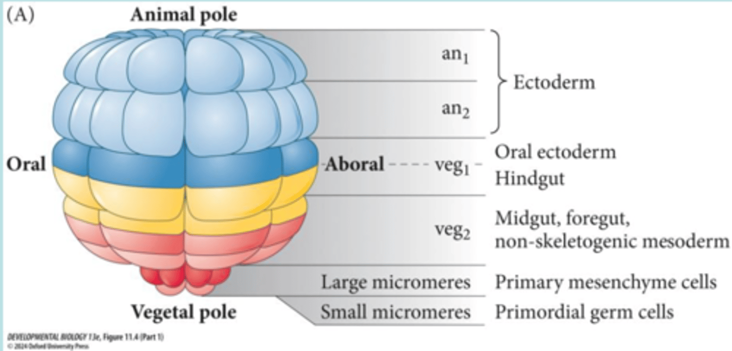
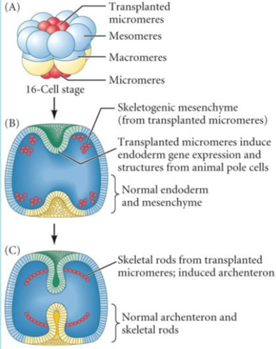
What happens to blastomeres and skeletogenic micromeres in terms of cell fate specification at the 60-cell stage?
Most blastomeres are conditionally specified, only skeletogenic micromeres are autonomously specified.
Autonomous specification
A mode of cell commitment in which the blastomere inherits a determinant, usually a set of transcription factors from the egg cytoplasm, and these transcription factors regulate gene expression to direct the cell into a particular path of development. It develops independent from its environment.
Conditional specification
The ability of cells to achieve their respective fates by interactions with other cells. What a cell becomes is in large measure specified by paracrine factors secreted by its neighbors.
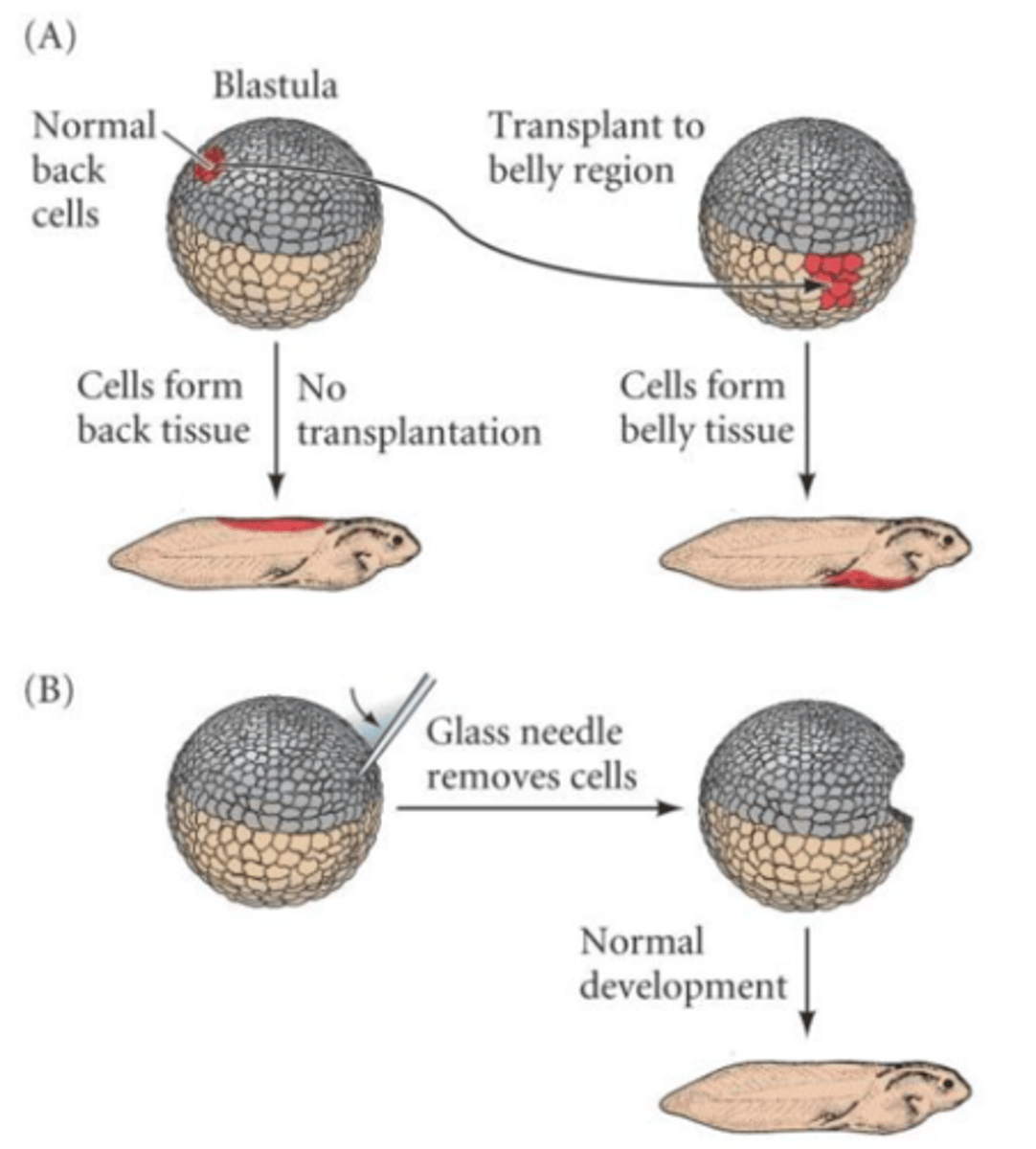
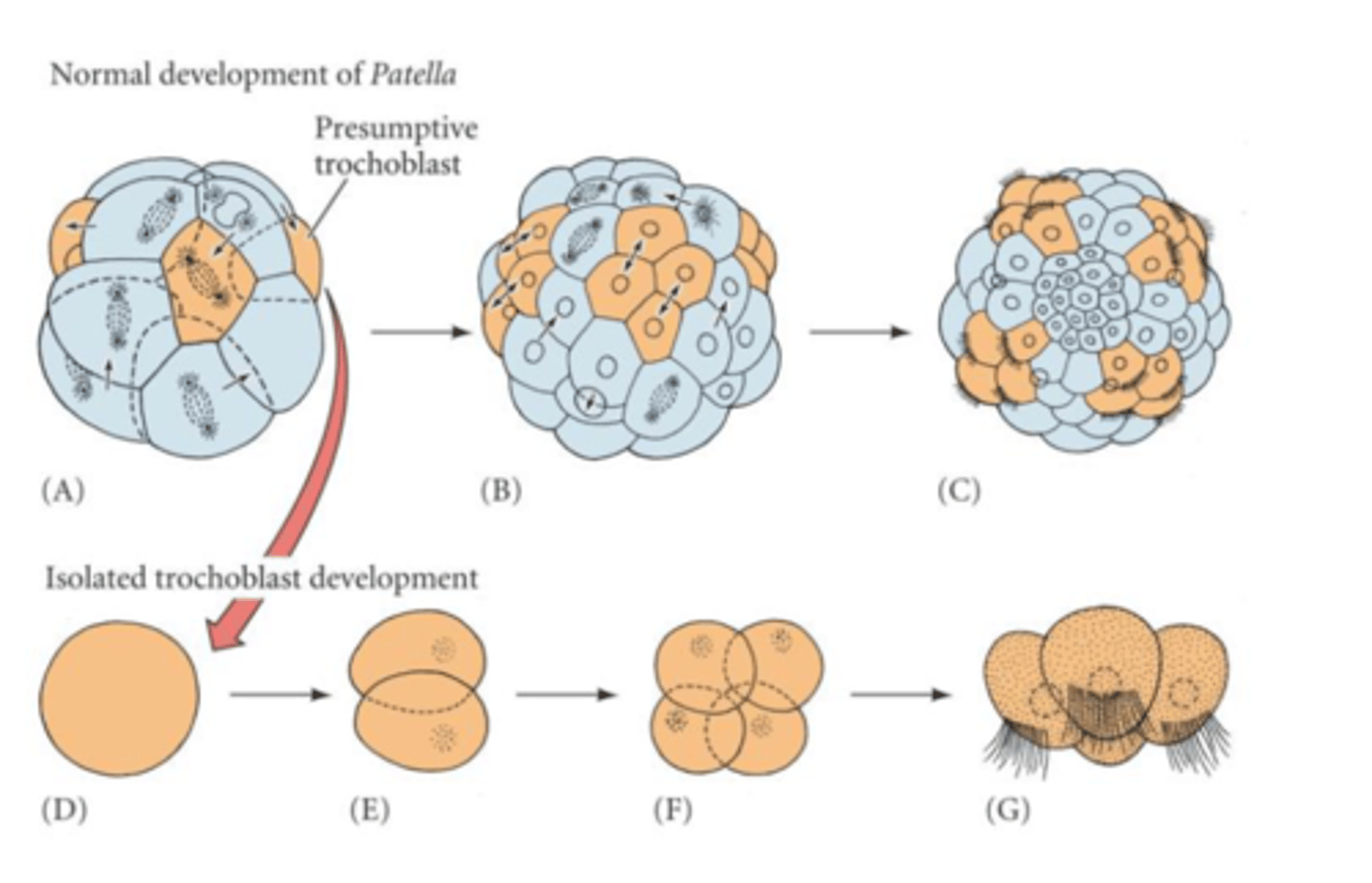
How can specification be revealed experimentally?
Micromeres can induce a secondary axis, as they are autonomously specified.
How does the specification of micromeres work?
- The initial regulatory input are the transcription factors
Disheveled and b catenin which specify micromeres.
- During oogenesis, Disheveled becomes localized to the
vegetal cortex, where it prevents degradation of b catenin.
- b-catenin accumulates in the nucleus of cells determined to
form endoderm or mesoderm.
- Nuclear accumulation is autonomous and specifies the
vegetal part of the embryo.
Which pathway is active in specifying the vegetal part of the sea urchin embryo?
The canonical Wnt pathway.
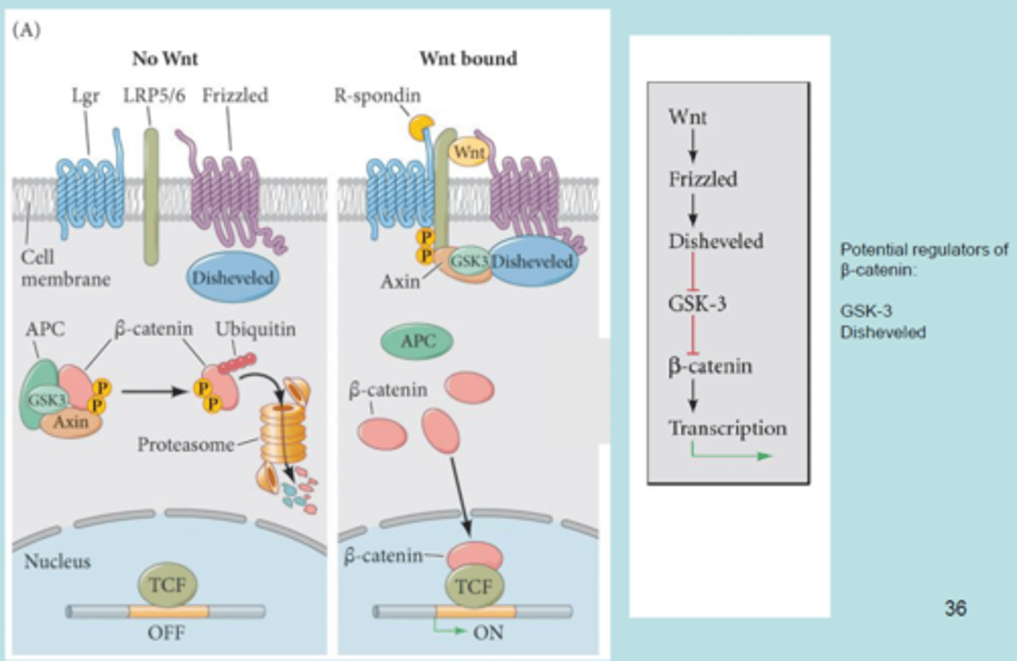
What are the regulators of b-catenin?
GSK-3 and Disheveled.
What is the role of b-catenin in specifying the vegetal cells of the sea urchin embryo?
The role of β-catenin in specifying vegetal cells of the sea urchin embryo involves its activation on the future vegetal side, promoting the expression of specific genes that contribute to endoderm development. This activation is crucial for establishing axial patterning and cell fate determination during early embryogenesis.
What happens if b-catenin is prevented from entering the nuclei?
The vegetal cell fates are not specified and the entire embryo develops into a ciliated ectodermal ball.
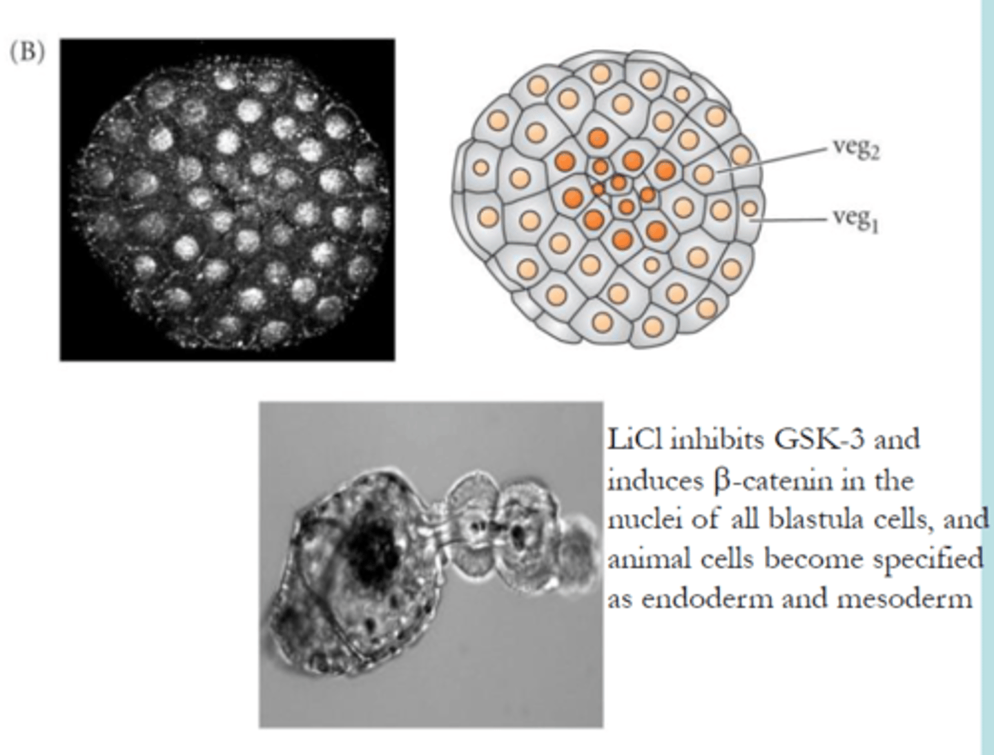
What happens to b-catenin during early cleavage?
It is initially expressed at similar lvls by all blastomeres, but at later stages the lvls decline drastically in animal blastomeres.
The loss of b-catenin in animal blastomeres is dependent on GSK3b, which phosphorylates b-catenin and targets the proteins for degradation.
What happens to GSK3 during early cleavage?
GSK3 is uniformly stable along the animal-vegetal axis.
Where is disheveled localized?
In the vegetal part of the embryo, which is where it has been pre-fertilization and where it remains.
Why is disheveled important?
Disheveled function is needed for endomesoderm specification and for the accumulation of b-catenin in the nuclei of vegetal blastomeres.
Which steps occur in gastrulation of sea urchins? (first 3)
1) First, primary skeletogenic mesenchyme cells ingress by
EMT and localize by use of filopodia.
Then, the primitive gut or "archenteron” forms in three phases
First, second and third stages of invagination
2) The vegetal plate initiates primary invagination into the
blastocoel to form the beginnings of the endodermal
archenteron about one fourth to one half of the way into
the blastocoel
3) Involution (and to some extent epiboly ) of cells over the lips of the forming blastopore (=> anus), changes in cell shape ,
growth and adhesion
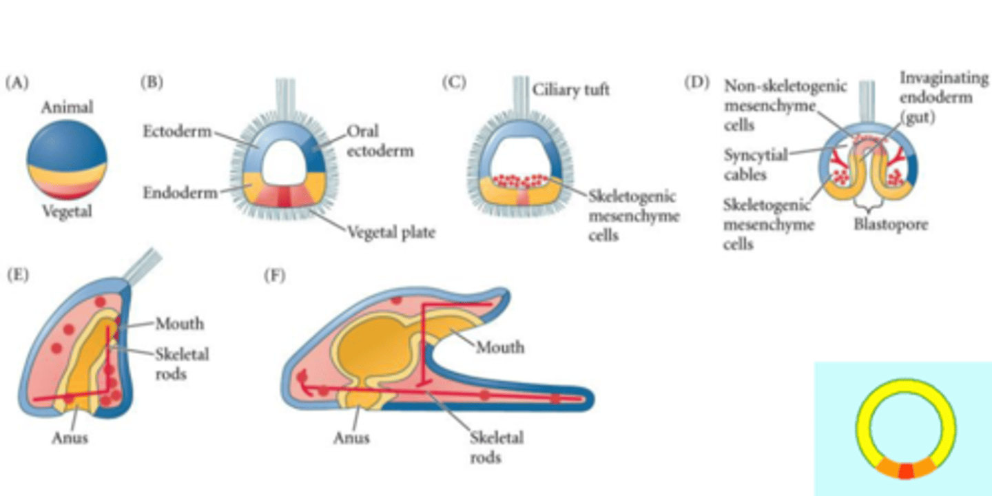
Which steps occur in gastrulation of sea urchins? (last 3)
4) The second phase of invagination of archenteron
includes rapid convergent extension towards the animal
pole , which has thickened to form the ” animal plate ”
with particularly long cilia (apical tuft).
5) The third phase of archenteron elongation is initiated by
non skeletogenic mesenchyme cells , which detach from
the tip of archenteron and guide archenteron towards a
patch of ectoderm called the target region.
6) Archenteron and oral ectoderm rupture in the contact
area and form the mouth

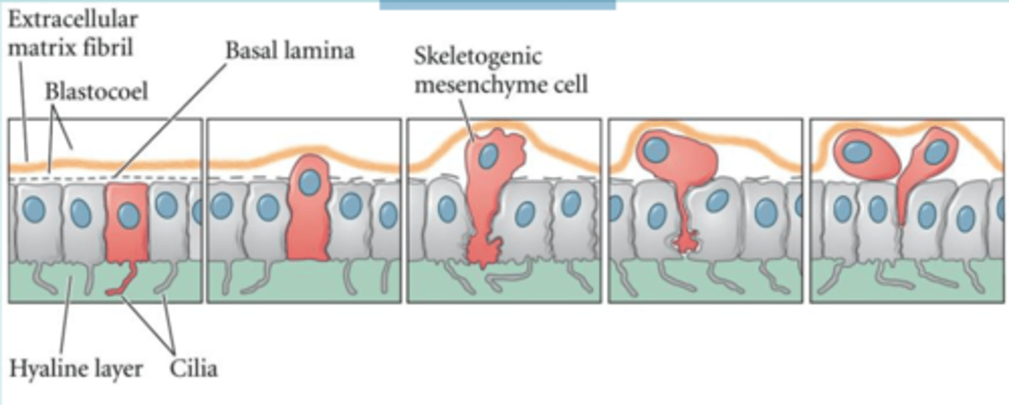
How does ingression of skeletogenic mesenchymal cells occur?
Initially all cells in the blastula are in contact with the hyalin on the outside, basal lamina on the inside and other cells laterally. Micromeres are firmly attached to each other and to hyalin, but less to the basal lamina.
During gastrulation the skeletogenic mesenchyme exhibits the opposite pattern.
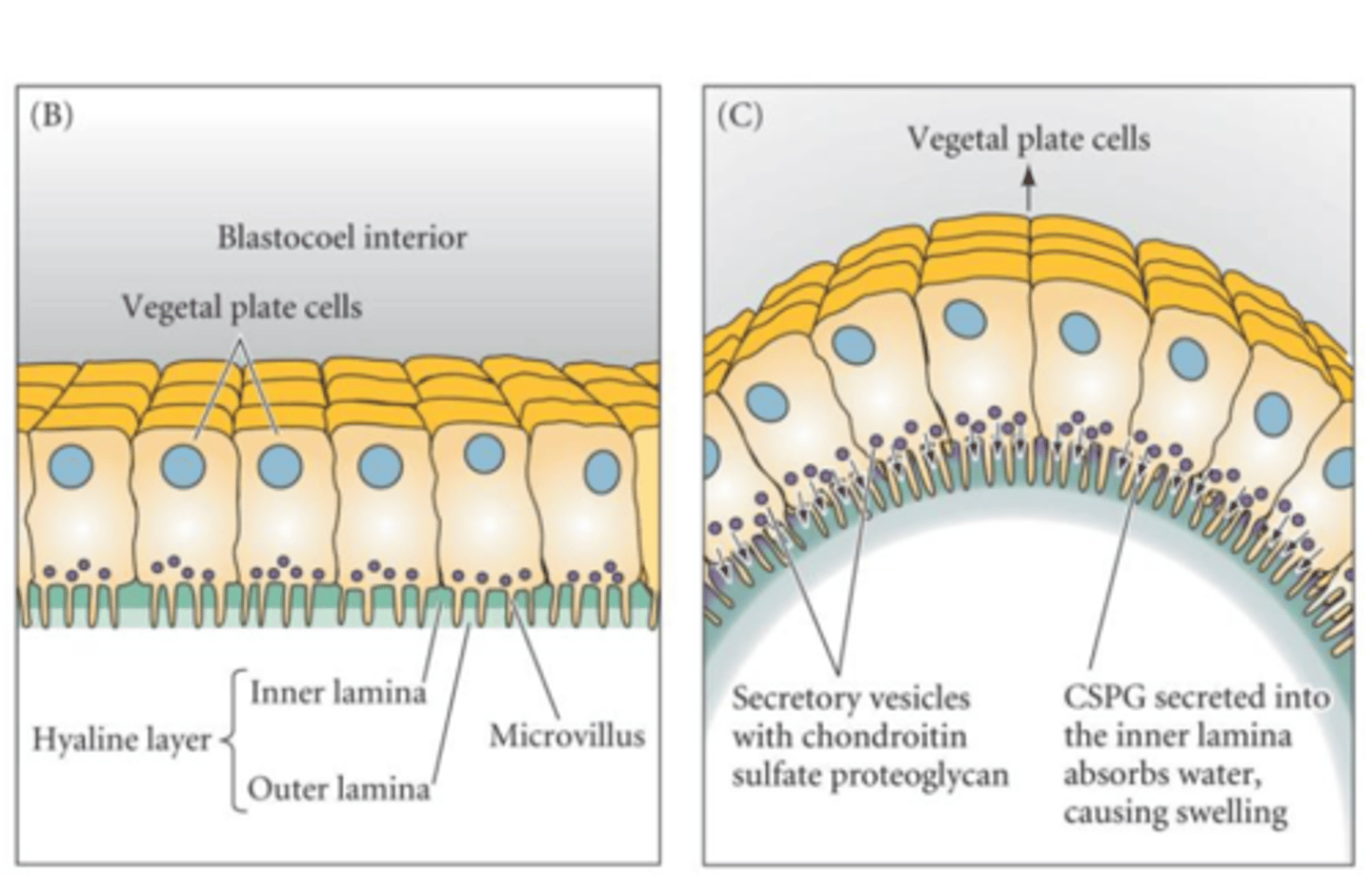
How does invagination of the skeletal plate occur?
The negatively charged proteoglycans in the vegetal cells are released into the hyalin layer, allowing it to swell and letting the layer fold inwards
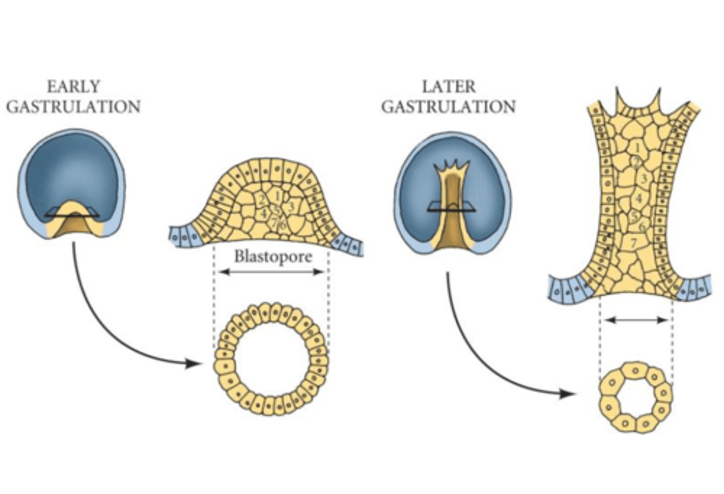
Convergent extension (archenteron)
A process in which the cells of a tissue layer rearrange themselves, so that the sheet of cells becomes narrower (converges) and longer (extends).
What are advantages of using sea urchins as a model organism?
• Easy to propagate, easy to synchronize, relatively rapid
embryogenesis, easy to manipulate gene expression
• Transparent embryo with simple structure
• Genomic resource available
• The sea urchin genome contains orthologs of human disease associated genes (neuro-degenerative disorders, tumorigenesis etc.)
• In vivo model for normal developmental processes relying on, for instance, Wnt signal transduction pathways and EMT, and for human diseases related to dysregulation of these processes
• Few if any ethical restrictions
What are the drawbacks of using sea urchins as model organisms?
• Not a vertebrate
• No inbred strains
• No immortalized cell lines available.