Finger and Hand Anatomy and Positioning
 Call Kai
 Learn
Learn Practice Test
 Spaced Repetition
 Match
Match Flashcards
 Knowt Play
1/77

There's no tags or description
Looks like no tags are added yet.
Name | Mastery | Learn | Test | Matching | Spaced | Call with Kai | Chat |
|---|
No analytics yet
Send a link to your students to track their progress
78 Terms
Digits
Each finger and thumb are known as what?

phalanges
Bones of the fingers and toes are known as what?

Phalanx
A singular bone of the finger or toe is known as what?
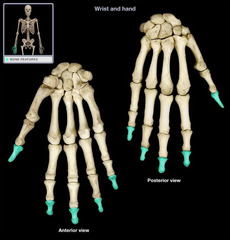
14
How many bones make up the phalanges?
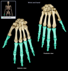
5
How many bones make up the metacarpals?
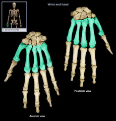
8
How many bones make up the carpals?

27
How many bones are in each hand? (add all bones together!)
3 (proximal, middle, distal)
How many phalanges make up each of the 2nd-5th digits?

Thumb (pollex)
Each digit is assigned a number. In anatomical digit numbering, which digit is designated as the 1st?

Pollex
Special name for the thumb?
Pinky
Each digit is assigned a number. In anatomical digit numbering, which digit is designated as the 5th?
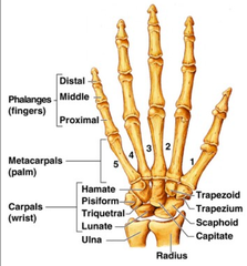
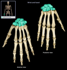
Carpals
wrist bones (8 total)
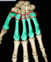
carpometacarpal joint (CMC)
Articulations between carpals and metacarpals
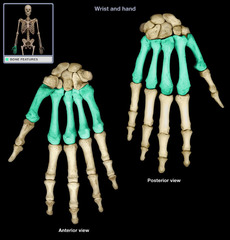
Metacarpals
the five bones that form the palms of the hand
Base of metacarpal
Expanded proximal end that articulates with carpals
body/shaft of metacarpal
Long curved portion
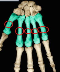
head of metacarpal
Distal round portion
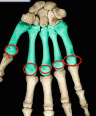
Metacarpophalangeal joint (MCP)
Connects metacarpals to the proximal phalanges
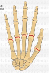
Base of phalanx
Located on the proximal end; articulates with the distal heads of the metacarpals (AKA Metacarpophalangeal joint). (proximal end)
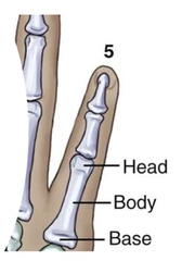
Body/shaft of phalanx
Middle part of the phalanx
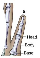
head of phalanx
distal end
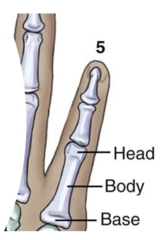
Proximal phalanges
the phalanges closest to the metacarpals

Proximal interphalangeal joint (PIP)
between proximal and middle phalanges
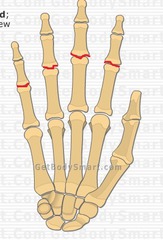
Middle phalanges
articulate with the proximal and distal phalanges
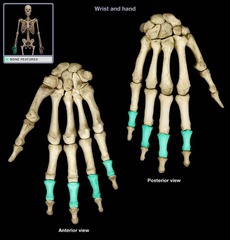
distal interphalangeal joint (DIP)
joint between middle and distal phalanges

Distal phalanges
the phalanges furthest from the wrist

Interphalangeal joint (IP)
Located between the proximal and distal phalanges
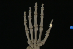
2 (proximal and distal)
How many phalanges make up the thumb?
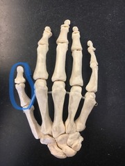
phalanges
A?
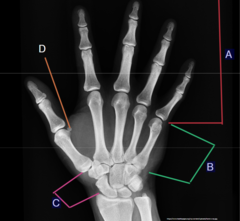
Metacarpals
B?
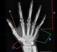
Carpals
C?
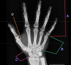
sesamoid bones
D?

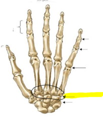
1st carpometacarpal joint (CMC)
A?
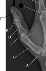
Body/shaft of phalanx (1st metacarpal)
B?
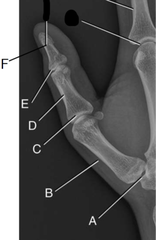
1st Metacarpophalangeal joint (MCP)
C?
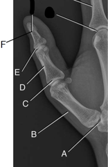
Body/shaft of phalanx (proximal phalanx)
D?
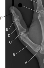
Interphalangeal joint (IP)
E?
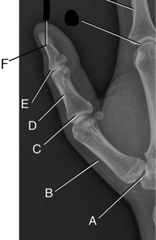
Distal phalanx
F?
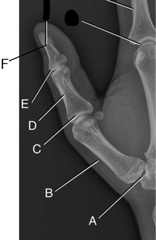
2nd Metacarpophalangeal joint (MCP)
G?
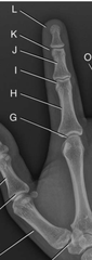
Body/shaft of phalanx (proximal phalanx)
H?

2nd Proximal interphalangeal joint (PIP)
I?
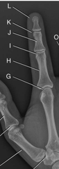
Body/shaft of phalanx (Middle Phalanx)
J?
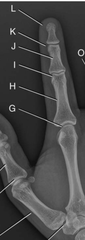
2nd Distal interphalangeal joint (DIP)
K?

head of 2nd pharynx (distal Phalanx)
L?

Head of Phalanx
A?

Body/shaft of Phalanx
B?
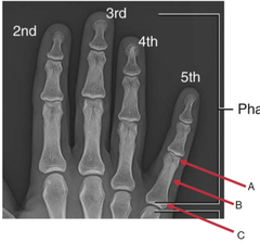
Base of Phalanx
C?
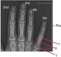
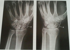
Scaphoid Fat Stripe
Visualized on PA and oblique projections, Elongated and slightly convex in shape, Located between the radial collateral ligament and adjoining muscle tendons lateral to scaphoid
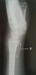
Pronator Fat Stripe
Visualized on lateral view, Located ¼ of an inch anterior to the radius
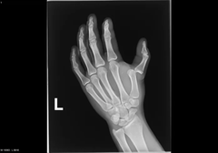
dislocation
displacement of a bone from its joint
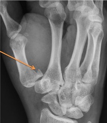
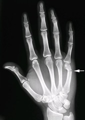
Bennett fracture (fx)
fracture through base of the first metacarpal bone, extending into the CMC joint with subluxation w/ posterior displacement
Boxer fracture (fx)
Transverse fracture that extends through the metacarpal neck (commonly seen on 5th metacarpal)
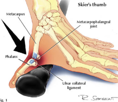
Skiers thumb
Sprain or tear of the ulnar collateral ligament (occurs from falling on outstretched arm and hand, thumb bends back toward hand)
Osteoarthritis
degenerative joint disease

rheumatoid arthritis
Chronic systemic disease with inflammatory changes throughout connective tissues
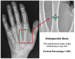
Osteoporosis
A condition in which the body's bones become weak and break easily.
-kVp: low to medium (60-80, typically 55-65 for fingers)
-Grid: No (unless bigger than 10cm or 4in)
-SID: 40in
what are the technical factors needed for a good hand x-ray (kVp, grid, SID)?
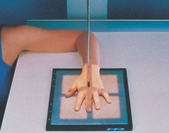
long hand=long board (hand perpendicular with IR)
What's the positioning rule for IR/hand positioning?
Correctly exposed images of the upper limbs should reveal soft tissue margins for fat pad visualization and fine trabecular markings of all bones being radiographed (good contrast)
What makes a good hand X-ray?
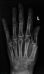
Yes, it won't take much technique to get through the bone
When X-raying geriatric patients with osteoporosis, should you lower your technical factors?
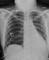
radiopaque objects, clothing removed placed in gown , long hair, oxygen tubing, and pacemaker leads to avoid reduced detail/scatter
During patient preparation, what must be removed?
1. Limit the number of exposures
2. Collimation
3. shielding (led apron)
4. correct positioning
What 4 things help protect you and the patient against radiation?
BEFORE the examination
When must the anatomical marker be placed?
PA
PA oblique
Lateral "FAN"
What are the routine projections of the hand?

What is the evaluation criteria and procedure for a PA Projection of the Hand X-ray?
SID: 40inches
Grid: No
kVp: 60-80
CR: center to 3rd metacarpal joint
Patient position: Patient seated sideways at end of table,
Height of table near shoulder height, palmar surface on IR, spread fingers slightly
Evaluation criteria: Entire hand and carpals demonstrated, Center of field at third MCP joint, No rotation of phalanges or metacarpals, Exposure factors

What is the evaluation criteria and procedure for a PA Oblique projection
X-ray?
SID: 40inches
Grid: No
kVp: 60-80
CR: center to 3rd metacarpal joint
Patient position: Patient seated sideways at end of table,
Height of table near shoulder height, Rotate entire hand and wrist laterally 45˚ with digits parallel to IR, spread fingers slightly
Evaluation criteria: Entire hand and carpals demonstrated, Center of field at third MCP joint, Joints open, Adequate separation of phalanges and metacarpals, Exposure factors
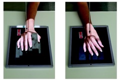
What is the evaluation criteria and procedure for a "Fan" Lateral Projection
X-ray?
SID: 40inches
Grid: No
kVp: 60-80
CR: center to 2nd metacarpal joint
Patient position: Patient seated sideways at end of table,
Height of table near shoulder height, Rotate hand and wrist into lateral position with thumb side up, Spread fingers and thumb into a "fan" position
Evaluation criteria: Entire hand and carpals demonstrated, Center of field at second MCP joint, Fingers equally separated Exposure factor
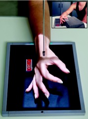
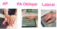
AP
PA Oblique
Lateral
What are the routine projections of the 1st digit?
the 1st metacarpal and trapezium (carpal articulating with 1st metacarpal)
During a thumb x-ray, what must you include?
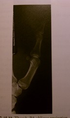
What is the evaluation criteria and procedure for a AP projection of the 1st digit?
SID: 40inches
Grid: No
kVp: 55-65
CR: center to 1st metacarpal joint
Patient position: Patient seated sideways at end of table,
Height of table near shoulder height, Rotate arm internally to supinate thumb
Evaluation criteria: Entire thumb demonstrated (including first CMC joint), Center field at first MCP joint, No rotation of phalanges, Exposure factors
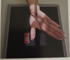
PA thumb
If the patient can't rotate their arm/hand for an AP projection of the thumb, what projection must be done?
What is the evaluation criteria and procedure for a PA projection of the 1st digit?
SID: 40inches
Grid: No
kVp: 55-65
CR: center to 1st metacarpal joint
Patient position: Place hand in pronation position, Rest thumb on sponge support block that is high enough to keep the thumb from rotating
-PA is not advisable b/c of loss of definition caused by increased OID
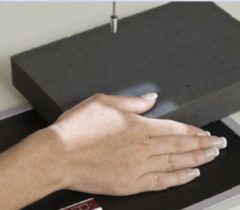
What is the evaluation criteria and procedure for a PA Oblique projection 1st digit X-ray?
SID: 40inches
Grid: No
kVp: 55-65
CR: center to 1st metacarpal joint
Patient position: Patient seated sideways at end of table,
Height of table near shoulder height, Abduct thumb slightly with palmar surface of hand in contact with IR to naturally places thumb in 45˚ oblique
Evaluation criteria: Entire thumb demonstrated, Joints partially open as in 45° oblique, Center of field at first MCP joint ,Exposure factors
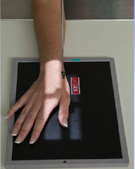
What is the evaluation criteria and procedure for a Lateral Projection of the 1st Digit?
SID: 40inches
Grid: No
kVp: 55-65
CR: center to 1st metacarpal joint
Patient position: Patient seated sideways at end of table,
Height of table near shoulder height, Start with hand pronated and thumb abducted, Fingers slightly arched
Evaluation criteria: Entire thumb demonstrated, Center of field at first MCP joint, No rotation from lateral evident, Exposure factors
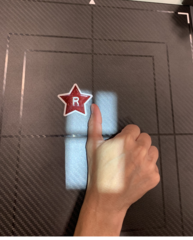
PA
PA Oblique
Lateral
Routine projections for digits 2-5?
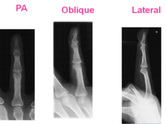
What is the evaluation criteria and procedure for a PA Projection digits 2-5
X-ray?
SID: 40inches
Grid: No
kVp: 55-65
CR: center to PIP joint
Patient position: Patient seated sideways at end of table,
Height of table near shoulder height, Pronate hand with fingers extended
Evaluation criteria: Entire finger and minimum ⅓ of MCP demonstrated, Center field at PIP joint, No rotation of phalanges, Exposure factors
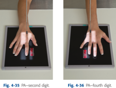
What is the evaluation criteria and procedure for a PA Oblique Projection digits 2-5 X-ray?
SID: 40inches
Grid: No
kVp: 55-65
CR: center to PIP Joint
Patient position: Patient seated sideways at end of table,
Height of table near shoulder height, rotate hand laterally 45° for digits 3-5, rotate medially 45° for 2nd digit
Evaluation criteria: Entire phalanx and MCP joint demonstrated, IP and MCP joints open, Center field at PIP, Exposure factors
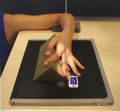
What is the evaluation criteria and procedure for a Lateral Projection digits 2-5 X-ray?
SID: 40inches
Grid: No
kVp: 55-65
CR: center to PIP Joint
Patient position: Patient seated sideways at end of table,
Height of table near shoulder height,
Lateromedial: 3-5th digits Place hand in lateral position (thumb side up) with finger to be examined fully extended then Flex unaffected fingers Finger is parallel to IR
Mediolateral: 2nd digit Rotate hand medially till thumb side is down and second digit is touching IR then Flex unaffected fingers
Evaluation criteria: Entire phalanx and MCP joint demonstrated, Center field at PIP, True lateral position, Digit parallel to IR, Exposure factors
