Unit 2 - Cells and Immunity
5.0(1)
Studied by 23 peopleCard Sorting
1/130
Last updated 9:11 AM on 3/31/25
Name | Mastery | Learn | Test | Matching | Spaced | Call with Kai |
|---|
No analytics yet
Send a link to your students to track their progress
131 Terms
1
New cards
\[3.2.1.1\] What is a **EUKARYOTIC** cell?
Cells which have a **membrane-bound nucleus** and **membrane-bound organelles**.
2
New cards
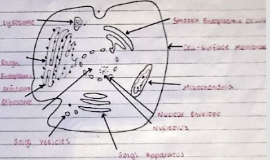
\[3.2.1.1\] What does a typical **animal** cell look like?
3
New cards
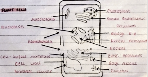
\[3.2.1.1\] What does a typical **plant** cell look like?
4
New cards
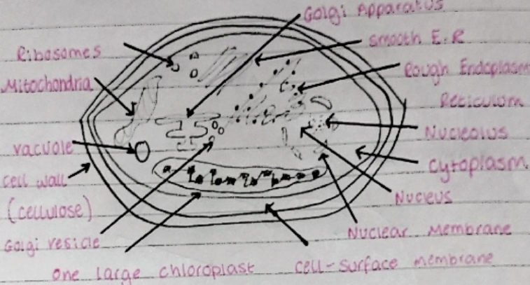
\[3.2.1.1\] What does a typical **algal** cell look like?
Algal cells can be **unicellular** or **multicellular**.
5
New cards
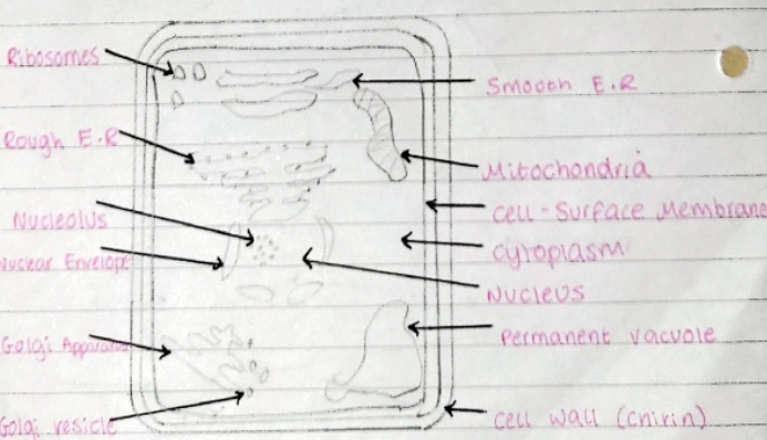
\[3.2.1.1\] What does a typical **fungal** cell look like?
6
New cards
\[3.2.1.1\] What is the **CELL-SURFACE MEMBRANE**?
A membrane found on the surface of animal cells and below the cell wall of other cells.
It has a ***fluid-mosaic*** structure.
* Contains a **phospholipid bilayer**.
It has a ***fluid-mosaic*** structure.
* Contains a **phospholipid bilayer**.
7
New cards
\[3.2.1.1\] What is the **NUCLEUS**?
* The Nucleus is a membrane bound structure that contains the cell’s hereditary information and controls the cell’s growth and reproduction.
\
* The nuclear envelope is a double membrane that separates the nucleus from the cytoplasm. The nuclear envelope is perforated with nuclear pores.
\
* When a cell is not dividing, the chromosomes are organised into long entangled structures called chromatin and not into individual chromosomes.
\
* Nucleoplasm is the gelatinous substance within the nuclear envelope.
\
* The nucleolus is not surrounded by a membrane.
\
* The nuclear envelope is a double membrane that separates the nucleus from the cytoplasm. The nuclear envelope is perforated with nuclear pores.
\
* When a cell is not dividing, the chromosomes are organised into long entangled structures called chromatin and not into individual chromosomes.
\
* Nucleoplasm is the gelatinous substance within the nuclear envelope.
\
* The nucleolus is not surrounded by a membrane.
8
New cards
[3.2.1.1] What is the function of the nucleus?
* It is responsible for protein synthesis, cell division, growth and differentiation.
\
* It is a site for the transcription process in which mRNA is produced for protein synthesis.
\
* Regulates the integrity of genes and gene expression.
\
* It is a site for the transcription process in which mRNA is produced for protein synthesis.
\
* Regulates the integrity of genes and gene expression.
9
New cards
\[3.2.1.1\] What are **LYSOSOMES**?
* Round organelles surrounded by a membrane, with no clear internal structure.
* Contain hydrolytic enzymes called lysozymes.
→ Used to digest invading cells or break down components of the cell.
* Contain hydrolytic enzymes called lysozymes.
→ Used to digest invading cells or break down components of the cell.
10
New cards
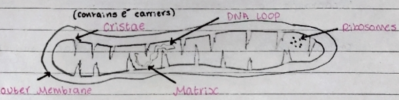
\[3.2.1.1\] What is the **MITOCHONDRIA**?
* The site of **aerobic** **respiration** where **ATP** is produced.
\
* Mitochondria have a double membrane - the inner one is highly folded into **cristae** which contain **electron carrier** proteins.
\
* Inside is the **matrix**, which contains **enzymes** involved in **respiration**.
\
* Contains its own **DNA** and **ribosomes** in order to duplicate in cell division.
\
* Mitochondria have a double membrane - the inner one is highly folded into **cristae** which contain **electron carrier** proteins.
\
* Inside is the **matrix**, which contains **enzymes** involved in **respiration**.
\
* Contains its own **DNA** and **ribosomes** in order to duplicate in cell division.
11
New cards
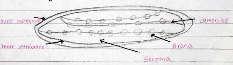
\[3.2.1.1\] What are **CHLOROPLASTS**?
* Small, flattened structures found in plant and algal cells which are the site of **photosynthesis**.
\
* They are surrounded by a double membrane, with an inner membrane called the **thylakoid** **membrane**.
\
* Thylakoid membranes are stacked up to form **grana**, which are linked together by **lamellae**.
\
* The fluid inside is the **stroma**.
\
* They are surrounded by a double membrane, with an inner membrane called the **thylakoid** **membrane**.
\
* Thylakoid membranes are stacked up to form **grana**, which are linked together by **lamellae**.
\
* The fluid inside is the **stroma**.
12
New cards
\[3.2.1.1\] What are **RIBOSOMES**?
* Very small organelles found attached to the **rough endoplasmic reticulum** or **free** in the cytoplasm.
\
* They are made from **proteins** and **RNA**.
\
* They do not have a membrane.
\
__***RESPONSIBLE FOR PROTEIN SYNTHESIS***__.
\
* They are made from **proteins** and **RNA**.
\
* They do not have a membrane.
\
__***RESPONSIBLE FOR PROTEIN SYNTHESIS***__.
13
New cards
[3.2.1.1] What is the ENDOPLASMIC RETICULUM?
* A series of thin intricate channels with a folded membrane enclosing a fluid filled space.
\
→ **Rough Endoplasmic Reticulum** have ribosomes on the surface and are responsible for *folding and processing proteins*.
\
→ **Smooth Endoplasmic Reticulum** are responsible for *lipid and steroid synthesis*.
\
→ **Rough Endoplasmic Reticulum** have ribosomes on the surface and are responsible for *folding and processing proteins*.
\
→ **Smooth Endoplasmic Reticulum** are responsible for *lipid and steroid synthesis*.
14
New cards
\[3.2.1.1\] What is the **CELL WALL**?
* A **rigid** structure which surrounds cells. Made from **cellulose** in plants and algae and made from **chitin** in fungi.
\
* It supports the cell and prevents change of shape.
\
* It supports the cell and prevents change of shape.
15
New cards
[3.2.1.1] What is the VACUOLE?
A membrane-bound organelle found in the cytoplasm that contains **cell sap** (a *weak solution of sugars and salts*). The surrounding membrane is called the **tonoplast**.
* **Maintains interior pressure**.
* Keeps cell **rigid**.
* **Isolates unwanted chemicals**.
* **Maintains interior pressure**.
* Keeps cell **rigid**.
* **Isolates unwanted chemicals**.
16
New cards
\[3.2.1.1\] What is the **GOLGI APPARATUS**?
A network of fluid-filled, membrane-bound flattened sacs which ***processes and packages new lipids and proteins into vesicles for secretion from the cell***.
17
New cards
\[3.2.1.1\] What are **GOLGI VESICLES**?
Small fluid-filled sacs in the cytoplasm, surrounded by a membrane. They ***store lipids and proteins and transport them out of the cell***.
18
New cards
[3.2.1.1] How do cells become specialised?
The cell **expresses different genes** which produces different proteins.
19
New cards
[3.2.1.1] How are epithelial cells adapted to their function?
* Many **microvilli** to **increase surface area**.
\
* Many mitochondria to produce **ATP** for **active transport**.
\
* Lots of golgi or rough endoplasmic reticulum in order to ***produce carrier and channel proteins for facilitated diffusion and active transport***.
\
* Many mitochondria to produce **ATP** for **active transport**.
\
* Lots of golgi or rough endoplasmic reticulum in order to ***produce carrier and channel proteins for facilitated diffusion and active transport***.
20
New cards
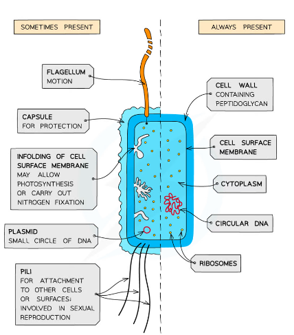
\[3.2.1.2\] What is a **prokaryotic** cell?
Cells which ***do not have a nucleus or any membrane-bound organelles***.
21
New cards
\[3.2.1.2\] What does a typical **prokaryote** look like?
22
New cards
\[3.2.1.2\] What is the **CAPSULE**?
An organelle made from secreted slime that protects the cell from an immune response.
23
New cards
[3.2.1.3] What are the two types of microscope?
Optical and Electron.
24
New cards
\[3.2.1.3\] What is meant by '**magnification**'?
How large an image is compared to real life.
25
New cards
\[3.2.1.3\] What is the equation for **magnification**?
==**Magnification**== = %%**Image Size**%% / ^^**Actual Size**^^
26
New cards
\[3.2.1.3\] What is meant by the term '**resolution**'?
The ability of a microscope to distinguish detail.
27
New cards
\[3.2.1.3\] What are **OPTICAL** microscopes?
Microscopes that use light to view objects.
\
* Maximum resolution of **0.2** micrometres.
(Cannot view ribosomes, endoplasmic reticulum or lysosomes).
* Maximum useful magnification of **x1500**.
\
* Maximum resolution of **0.2** micrometres.
(Cannot view ribosomes, endoplasmic reticulum or lysosomes).
* Maximum useful magnification of **x1500**.
28
New cards
\[3.2.1.3\] What are **ELECTRON** microscopes?
Electron microscopes use a __beam of electrons that are focused by electromagnets inside a vacuum environment__.
\
* Maximum resolution of **0.0002** micrometres.
* Maximum useful magnification of **x1500000**.
* Produces **black and white** images.
\
* Maximum resolution of **0.0002** micrometres.
* Maximum useful magnification of **x1500000**.
* Produces **black and white** images.
29
New cards
\[3.2.1.3\] Why do electron microscopes use a **vacuum** environment?
So that particles in the air *do not deflect electrons* out of the beam alignment.
30
New cards
[3.2.1.3] What are the two types of electron microscope?
**Transmission** and **Scanning**.
31
New cards
\[3.2.1.3\] What are **TRANSMISSION** electron microscopes?
A beam of electrons passes through a __**THIN**__ section of specimen.
\--> Areas that **absorb** the electrons appear darker on the electron micrograph that is produced.
\--> Areas that **absorb** the electrons appear darker on the electron micrograph that is produced.
32
New cards
\[3.2.1.3\] What are **SCANNING** electron microscopes?
A beam of electrons passes across the surface and **scatters**.
\--> The pattern of scattering builds up a **3D** image depending on the contours of the specimen.
\
*LOWER RESOLVING POWER THAN TRANSMISSION ELECTRON MICROSCOPES*.
\--> The pattern of scattering builds up a **3D** image depending on the contours of the specimen.
\
*LOWER RESOLVING POWER THAN TRANSMISSION ELECTRON MICROSCOPES*.
33
New cards
[3.2.1.3] What are the limitations to electron microscopes?
* The whole system must be in a __**vacuum**__, so living specimens __cannot__ be observed.
* A complex straining process is required which may introduce **artefacts** into the image.
* Specimens have to be __very thin__.
* A complex straining process is required which may introduce **artefacts** into the image.
* Specimens have to be __very thin__.
34
New cards
\[3.2.1.3\] What is an **ARTEFACT**?
Artefacts look like part of the microscope sample but are actually a side-effect of sample preparation or the conditions in the microscope.
35
New cards
\[3.2.1.3\] What is step one of **CELL FRACTIONATION**?
**HOMOGENISATION**.
\
This process releases the organelles by breaking open the cell membranes without damaging them. This is done with a __homogeniser__ and it produces a ***homogenate*** (a liquid of broken up cells).
\
This process releases the organelles by breaking open the cell membranes without damaging them. This is done with a __homogeniser__ and it produces a ***homogenate*** (a liquid of broken up cells).
36
New cards
\[3.2.1.3\] What are the characteristics of the solution in which **homogenisation** is carried out?
* **ICE COLD**
→ Reduces enzyme activity.
* **ISOTONIC**
→ Same **water** **potential** as the organelles so water is not taken in or lost by **osmosis**.
* **BUFFERED**
→ Maintains **pH**.
→ Reduces enzyme activity.
* **ISOTONIC**
→ Same **water** **potential** as the organelles so water is not taken in or lost by **osmosis**.
* **BUFFERED**
→ Maintains **pH**.
37
New cards
\[3.2.1.3\] What is step two of **CELL FRACTIONATION**?
**Filtration** of large cell debris.
38
New cards
\[3.2.1.3\] What is step three of **CELL FRACTIONATION**?
**Ultracentrifugation**.
\
Cell Fractionation involves spinning samples at *progressively higher speeds* (**differential ultracentrifugation**) to separate the organelles.
\
Different types of organelles have different **masses** or **densities**.
\
When the **homogenate** is spun in a **centrifuge** the heaviest organelle will sink to the bottom. The density of the organelle relates to the speed it falls.
\
Cell Fractionation involves spinning samples at *progressively higher speeds* (**differential ultracentrifugation**) to separate the organelles.
\
Different types of organelles have different **masses** or **densities**.
\
When the **homogenate** is spun in a **centrifuge** the heaviest organelle will sink to the bottom. The density of the organelle relates to the speed it falls.
39
New cards
\[3.2.1.3\] What is the **SUPERNATANT**?
The **fluid above the pellet** which may contain less dense material.
40
New cards
\[3.2.1.3\] What is the **PELLET/FRACTION**?
The ***mass of dense material*** at the **bottom of the centrifuge** tube after spinning.
41
New cards
\[3.2.2\] What are **autosomes**?
**Autosomes** are ***chromosomes which are* __*not*__ *sex chromosomes***.
42
New cards
\[3.2.2\] How are **chromosomes** formed?
**Chromosomes** are formed when **DNA** winds around **histone proteins** to form **nucleosomes**. They then **supercoil** to form the dense chromosomal structure. This then replicates to form a **double-stranded chromatid**.
43
New cards
\[3.2.2\] What are **SISTER CHROMATIDS**?
**Sister** **chromatids** are formed when two identical chromatids are joined together at the **centromere**.
44
New cards
\[3.2.2\] What is the **centromere**?
The centromere is a **constricted** **region** where two sister chromatids are in contact with each other.
45
New cards
\[3.2.2\] What is **chromatin**?
**Chromatin** is the material that chromosomes are made up of.
46
New cards
\[3.2.2\] What is meant by the term ‘**haploid**’?
Cells with only **23 chromosomes** (e.g. gametes).
47
New cards
\[3.2.2\] What is meant by the term ‘**diploid**’?
Cells which have **46 chromosomes** (23 from each parent).
48
New cards
\[3.2.2\] What is **MITOSIS**?
Mitosis is the division of a diploid cell to produce two genetically identical daughter cells which have the same number of chromosomes as eachother.
49
New cards
\[3.2.2\] Why do cells need to divide?
* **Growth** (*all over in animals, in the meristems of plants*)
\
* **Repair** of Damaged Tissues
\
* **Asexual** **Reproduction** (*genetically identical offspring*)
\
* **Maintaining** the number of **Chromosomes**
\
* **Repair** of Damaged Tissues
\
* **Asexual** **Reproduction** (*genetically identical offspring*)
\
* **Maintaining** the number of **Chromosomes**
50
New cards
\[3.2.2\] What are the stages of **mitosis**?
I - Interphase
P - Prophase
M - Metaphase
A - Anaphase
T - Telophase
(c) - Cytokinesis
P - Prophase
M - Metaphase
A - Anaphase
T - Telophase
(c) - Cytokinesis
51
New cards
\[3.2.2\] What is the first stage of **interphase**?
__**G1 (Growth or Gap)**__
\
→ Cell **Grows** and **Develops** whilst undergoing normal cell processes.
\
→ Makes **proteins** for cell division.
\
→ **ATP** is produced and stored.
\
→ Cell **Grows** and **Develops** whilst undergoing normal cell processes.
\
→ Makes **proteins** for cell division.
\
→ **ATP** is produced and stored.
52
New cards
\[3.2.2.\] What is the second stage of **interphase**?
__**S (Synthesis)**__
\
→ **DNA Replication.**
\
→ Chromosomes form chromatin.
\
→ **DNA Replication.**
\
→ Chromosomes form chromatin.
53
New cards
\[3.2.2\] What is the third stage of **interphase**?
__**G2 (Gap or Growth)**__
\
→ **ATP** is produced.
\
→ **Organelles** **duplicate**.
\
→ **More cytoplasm**.
\
→ DNA is **error-checked**, the cell will **self-terminate** if something is massively wrong.
\
→ **ATP** is produced.
\
→ **Organelles** **duplicate**.
\
→ **More cytoplasm**.
\
→ DNA is **error-checked**, the cell will **self-terminate** if something is massively wrong.
54
New cards
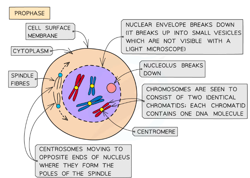
\[3.2.2.\] What is **prophase**?
* The **Nucleolus** breaks down and the **Nuclear** **Envelope** disappears.
\
* Chromosomes coil and become visible under a microscope.
\
* Chromosomes consist of **two sister chromatids** joined at the **centromere**.
\
* **Centrosomes** move to **opposite** **poles**.
\
* **Spindle Fibres** (***protein*** ***microtubules***) emerge from **centrosomes** (two **centrioles**).
\
* Chromosomes coil and become visible under a microscope.
\
* Chromosomes consist of **two sister chromatids** joined at the **centromere**.
\
* **Centrosomes** move to **opposite** **poles**.
\
* **Spindle Fibres** (***protein*** ***microtubules***) emerge from **centrosomes** (two **centrioles**).
55
New cards
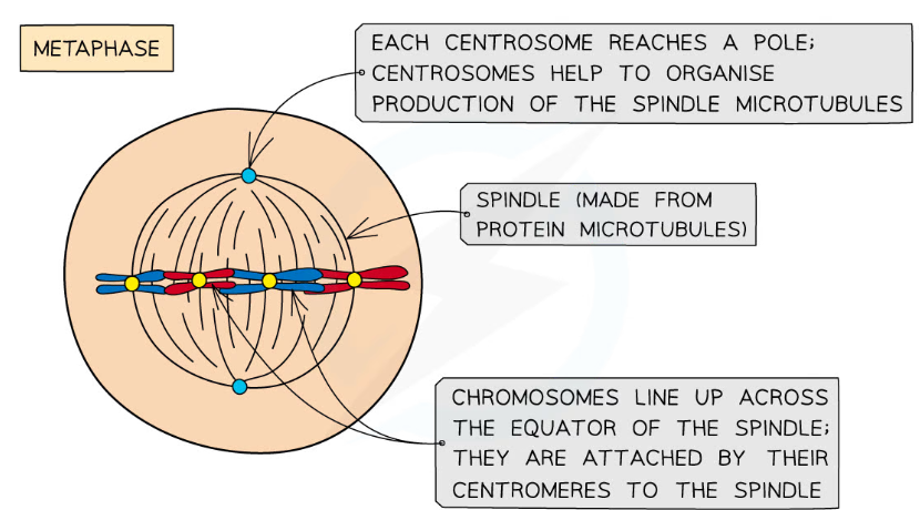
\[3.2.2.\] What is **metaphase**?
* The **spindle** is completely formed.
\
* Chromosomes line up along the **equator** of the spindle so that they are **equidistant** to the centrosome poles.
* *The microtubules attach to the centromere of each chromatid*.
\
* Chromosomes line up along the **equator** of the spindle so that they are **equidistant** to the centrosome poles.
* *The microtubules attach to the centromere of each chromatid*.
56
New cards
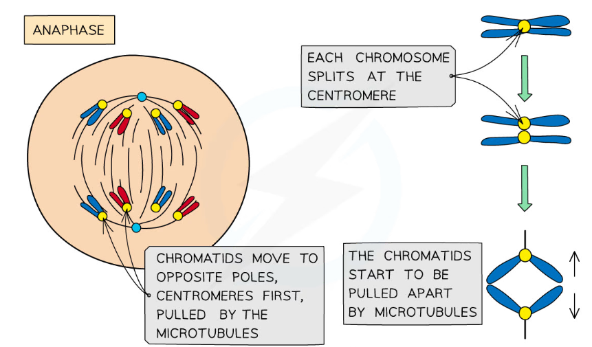
\[3.2.2\] What is **anaphase**?
* The sister chromatids **divide** at the **centromere**.
\
* The spindle fibres **contract** and pull the sister chromatids to opposite poles of the spindle (they are now called **chromosomes**).
\
* The spindle fibres **contract** and pull the sister chromatids to opposite poles of the spindle (they are now called **chromosomes**).
57
New cards
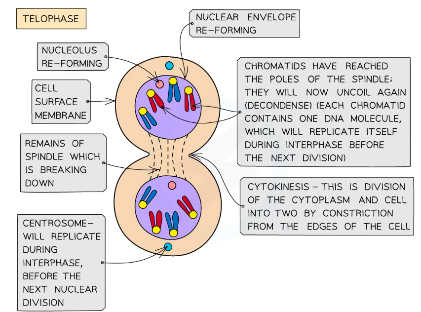
\[3.2.2\] What is **telophase**?
* Chromosomes **decondense** (they become long and thin again and can no longer be seen clearly under a microscope).
\
* New Nuclear Envelopes form around each set of chromosomes, but in the same cytoplasm space.
\
* The spindle begins to break down.
\
* The nucleolus reforms.
\
* **Cytokinesis** occurs.
\
* New Nuclear Envelopes form around each set of chromosomes, but in the same cytoplasm space.
\
* The spindle begins to break down.
\
* The nucleolus reforms.
\
* **Cytokinesis** occurs.
58
New cards
\[3.2.2\] What is **cytokinesis**?
The **division of the cytoplasm** by constriction from the edges of the cell.
\
* In **ANIMAL CELLS**:
→ The plasma membrane is *pulled inwards* around the *equator* to form a **cleavage furrow**. This is done using a **contractile ring of protein** of **actin** and **myosin** **fibres**.
\
* In **PLANT CELLS**:
→ A new cell wall is formed across the **equator**.
→ A **middle lamella** forms and **cellulose** is deposited.
\
* In **ANIMAL CELLS**:
→ The plasma membrane is *pulled inwards* around the *equator* to form a **cleavage furrow**. This is done using a **contractile ring of protein** of **actin** and **myosin** **fibres**.
\
* In **PLANT CELLS**:
→ A new cell wall is formed across the **equator**.
→ A **middle lamella** forms and **cellulose** is deposited.
59
New cards
\[3.2.2\] What is the formula for **mitotic index**?
**NUMBER OF CELLS UNDERGOING MITOSIS**
----------------------------------------------------
**TOTAL NUMBER OF CELLS IN THE FIELD OF VIEW**
----------------------------------------------------
**TOTAL NUMBER OF CELLS IN THE FIELD OF VIEW**
60
New cards
\[3.2.2\] What causes **cancer**?
**Uncontrollable Cell Division**.
61
New cards
\[3.2.2\] What is a **tumour**?
A large group of cells that are **dividing uncontrollably**.
62
New cards
\[3.2.2\] How can **drugs treat cancer** by ***blocking part of the cell cycle***?
* **Chemotherapy** prevents the **synthesis of enzymes** needed for **DNA Replication**. This means that cells cannot enter the **S phase** and therefore self-destruct.
\
* **Radiation** and **Drugs** **damage** **DNA** so much that the cell cannot fix it which means the cell has to self-destruct.
\
* **Radiation** and **Drugs** **damage** **DNA** so much that the cell cannot fix it which means the cell has to self-destruct.
63
New cards
\[3.2.2\] Why can drugs that are used to treat **cancer** cause **hair loss**?
Rapidly dividing cells like hair cells are killed which leads to hair loss as there is nothing to keep producing the proteins needed for hair growth.
64
New cards
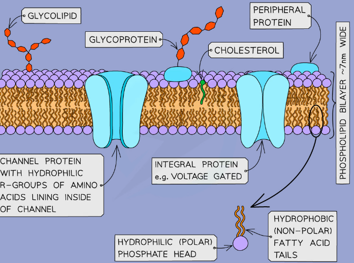
\[3.2.3\] Why is the **cell membrane** described as ‘**fluid-mosaic**’?
The molecules can **move** within the membrane and there are other molecules (such as **proteins**) embedded within the **phospholipid bilayer**.
65
New cards
\[3.2.3\] What is the structure of the cell membrane?
66
New cards
\[3.2.3\] What is the **phospholipid bilayer**?
A **phospholipid** **bilayer** is composed of two layers of polar phospholipids; their **hydrophobic** **tails** facing inwards and **hydrophilic** **heads** outwards.
\
* They *regulate the movement of substances* through the membrane (which is **selectively permeable**).
\
* Internally, **membrane-bound compartments** formed from phospholipid bilayers provide the basic structure of organelles. An example of a membrane-bound organelle is the **lysosome**. The **lysozymes** need to be kept **compartmentalised** otherwise they would breakdown most of the cellular components.
\
* They *regulate the movement of substances* through the membrane (which is **selectively permeable**).
\
* Internally, **membrane-bound compartments** formed from phospholipid bilayers provide the basic structure of organelles. An example of a membrane-bound organelle is the **lysosome**. The **lysozymes** need to be kept **compartmentalised** otherwise they would breakdown most of the cellular components.
67
New cards
\[3.2.3\] What is **cholesterol**?
Cholesterol is a type of **lipid**.
\
* Cholesterol binds to **hydrophobic** tails of phospholipids, causing them to pack together. This restricts movement, making the membrane ***less fluid and more rigid***.
\
* It has **hydrophobic** regions so acts as a **barrier to polar substances**.
\
* Gives the membrane **stability**.
\
* **Maintains the shape** of animal cells.
\
* Cholesterol binds to **hydrophobic** tails of phospholipids, causing them to pack together. This restricts movement, making the membrane ***less fluid and more rigid***.
\
* It has **hydrophobic** regions so acts as a **barrier to polar substances**.
\
* Gives the membrane **stability**.
\
* **Maintains the shape** of animal cells.
68
New cards
\[3.2.3\] How does **TEMPERATURE** affect the **permeability** of cell membranes?
__**BELOW 0°C:**__
\
* Phospholipids **don’t have much energy** so are unable to move and they are **packed** tightly together, membrane is **rigid**.
* Channel and carrier proteins **denature**, **increasing** **permeability**.
* **Ice** **Crystals** may form and **pierce** the membrane, **increasing** **permeability** once it is melted.
\
__**BETWEEN 0°C AND 45°C:**__
\
* Phospholipids can **move** around and aren’t as packed, the membrane is **selectively permeable**.
* ==*Increased Temperature = Increased Movement = Increased Permeability*==
\
__**ABOVE 45°C:**__
\
* The phospholipid bilayer begins to **break** **down**, **increasing** **permeability**.
* Water expands, increasing the **pressure**.
* Channel and carrier proteins **denature**, **increasing permeability**.
\
* Phospholipids **don’t have much energy** so are unable to move and they are **packed** tightly together, membrane is **rigid**.
* Channel and carrier proteins **denature**, **increasing** **permeability**.
* **Ice** **Crystals** may form and **pierce** the membrane, **increasing** **permeability** once it is melted.
\
__**BETWEEN 0°C AND 45°C:**__
\
* Phospholipids can **move** around and aren’t as packed, the membrane is **selectively permeable**.
* ==*Increased Temperature = Increased Movement = Increased Permeability*==
\
__**ABOVE 45°C:**__
\
* The phospholipid bilayer begins to **break** **down**, **increasing** **permeability**.
* Water expands, increasing the **pressure**.
* Channel and carrier proteins **denature**, **increasing permeability**.
69
New cards
\[3.2.3\] How do **SOLVENTS** affect membrane **permeability**?
Surrounding cells in an **increasing concentration** of a solvent **increases membrane permeability** because the **solvent dissolves the lipids**, causing a loss of structure.
70
New cards
\[3.2.3\] What are the four ways that molecules can be transported across a membrane?
* Diffusion
* Facilitated Diffusion
* Active Transport
* Osmosis
* Facilitated Diffusion
* Active Transport
* Osmosis
71
New cards
\[3.2.3\] What is the definition of **diffusion**?
The **net movement** of molecules or ions from a **region of high concentration** to a region of **low concentration** until they are **equally distributed**. It is a **random** and **passive** process.
72
New cards
\[3.2.3\] What type of molecules are transported through the phospholipid bilayer by **simple diffusion**?
**Small** molecules or **uncharged particles**.
\
* e.g. H₂O, CO₂, O₂
\
* e.g. H₂O, CO₂, O₂
73
New cards
\[3.2.3\] What are the factors which affect the rate of diffusion across membranes?
* **Steepness** of the **Concentration Gradient**
\
* **Distance** over which diffusion occurs (**DIFFUSION PATHWAY**)
\
* **Surface Area** between the two regions.
→ an increased surface area = an increased rate of diffusion
\
* **Temperature**
→ an increased temperature increases the kinetic energy of the molecules so they move faster across the membrane
\
* **Distance** over which diffusion occurs (**DIFFUSION PATHWAY**)
\
* **Surface Area** between the two regions.
→ an increased surface area = an increased rate of diffusion
\
* **Temperature**
→ an increased temperature increases the kinetic energy of the molecules so they move faster across the membrane
74
New cards
\[3.2.3\] What is the definition of **facilitated diffusion**?
The transport of molecules from a **region of high concentration** to a **region of low concentration** across a membrane using **channel** and **carrier proteins**. It relies on the **inbuilt motion (kinetic energy) of diffusing molecules** and occurs **down a concentration gradient**.
75
New cards
\[3.2.3\] What particles are transported via facilitated diffusion in **CHANNEL proteins**?
**Charged particles** (**ions**).
\
* e.g. Na⁺, PO₄³⁻, O²⁻
\
* e.g. Na⁺, PO₄³⁻, O²⁻
76
New cards
\[3.2.3\] How do **channel proteins** work in facilitated diffusion?
Particles can diffuse in/out of the cell dependent on the *direction* of the **concentration gradient**.
\
* Some channel proteins may open or close in response to a **specific messenger** or a **change in voltage** (*gated channel proteins*).
\
* Some channels are always open but others open when a ***hormone*** is attached.
\
* Some channel proteins may open or close in response to a **specific messenger** or a **change in voltage** (*gated channel proteins*).
\
* Some channels are always open but others open when a ***hormone*** is attached.
77
New cards
\[3.2.3\] What particles are transported via facilitated diffusion in **CARRIER proteins**?
**Large Molecules**.
\
* e.g. Glucose and Amino Acids.
\
* e.g. Glucose and Amino Acids.
78
New cards
\[3.2.3\] How do **carrier proteins** work in facilitated diffusion?
The particle binds to the protein, which then **changes shape** and transports the particle across the membrane.
79
New cards
\[3.2.3\] What are the types of **passive** transport?
Osmosis, Diffusion, Facilitated Diffusion.
80
New cards
\[3.2.3\] What are the types of **active** transport?
Active Transport, Bulk Transport (e.g. *Endocytosis*).
81
New cards
\[3.2.3\] What is the definition of **active transport**?
The **active movement** of molecules or ions from a **region of low concentration** to a region of **high concentration** using **energy** and **intrinsic carrier molecules**. This occurs **AGAINST** a **concentration gradient**.
82
New cards
\[3.2.3\] How does **direct active transport** work?
* A **phosphate group** from an **ATP** molecule attaches to the carrier protein, causing it to change shape.
\
* As it changes shape, it *pumps the molecule into the cell*.
\
* The phosphate group is then released and the protein reverts back to its original shape.
\
* As it changes shape, it *pumps the molecule into the cell*.
\
* The phosphate group is then released and the protein reverts back to its original shape.
83
New cards
\[3.2.3\] What do cells that carry out active transport need many of?
**Mitochondria** to produce **ATP** by **aerobic respiration**.
84
New cards
\[3.2.3\] Which factors affect the **rate of active transport**?
* The **speed** of individual carrier proteins.
→ *The faster they work, the faster the rate of active transport.*
\
* The **number** of carrier proteins.
→ *More carrier proteins = faster rate of active transport.*
\
* **Rate of Respiration**
→ *If respiration is inhibited, active transport doesn’t occur.*
→ *The faster they work, the faster the rate of active transport.*
\
* The **number** of carrier proteins.
→ *More carrier proteins = faster rate of active transport.*
\
* **Rate of Respiration**
→ *If respiration is inhibited, active transport doesn’t occur.*
85
New cards
\[3.2.3\] What is **co-transport**?
**Co-transport** is the **coupled** movement of substances across a cell membrane via **carrier proteins**.
86
New cards
\[3.2.3\] How does **glucose** enter cells by co-transport?
1) Sodium Ions are **actively transported out of the epithelial cells** and into the blood.
→ *The concentration of Na⁺ in the cell* ***decreases****. There is now a* ***higher*** *concentration of Na*⁺ *in the intestine than in the epithelial cell.*
\
2) Na⁺ ions can now diffuse into the cell through the **sodium-glucose cotransporter protein**. *[They carry the glucose with them.]*
\
3) Glucose is transported into the cell **against its concentration gradient** by the cotransporter.
\
4) The concentration of glucose in the epithelial cell is **higher** than in the blood. Glucose diffuses out of the cell by **facilitated diffusion** through a carrier protein.
87
New cards
\[3.2.3\] How is the **ileum** adapted for absorption?
* **Villi** increases **surface area**.
\
* **Very thin walls** so there is a **short diffusion pathway**.
\
* Walls contain **muscle** so can move.
\
* **Good blood supply** (*maintains concentration gradient*).
\
* **Microvilli** increase **surface area to volume ratio**.
\
* **Very thin walls** so there is a **short diffusion pathway**.
\
* Walls contain **muscle** so can move.
\
* **Good blood supply** (*maintains concentration gradient*).
\
* **Microvilli** increase **surface area to volume ratio**.
88
New cards
\[3.2.3\] What is **osmosis**?
**Osmosis** is the **net movement of water molecules** from a region of **less negative water potential** to a region of **more negative water potential** through a **partially permeable membrane**. It is a **random** and **passive** process.
89
New cards
\[3.2.3\] What is **water potential**?
The **tendency of water molecules** to **leave** a solution via **osmosis**.
90
New cards
\[3.2.3\] What is the **water potential scale**?
The water potential of **pure water** is **0KPa**, which is the highest value.
→ *This is because adding a solute makes it harder for water molecules to move, reducing the water potential (making it more negative)*
\
\[***A less negative water potential = a higher water potential = a lower concentration of solute]***
\
\[***A more negative water potential = a lower water potential = a higher concentration of solute]***
91
New cards
\[3.2.3\] What is a **hypotonic solution**?
Solutions with a **less negative** water potential compared with the inside of the cell.
\
* Animal Cells in hypotonic solutions undergo cell **lysis**.
* Plant Cells in undergo **turgor** (become **turgid**).
\
* Animal Cells in hypotonic solutions undergo cell **lysis**.
* Plant Cells in undergo **turgor** (become **turgid**).
92
New cards
\[3.2.3\] What is an **isotonic solution**?
Solutions with the **same water potential** as the cell.
\
* Animal Cells and Plant Cells have no net movement of water.
\
* Animal Cells and Plant Cells have no net movement of water.
93
New cards
\[3.2.3\] What is a **hypertonic solution**?
Solutions with a **more negative** water potential than the cell.
\
* Animal Cells undergo **crenation**.
* Plant Cells undergo **plasmolysis**.
\
* Animal Cells undergo **crenation**.
* Plant Cells undergo **plasmolysis**.
94
New cards
\[3.2.4\] What is a **pathogen**?
A **microorganism** that **causes disease**.
95
New cards
\[3.2.4\] What are the body’s **defense mechanisms**?
__**NON-SPECIFIC:**__
\
* **Physical** **Barrier** (e.g. Skin, Mucus, Stomach Acid, Cilia)
* **Phagocytosis**
\
__**SPECIFIC:**__
\
* **Cell** **Mediated** Response (*T-Lymphocytes*)
* **Humoral** Response (*B-Lymphocytes*)
\
* **Physical** **Barrier** (e.g. Skin, Mucus, Stomach Acid, Cilia)
* **Phagocytosis**
\
__**SPECIFIC:**__
\
* **Cell** **Mediated** Response (*T-Lymphocytes*)
* **Humoral** Response (*B-Lymphocytes*)
96
New cards
\[3.2.4\] What is an **antigen**?
Molecules (usually *proteins*) which trigger an **immune response**, i.e. trigger the production of antibodies. They are found on the surface of body cells and pathogens.
97
New cards
\[3.2.4\] What do antigens allow our immune systems to identify?
* Bacterium
* Viruses
* Transplanted Cells
* Abnormal Cells
* Viruses
* Transplanted Cells
* Abnormal Cells
98
New cards
\[3.2.4\] How does **phagocytosis** work?
* The phagocyte surrounds the pathogen and **engulfs** it, forming a **phagocytic vacuole**.
\
* The phagocyte’s **lysosomes** fuse with the phagocytic vacuole and the **lysozymes** digest the pathogen.
\
* **Antigens** from the pathogen appear on the surface of the body cell, meaning it has become an **antigen-presenting cell**.
\
* The phagocyte’s **lysosomes** fuse with the phagocytic vacuole and the **lysozymes** digest the pathogen.
\
* **Antigens** from the pathogen appear on the surface of the body cell, meaning it has become an **antigen-presenting cell**.
99
New cards
\[3.2.4\] What are **T-Lymphocytes**?
A type of white blood cell which have receptors on their membrane which are complementary to the shape of a specific antigen. The T-cell will become activated when an antigen binds to the receptor.
100
New cards
\[3.2.4\] How do **Helper T Cells** work?
* Receptors on the surface bind to antigens on antigen-presenting cells.
→ This activates helper T cells to replicate via **mitosis**.
→ Some of these T cells then differentiate into other cells.
→ Some remain as T cells which active **B Cells**.
→ Some activate **macrophages** to perform **phagocytosis**.
→ Some form **cytotoxic T cells**.
→ This activates helper T cells to replicate via **mitosis**.
→ Some of these T cells then differentiate into other cells.
→ Some remain as T cells which active **B Cells**.
→ Some activate **macrophages** to perform **phagocytosis**.
→ Some form **cytotoxic T cells**.