INTRO to NEURO
 Call Kai
 Learn
Learn Practice Test
 Spaced Repetition
 Match
Match Flashcards
 Knowt Play
1/73

Earn XP
Description and Tags
Name | Mastery | Learn | Test | Matching | Spaced | Call with Kai | Chat |
|---|
No analytics yet
Send a link to your students to track their progress
74 Terms
Contralateral
Anatomical term for relating to the opposite side of the body; Opposite of ipsilateral (same side of the body)
Ipsilateral
Anatomical term for relating to the same side of the body; opposite of contralateral (opposite side of the body)
Posterior
Anatomical term for toward the back of the brain or toward the back side of the spinal cord; opposite of anterior (front side)
Lateral
Anatomical term for toward the edge or side of the brain; opposite of medial (toward the midline of the brain)
Anterior
Anatomical term for toward the front of the brain or toward the front surface of the spinal cord; opposite of posterior (back side)
Medial
Anatomical term for toward the midline of the brain when viewed from the midsagittal plane

Rostral
Anatomical term for towards the nose or front of the head in regard to the brain or upward toward the brainstem in regard to the spinal cord; Means beak in Latin
Caudal
Anatomical term for towards the tail or away from the head end of the body
Dorsal
Anatomical term for towards top of head in regard to the brain or toward back (posterior) side in regard to the spinal cord
Ventral
Anatomical term for underside (belly) for the brain or toward the anterior (front) of the spinal cord
Systems Neuroscience
Investigates brain cells collections that work together to complete a common function
Cognitive Neuroscience
Investigates highest-level of human thinking (i.e. language, emotion, attention, and consciousness)
Behavioral Neuroscience
Investigates how brain systems work together to generate behavior (i.e. daily rhythms of wake and sleep)
Molecular Neuroscience
Investigates smallest units of cell-to-cell communication in the brain, the conductors guiding brain development, and the sentries controlling access to neurons.
Cellular Neuroscience
Investigates ways molecules interact to give neurons their special properties and different types of brain cells.
Sagittal Plane
Vertical anatomical plane that runs front → back and divides the body and brain into left and right sections; towards perpendicular to coronal (frontal) plane

Coronal Plane
Vertical anatomical plane that runs left → right and divides the body and brain into anterior (front) and posterior (back) sections; perpendicular to the floor, parallel to body’s long axis
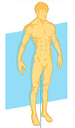
Horizontal Plane
Transverse or axial plane that divides the brain and body into superior (upper) and inferior (lower) portions


…Similarities between the organization of the rodent and human brain
Symmetrical paired hemispheres, positioning of major divisions, and a strong correlation between structures and functions are all…
…Differences between the organization of the rodent and human brain
More folding—which = more brain matter in the human brain; smaller olfactory bulbs for humans because they are more important for rodents; Ram’s Horn pattern in the human brain (folds in itself); Human brain having multiple lobes are all…
…Similarities between the cerebral cortex of vertebrates and mammals
Parallel-running cortical neurons are arranged in layers; No neuron cell bodies in layer closer to the surface of the brain surface (layer #1); and one layer having cells with large dendrites that project up to layer one are all…
…Difference between the cerebral cortex of vertebrates and mammals
Mammals having a multi-layer neocortex, which increased the association cortex (not with sensorimotor or secondary sensory functions)
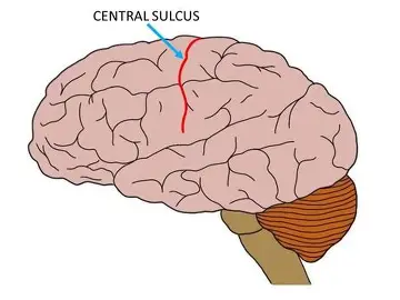
Central Sulcus
Separates the Frontal lobe and the Parietal lobe
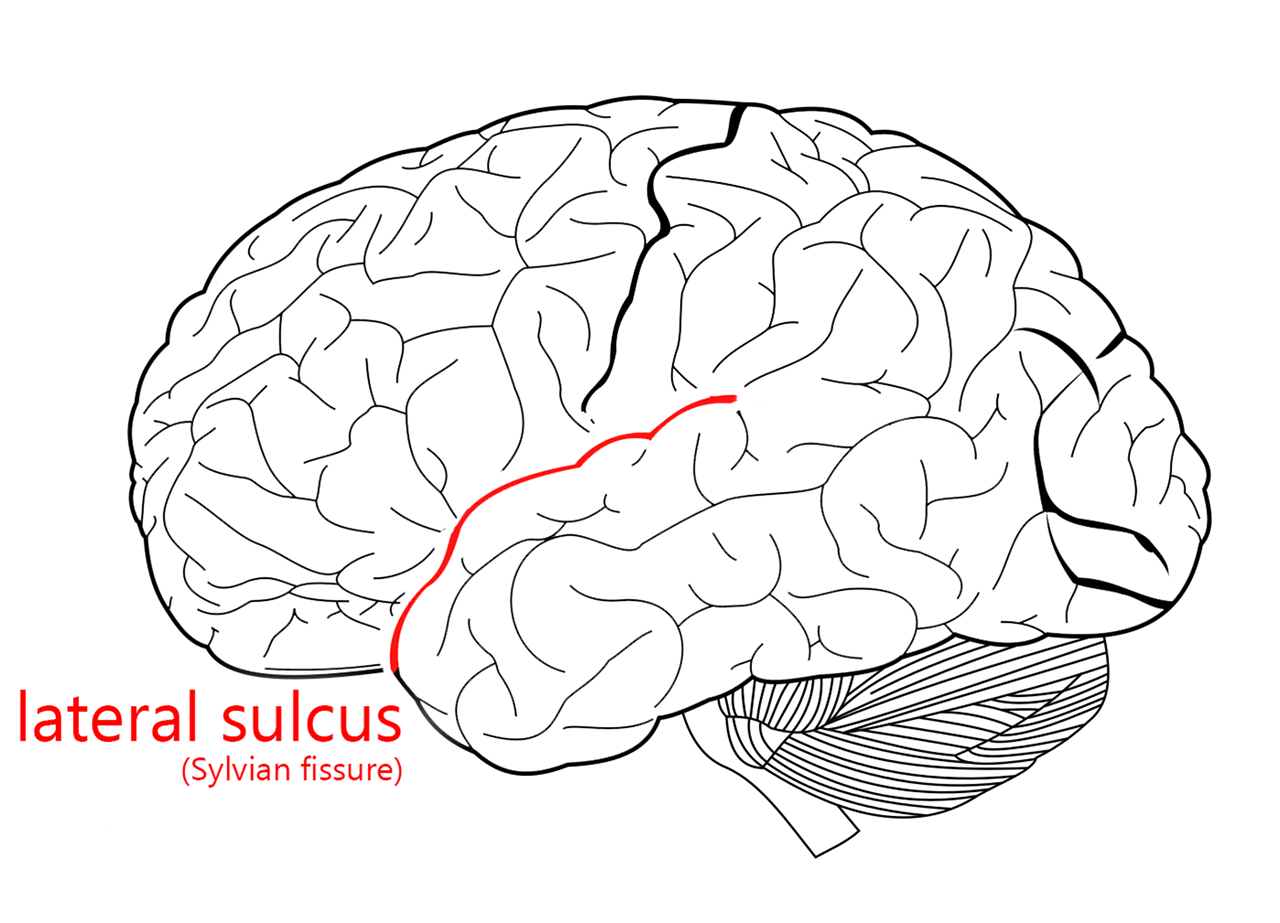
Lateral Fissure
Separates the Temporal lobe, Frontal lobe, and the Parietal lobe.
Cytoarchitecture
Varying cell compositions of brain regions
Molecular Layer
Layer #1 of the Neocortex, contains no neuronal cell bodies, and has at least one layer extending pyramidal cells with large apical dendrites up to it (intra- and inter-region connections)
Apical Dendrites
…
External Granular Layer
Layer #2 of the Neocortex, contains small pyramidal and small stellate cells, receives inputs from Layer #4, and sends outputs to Layer # 5
External Pyramidal Layer
Layer #3 of the Neocortex, contains med-size pyramidal cells, receives inputs from Layer #4, and sends outputs to Layer #5
Internal Granular Layer
Layer #4 of the Neocortex, contains small stellate and pyramidal cells, receives sensory inputs, and varies substantially across the cortex
Internal Pyramidal Layer
Layer #5 of the Neocortex, contains large pyramidal cells with long apical dendrites, it is the primary output layer to other cortical areas and the corticospinal tract
Multiform Layer
Layer #6 of the Neocortex, contains multiple types of cells, and it is responsible for making reciprocal connections with the thalamus
Primary Sensory Cortex
Has a large layer #4 of the Neocortex (Internal Granular Layer)
Primary Motor Cortex
Has a large layer #5 of the Neocortex (Internal Pyramidal Layer), but very small layer #4 of the Neocortex (Internal Granular Layer)

Precentral Gyrus (bump)
Primary Motor Cortex
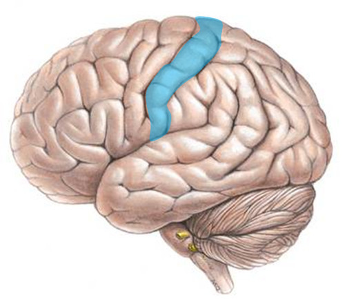
Postcentral Gyrus (bump)
Primary Sensory Cortex
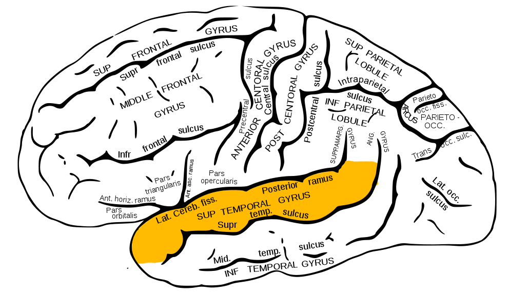
Superior Temporal Gyrus (bump)
Primary Auditory Cortex
STRATEGY
Level #1 of the Motor Control Hierarchy: identifying the goal of movement as an achievement (association areas of cortex, and basal ganglia)
TACTICS
Level #2 of the Motor Control Hierarchy: thinking about the sequence of muscle contractions needed to accomplish STRATEGY goal (motor cortex, and cerebellum)
EXECUTION
Level #3 of the Motor Control Hierarchy: the completion of the TACTIC motor movement (brainstem, cerebellum, and spinal cord)
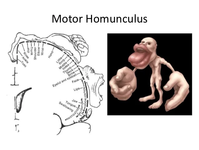
The Motor Homunculus
Homunculus = “Little Man”; Shows that the relative size of structure of a drawing indicates the amount of motor cortex dedicated to that structure.
…Contralateral
Control of voluntary movement in the motor cortex is
Decussation
Crossing the midline in the medulla to go to the other side
Horizontal Sound Localization
The auditory system’s ability to determine the azimuth (left–right position) of a sound source in the horizontal plane

Interaural Time Delay
One of two sources of information that contribute to horizontal sound localization. This means that sounds coming from one side of the ear will hit that side’s ear before the other ear. The # of spikes per response codes information on where the sound is coming from

Cingulate Gyrus
A Midsagittal Landmark that extends from just below the corpus callosum and an anterior portion of the cingulate all the way to the posterior portion of the cingulate
Perigenual/Subgenual Cingulate Gyrus
Right next to the genu of the corpus callosum and is involved in emotion processing and emotion control/regulation
Dorsal Anterior Cingulate Gyrus
Related to advanced cognitive controls (i.e. decision making, reward processing, controlling behavioral responses to stimuli)
Posterior Cingulate Gyrus
Involved in processes such as imagination and autobiographical memory
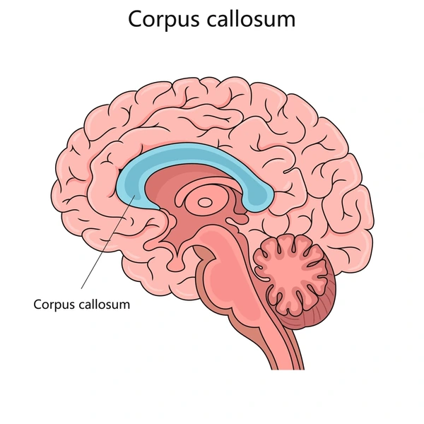
Corpus Callosum
A bundle of white matter tracks of axons that run between the two cerebral hemispheres, holds them together, and is related to interhemispheric communication of information

Fornix
White matter bundle that extends laterally to the hippocampus in each hemisphere and provides an output pathway for the hippocampus to the thalamus and hypothalamus.

Olfactory bulb
Associated with the sensation of smell
Optic Chiasm
Sits below the hypothalamus where optic nerves cross from the right side to the left side.
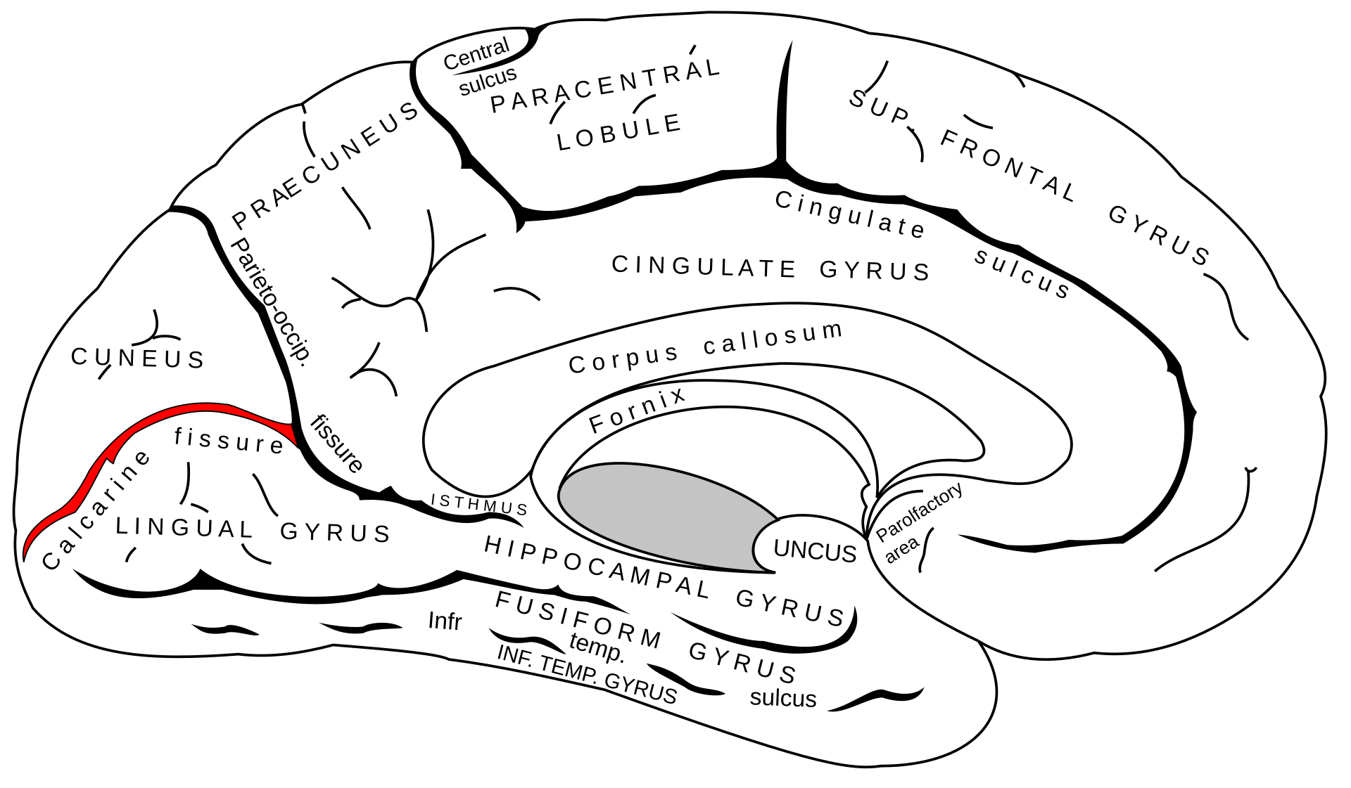
Calcarine Fissure
Primary Visual Cortex in the occipital lobe on both sides of the brain. Bisects the parietal occipital sulcus (grooves)
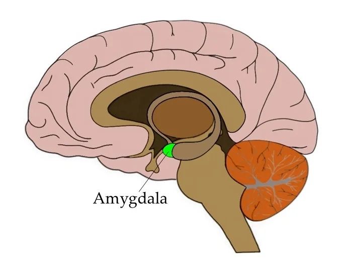
Amygdala
Sits beneath the overlying cortex (inside temporal lobe) and at the end of the hippocampus. Important for threat detection and emotion processing

Hippocampus
Sits beneath the overlying cortex (inside temporal lobe) and central to the formation of new memories

Thalamus
Sits at the center of the cerebrum.

Hypothalamus
“Hypo” = “Below.” Important for the autonomic nervous system (includes command of “fight or flight” mechanism) and motivational processes that are related in seeking out food or reproductive opportunities

Midbrain
Made up of the Tectum and Tegmentum. It is a conduit for information moving between the spinal cord and the cerebrum

Tectum
Latin for “Roof” (on top of the Tegmentum) includes the superior caliculus (receives inputs from the eyes and contributes to eye movements) and the inferior caliculus (serves as a relay station for auditory input from ear → thalamus).
Tectum to Detect ‘em

Tegmentum
Contributes primarily to voluntary movement. Includes the substantia nigra and the red nucleus. (TegMENTUM for MoMENTUM)

Pons
Transmits information between the cortex and the cerebellum, as well as between the cerebellum and the spinal cord. Projects to the opposite side of the cerebellum, which makes the cerebellum ipsilaterally organized for movement control

Medulla
Serves as a pathway for sending cortical axons (including the cortical spinal tract) and carries motor information down to the spinal cord and the cortical spinal tract decussates (crosses the midline to go to the other side of the spinal cord). Cochlear nucleus is in it (sensitive nuclei related to hearing). And it also contributes to touch, sensation, and taste (motor neurons help control the tongue)
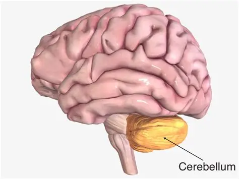
Cerebellum
“Little Brain” critical for accurate movements, receives input from pons and spinal cord to combine info regarding intended movement and body position in space, and is ipsilaterally organized. Important for sleep, working memory, and learning.
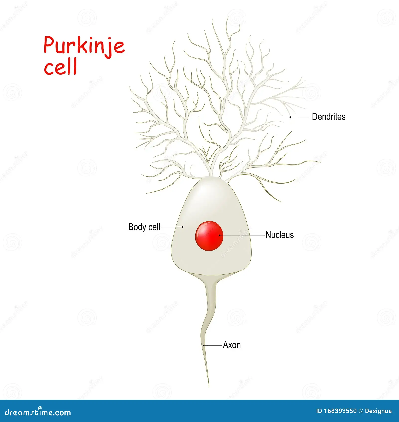
Purkinje cells
The fundamental unit of cerebellar function, and are only output cells (inhibitory), receive input from parallel fibers from pontine nuclei and from climbing fibers
Insula
Inside and behind the temporal lobe when looking at the brain from a lateral perspective. Important for processing social pain and integration of somatosensory
Caudate nucleus
Apart of the Basal Ganglia and is involved in the motor control of posture, reward processing, and approach or attachment behaviors
Putamen
Apart of the Basal Ganglia, helps with reward and cognitive function, and implicated in addiction in the use of substances. Posterior portion is important for motor control
Globus Pallidus
Apart of the Basal Ganglia that important for motor function

Basal Ganglia
…
Ventral Lateral Nucleus
Makes motor system projections to the precentral gyrus
Ventral Posterior Nucleus
Projects to the primary sensory system regions on the postcentral gyrus
Substantia Nigra
Apart of the midbrain, important component of the motor system and degeneration is implicated in Parkison’s disease
Subthalamus
Strongly connected to the Globus palladus