Ultrasound
1/29
Earn XP
Description and Tags
TTE, TEE, gastric and lung POCUS
Name | Mastery | Learn | Test | Matching | Spaced | Call with Kai |
|---|
No analytics yet
Send a link to your students to track their progress
30 Terms
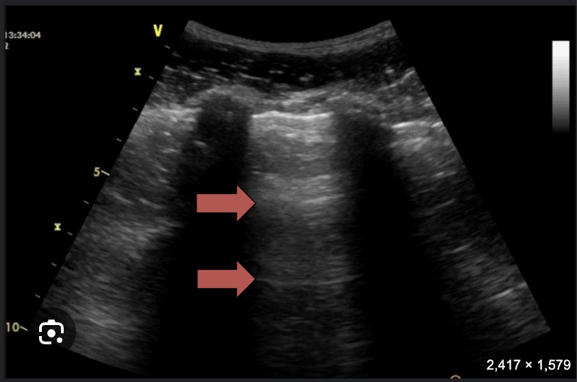
What US finding are the arrows pointing to? Is this normal?
A lines. Normal. Represents reverberation of the ultrasound beams on air in the lung parenchyma
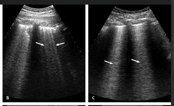
What US finding are the lines pointing to? Are these normal findings?
B lines. Can be normal (especially at the bases), but if more than 3 in multiple areas, need to consider pulmonary edema
If <3mm, then think about GGO. If <7mm then interlobular tissue thickening.
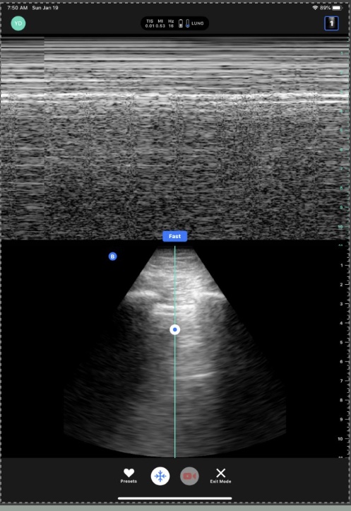
What sign is this?
Seashore sign. Indicates normal lung sliding
What sign is this?
Barcode sign. Pathognomonic for pneumothorax
Does the presence of A lines rule out pneumothorax?
No
What are possible explanations for lung point?
Pneumothorax
Pulmonary adhesions
Pleurodesis
What lung POCUS findings are c/w consolidation?
Hepatization, heterogenous appearance within the parenchyma. Can also see dynamic and static air bronchograms.
What is the ideal probe to use to look at lung pleura?
Linear probe
Which probe should be used to look at lung parenchyma?
Curvilinear or phased array
When performing lung POCUS, where should the marker face? What is the probe orientation?
Marker faces cephalad, sagittal orientation
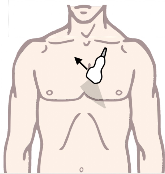
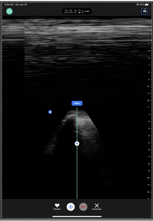
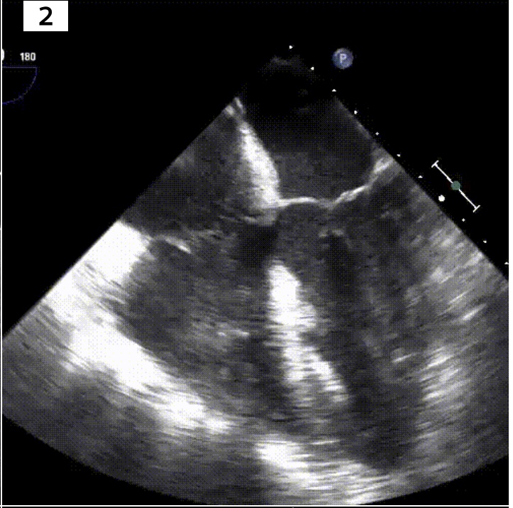
What view is this?
Parasternal long axis (PLAX)
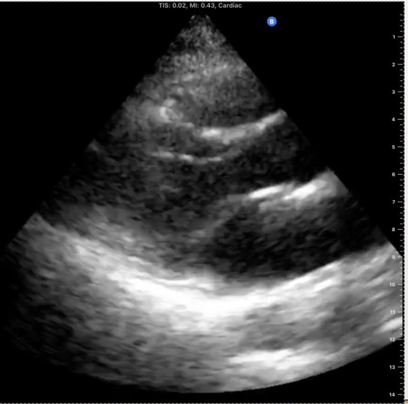
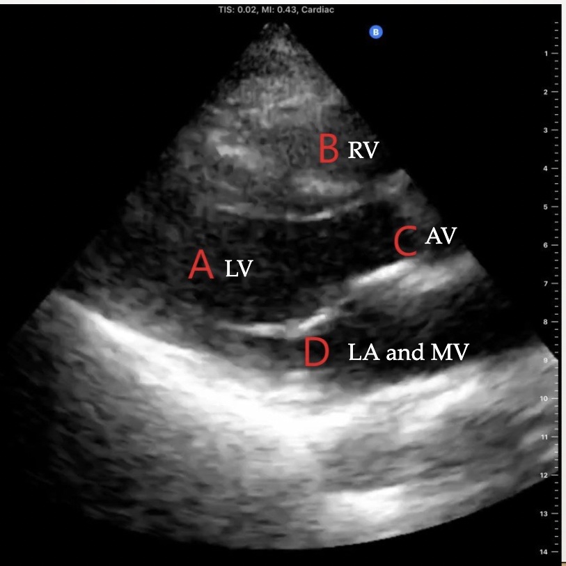
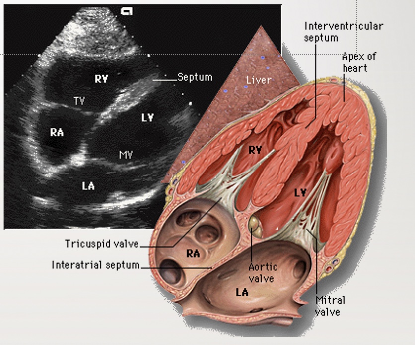
Identify the view and the structures on this echo
Parasternal long axis (PLAX)
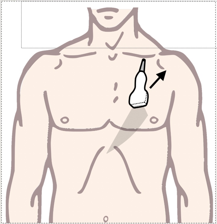
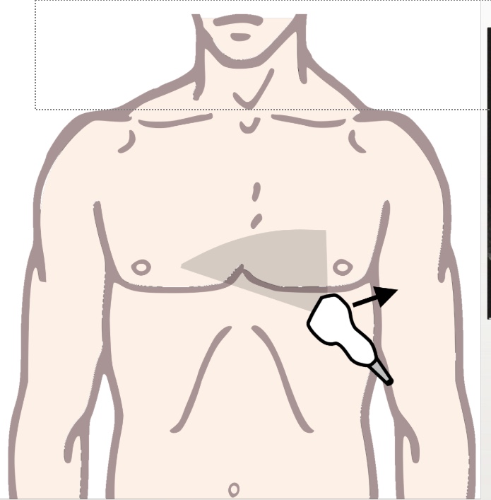
What echocardiographic view will this obtain?
Parasternal short axis (PSAX)

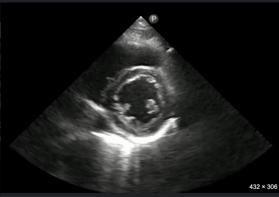
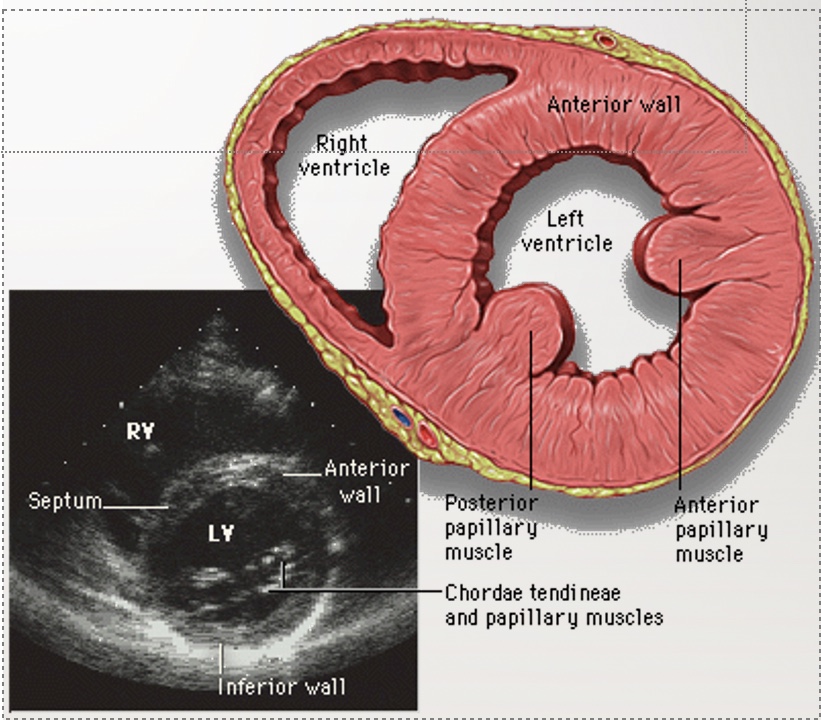
Identify structures and the view of this echo
PSAX
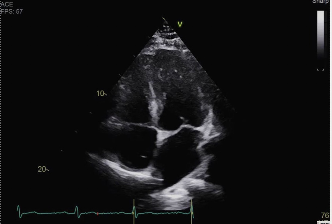
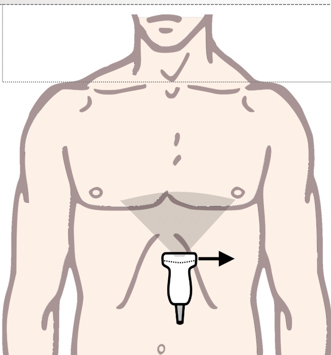
What echo view will this position give you?
Apical 4 chamber
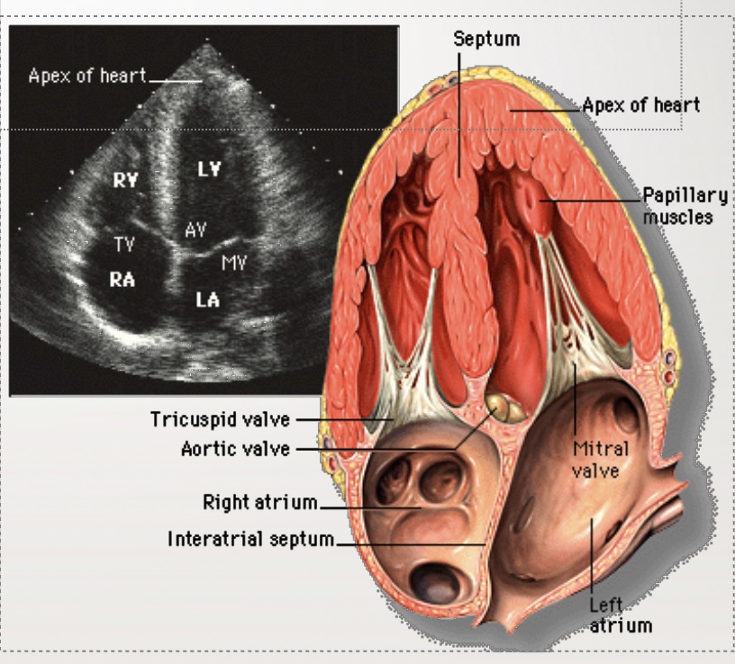
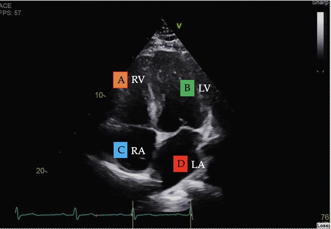
Identify the view and structures
Apical 4 chamber
What echo view will this give you?
Subcostal 4 chamber (SC4C)
What are the 3 primary positions in the GI tract used to obtain a comprehensive TEE?
Upper esophageal (UE), midesophageal (ME), and transgastric (TG)
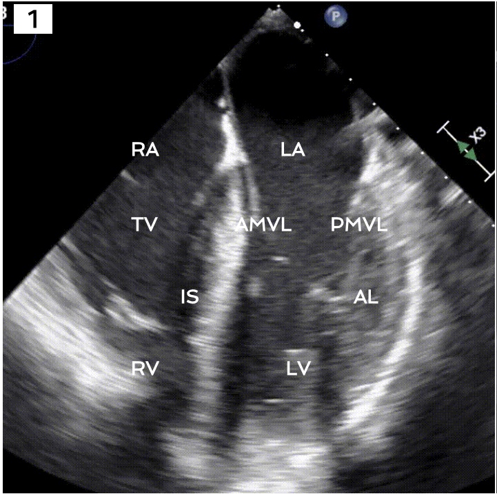
Identify the view and structures of interest
Midesophageal 4 chamber view.
What transducer angle do you need to get ME4C view?
0-10*
What is the difference between the ME4C and ME5C views?
ME5C gives you a look at the aortic valve
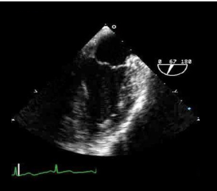
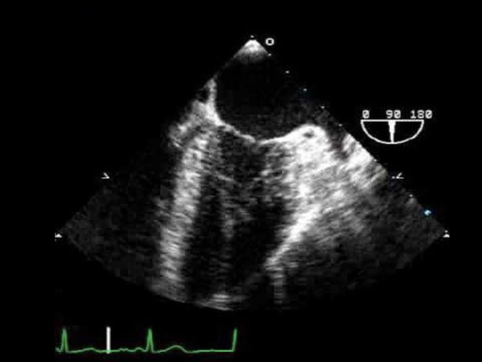
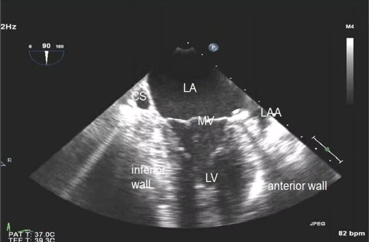
What TEE view is this? ID the structures
ME MV commissural view
LA & LV
Coronary sinus
Anterior and posterior leaflets of MV
Papillary muscles
Chordae tendinae
Are the ME2C view and the mitral commissural views the same?
No - ME2C is obtained around 90* (80-100*) and the commissural view around 50-70*. The ME2C is better for looking at LAA, while the commissural view looks more at the MV.
Which MV leaflets are examined on the commissural view?
P3-A2-P1
ID this view and the structures.
ME2C
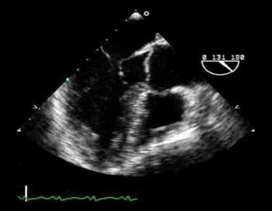
What view is this?
ME LAX
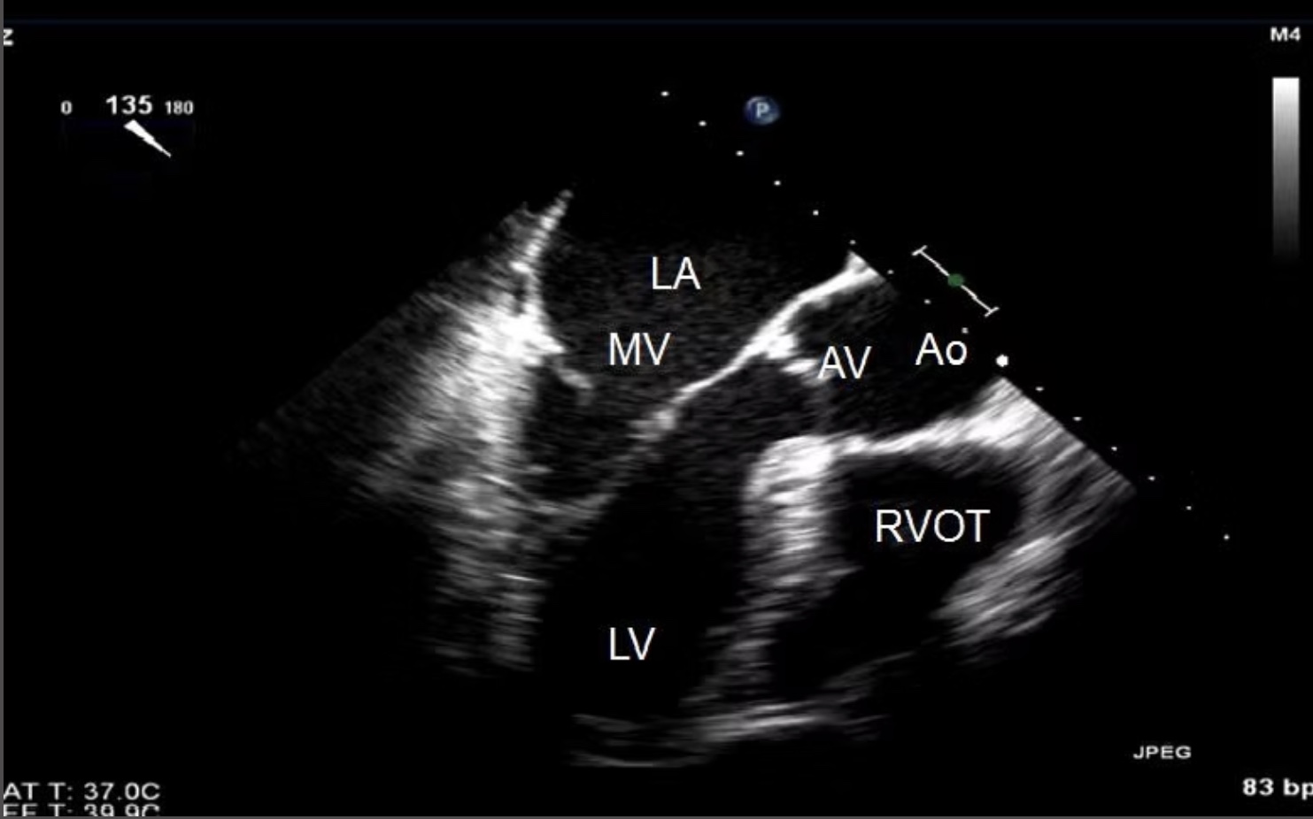
What transducer angle is used to obtain ME LAX?
120-140*
Which MV leaflets can be evaluated on the ME LAX view?
A2-P2
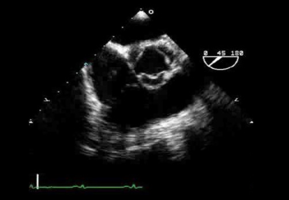
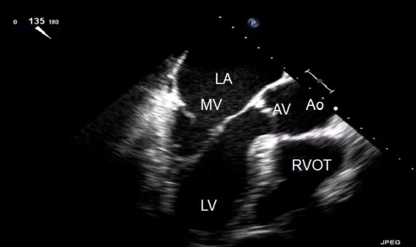
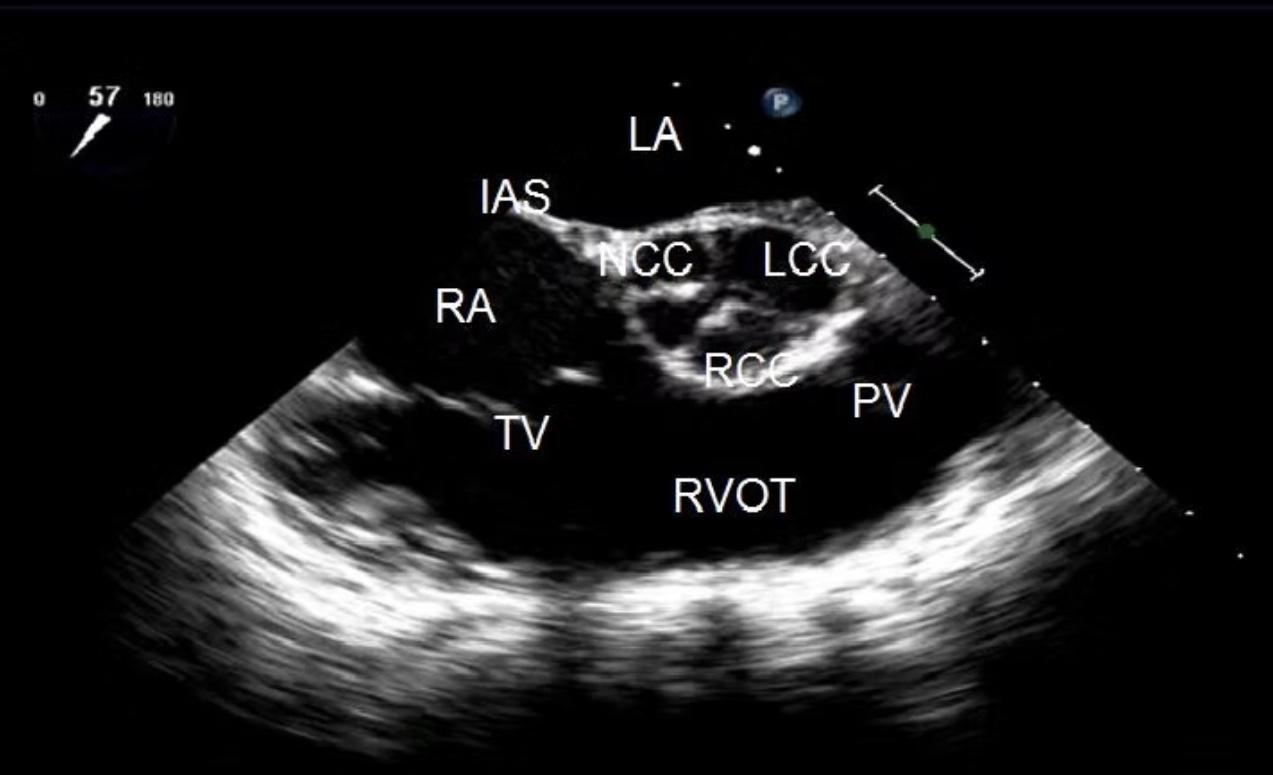
What view is this? ID the structures
ME AV SAX
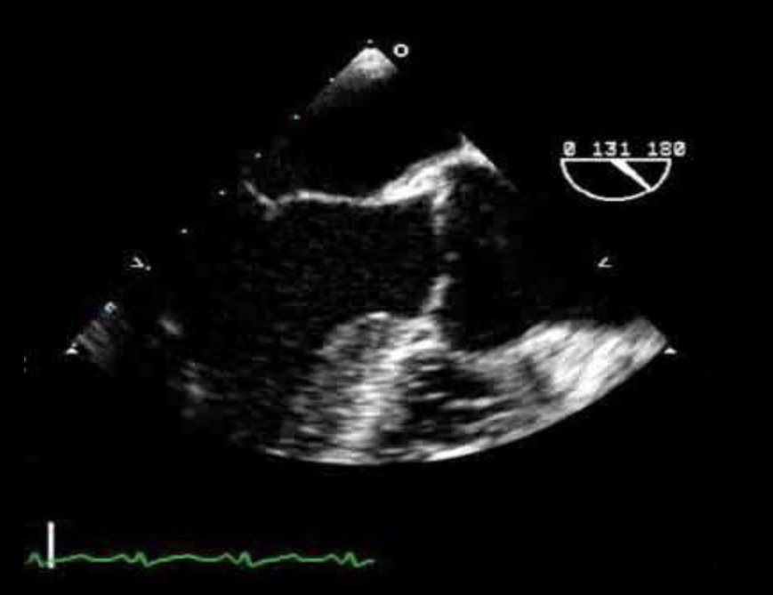
Identify the view and structures
ME AV LAX