BIO 213.01 Human Physiology Lecture : Exam 3 Review (F6) Muscles
1/42
There's no tags or description
Looks like no tags are added yet.
Name | Mastery | Learn | Test | Matching | Spaced |
|---|
No study sessions yet.
43 Terms
What is a sarcomere?
The functional contractile unit of a muscle, made up of thick (myosin) and thin (actin) filaments
What are the components of the sarcomere?
Thick Filaments (Myosin)
Thin Filaments (Actin, Tropomyosin, Troponin)
Z-Lines
A-Band
I-Band
H-Zone
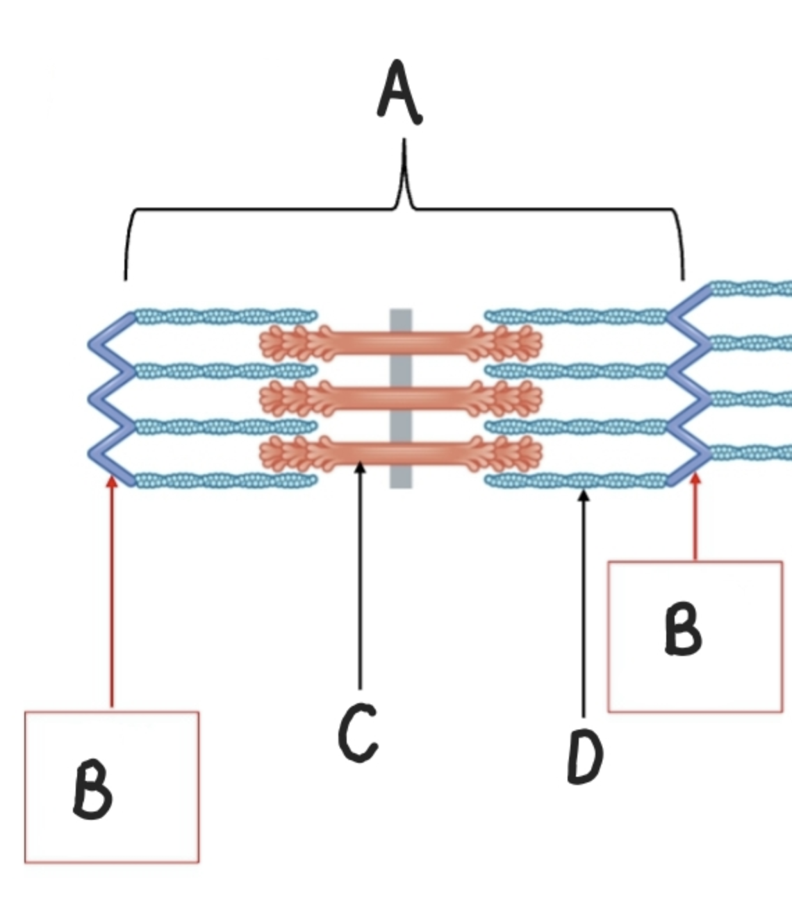
What is Label A?
Sarcomere

What is Label B?
Z-Line

What is Label C?
Thick Filament (Myosin)

What is Label D?
Thin Filament (Actin)

What is Label A?
Z-Line )Defines the boundaries of the sarcomere)

What is Label B?
H-Zone

What is Label D?
I Band

What is Label C?
A-Band
The A-Band on the sarcomere is what?
Thick Filament Length
What is the I-Band on the sacromere?
Thin Filament Length
What is the H-Zone on the sarcomere?
Portions of A-Band where thin filaments don’t reach.
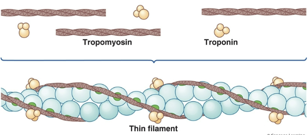
What 2 other micro-filaments cover the binding sites when the when the muscles is at rest?
Tropomyosin and Troponin
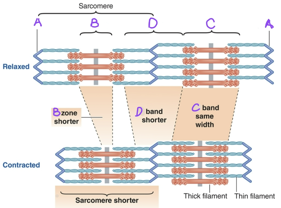
What sections of the sarcomere shorten during a contraction?
H-Zone Shortens
I-Band Shortens
Sarcomere Shortens
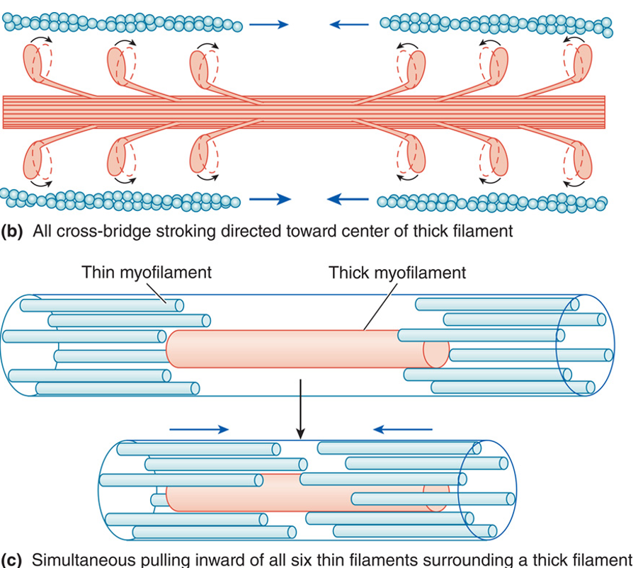
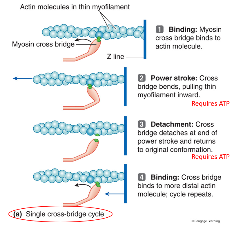
What is the Sliding Filament Mechanism?
When thin filaments slide along thick filaments; which is made possible because of cross-bridge activity.
What is the role of calcium, extracellularly at the neuromuscular junction (NMJ)?
Calcium influx triggers the release of (ACh) from the motor neuron.
Initiates muscle contractions
What is the role of calcium, intracellularly at the muscle fiber?
Calcium is stored at the sarcoplasmic reticulum.
When an action potential reaches the T-Tubules, it triggers the release of Ca²⁺ from the sarcoplasmic reticulum.
Ca²⁺ binds to troponin, causing tropomyosin to move and expose myosin-binding sites on actin, allowing cross-bridge formation and contraction.
What is the role of calcium in relaxation?
Ca²⁺ is pumped back into the sarcoplasmic reticulum by Ca²⁺ pumps (requires ATP).
Function (Regulatory Proteins): Troponin and Tropomyosin
Covers up the actin binding sites when muscle is at rest.
Function: T-Tubules
Carries action potential deep into muscle cell.
Function: Sarcoplasmic Reticulum
Stores and release Ca²⁺.

What is actin and what does it do?
Thin filament, anchored at the Z-Disc
Contains binding sites for myosin heads.
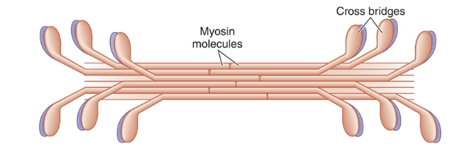
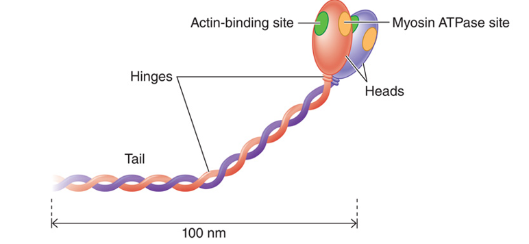
What is myosin and what does it do?
Thick filament with heads that form cross-bridges with actin.
Uses ATP to perform a power stroke, pulling actin toward the M-Line.
What is the levels of organization of skeletal muscles from largest to smallest?
Whole Muscle (Organ)
Fascicles (Bundle of Muscle Fibers)
Muscle Fibers (Cell)
Myofibrils (Specialized Organelle)
Myofilaments (Cellular Protein Responsible for Contraction)
Excitation Contraction Coupling Review
Axon (neuron) transmits an action potential (AP) to the axon terminal, synaptic vesicles release acetylcholine (ACh)
ACh diffuses across synaptic cleft, attaches to nicotinic receptors, opens Na⁺/K⁺ channels – produces an end plate potential (EPP)
End plate potential causes voltage Na⁺ channels to open, an action potential results in the muscle cell
Action potential is transmitted deep into muscle cell via T-tubules
Ca²⁺ (calcium) is released from sarcoplasmic reticulum
Calcium (Ca²⁺) binds to troponin changing its shape
Tropomyosin is moved to expose myosin binding sites on actin
Myosin heads attach to binding sites on actin forming a cross bridge
ATP binds to myosin, is broken down (hydrolyzed), returning the myosin heads to their “high energy” (cocked) shape – myosin heads are ready to form a new cross bridge
Cross bridges attach/detach as long as Ca²⁺ and ATP are present = muscle cell shortening (contraction)
Myosin heads pull on actin and each sarcomere shortens (actin & myosin slide past one another)
Nerve stimulus stops, AChE (acetylcholinesterase) degrades ACh, Ca²⁺ pumped back into sarcoplasmic reticulum – muscle relaxes
Know The Steps To The Sliding Filament Mechanism
Binding
Power Stroke
Detachment
Binding
What is the Role of Acetylcholinesterase?
Enzyme that breaks down ACh in the synaptic cleft
Prevents continuous muscle contraction by stopping the signal, allowing Ca²⁺ to be pumped back into the SR.
What is a motor unit?
One motor neuron and the muscle cells it innervates.
How do we get muscle contractions for varying force?
Recruit larger or smaller motor units and more or less of them.
What do we need for muscles for precision?
To produce precise, delicate, fine motor control
1 Motor unit size may only be as few as 12 muscle fibers.
What do we need for muscles for power?
To produce powerful, gross motor movements
1 Motor unit size may contain 1,500-2,000 muscle fibers.

Golgi Tendon Organs do what?
Detect tension in the tendon to prevent muscle damage
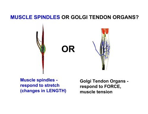
Muscle Spindles do what?
Monitor muscle stretch and initiate reflexive contractions to maintain muscle tone.
In the process of a muscle contracting and relaxing, when is ATP used?
Energizing the myosin head for cross-bridge formation.
Detaching the myosin head from actin.
Pumping Ca²⁺ back into the SR.

Type I (Slow-Twitch)
Efficient at using oxygen for sustained contractions.
Suited for endurance activities.
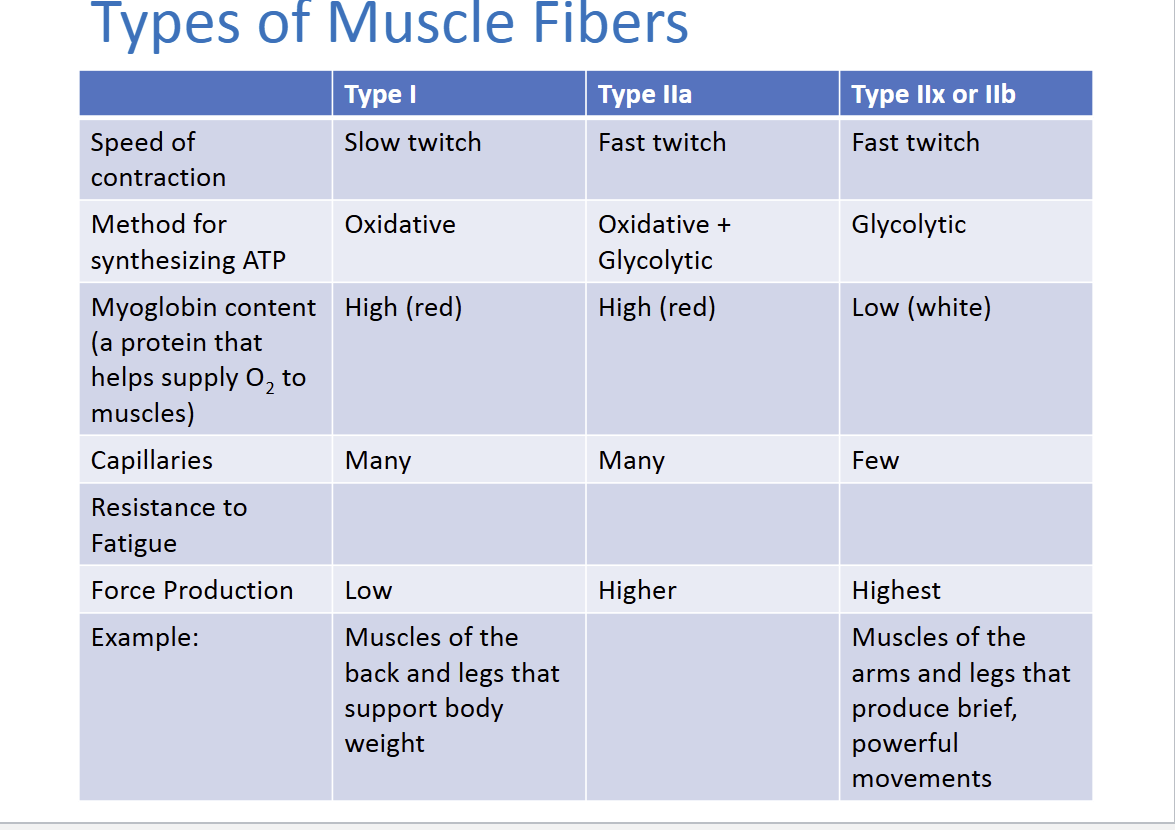
Type IIa (Fast-Twitch Oxidative)
Intermediate fibers with moderate endurance and strength.

Type IIb (Fast-Twitch Glycolytic)
Produce powerful contractions but fatigue quickly.

Overview of the Reflexes
Withdrawal Reflex (Flexion Reflex): This is a protective reflex that causes you to withdraw a body part (e.g., your leg) from a painful stimulus.
Crossed Extensor Reflex: This occurs simultaneously to maintain balance. When one leg withdraws, the opposite leg extends to support your body weight.

Steps in the Diagram
Stimulus: Pain receptors (nociceptors) in the foot detect a painful stimulus (like stepping on something sharp).
Afferent Pathway: Sensory neurons carry the pain signal to the spinal cord.
Integrating Center: The signal reaches the spinal cord, which processes it through interneurons. This results in both withdrawal and extension actions.
Efferent Pathway: Motor neurons send signals to the muscles of both legs to respond appropriately.

Withdrawal Reflex (Injured Limb)
On the side of the painful stimulus:
Flexor Muscles Contract: The muscles on the back of the leg (hamstrings) contract to pull the foot away from the painful object.
Extensor Muscles Relax: The quadriceps relax to allow the leg to bend.

Crossed Extensor Reflex (Opposite Limb)
On the opposite side to maintain balance:
Extensor Muscles Contract: The quadriceps in the opposite leg contract to support your body weight.
Flexor Muscles Relax: The hamstrings on the opposite leg relax, keeping the leg extended and stable.
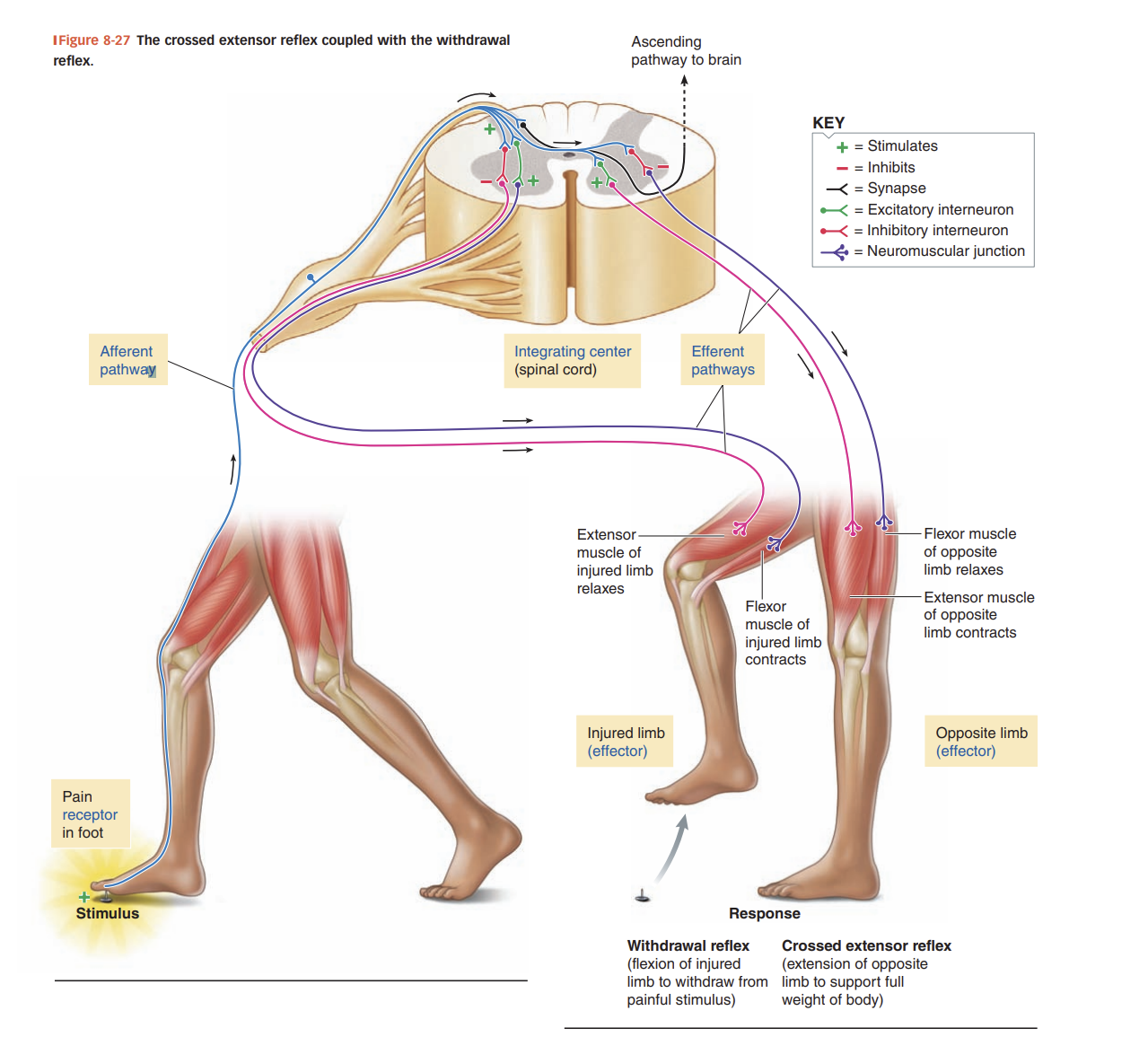
Key Components
Excitatory Interneurons: Activate motor neurons to contract the appropriate muscles.
Inhibitory Interneurons: Prevent contraction of opposing muscles to ensure coordinated movement.
Ascending Pathway: Some signals are sent to the brain for pain perception, though the reflex itself is spinal and immediate.