Lecture 9- Biophysics
 Call Kai
 Learn
Learn Practice Test
 Spaced Repetition
 Match
Match Flashcards
 Knowt Play
1/51

Earn XP
Description and Tags
Concepts of Mechanobiology
Name | Mastery | Learn | Test | Matching | Spaced | Call with Kai | Chat |
|---|
No analytics yet
Send a link to your students to track their progress
52 Terms
What are biological flows shape tissues and organs important for
inner ear and otolith formation, cardiovascular development, kidney morphogenesis and bone morphogenesis
What are some pathologies linked with abnormal flow cells interactions
embryonic heart and abnormal cilia
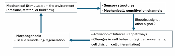
Mechanotransduction
Cells convert mechanical stimuli into electrochemical activity
Crucial for various physiological processes including touch, hearing, balance, and proprioception
Vital role in development, tissue repair, and the progression od diseasef
The majority of growth and remodelling of the vascular network takes place when
blood circulation has already initiated
What forces does flow velocity create
shear stress parallel to the tissue surface

What forces does pressure create
circumferential (tangential) and axial stress (along the long axis of the vessel)
What are potential flow mechanosensing complexes
flow sensitive membrane channels
caveolae
protruding cilia contain with mechanosensitive proteins
endothelial glycocalyx
What does glycocalyx experience
drag forces that are transmitted to the underlying cortical cytoskeleton as well as to distant integrin-dependent adhesions
Endothelial cell polarization, migration, differentiation
Culture ECs elongate along flow direction
However, ECs exposed to cyclic mechanical strain re-orient perpendicularly to the unidirectional shear stress generated by steady laminar flow
Density dependant: high confluent cells migrate against the flow direction where low-density (isolated) cells migrate with the flow
Endocardial cells: cells move towards-higher shear stress; endothelial mesenchymal transition leading valve formation
Why would you use a zebrafish to show the endothelial response to blood flow
Transparent: Flow can be imaged in vivo
Flow responsive genes can be image in vivo
It can live without blood flow: flow can be perturbed
in vivo
What are primary cilia, and how do they act as mechanosensors
Primary cilia are solitary, non-motile cilia found on most mammalian cells, projecting from the apical surface into the tissue lumen. They can detect mechanical stimuli such as:
shear stress from fluid flow
ECM distortion/compression in bone and cartilage
biological flows in brain and spinal cavities
When bent by these forces, they can trigger ionic fluxes, especially Ca²+ signalling, which can then alter cell behaviour such as angiogenesis.
What happens to endothelial cilia in zebrafish embryos
Endothelial cilia are present during angiogenesis and deflected by low flow forces
Cilia deflection leads to endothelial calcium increase as flow forces increase
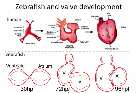
valvulogenesis
Complex embryonic process of developing heart valves from gelatinous endocardial cushions into think, functional fibrous leaflets (flow dependent)
conservation of momentum eqn
delta p = u delta² u
conservation of mass
delta u =0
boundary conditions from wall dynamics
u|omega = ub
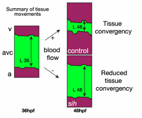
What shows tissue convergence is flow dependent
endocardial cells converge toward the area of high shear and oscillatory flow; not exactly the direction of net flow
How does direct imaging of mechanotransduction work
Shear stress is perturbed exogenously by injecting a bead
Calcium imaging using GcaMP reporters, confirms shear stress mechanotransduction
What are the main stages of cardiac valve development in the zebrafish atrioventricular canal (AVC)?
Cardiac valve development in the atrioventricular canal (AVC) happens in a sequence:
28–48 hpf: cellular volume regulation
54 hpf: cell protrusion and EndoMT (endothelial-to-mesenchymal transition) begin
72 hpf: VIC formation and delamination
126 hpf: elongation and cardiac cushion remodelling
Cell types involved
EdCs = endothelial/endocardial cells
MCs = myocardial cells
VICs = valve interstitial cells
ECM = extracellular matrix
how are the mechanical forces from oscillatory blood flow come from
detected by some sensory machinery
What do ECs experience a broad range of mechanical stimuli…
different flow profiles
shear stress magnitude and gradients
direction
temporal gradients and frequency content
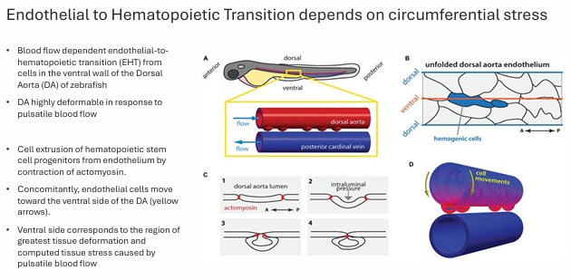
How does circumferential stress regulate endothelial-to-hematopoietic transition (EHT) in the dorsal aorta?
During zebrafish development, blood flow generates circumferential stress in the dorsal aorta (DA), especially on the ventral side. This mechanical deformation helps drive endothelial-to-hematopoietic transition (EHT), where endothelial cells become hematopoietic stem/progenitor cells.
Key points
EHT occurs from cells in the ventral wall of the dorsal aorta
Pulsatile blood flow causes the DA to deform strongly
This creates tissue stress, greatest at the ventral side
Endothelial cells move toward the ventral side
Stem/progenitor cells are then extruded from the endothelium
This extrusion depends on actomyosin contraction
How do cell–ECM and cell–cell interactions mediate mechanotransduction?
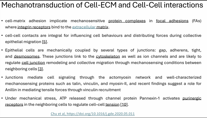
Mechanotransduction occurs through both cell–ECM adhesions and cell–cell junctions, which allow cells to sense, transmit, and respond to forces.
Cell–ECM interactions
Cells attach to the extracellular matrix (ECM) through integrins
Integrins connect to focal adhesions (FAs)
FAs contain mechanosensitive proteins that transmit force to the cytoskeleton
Cell–cell interactions
Neighboring cells are linked by gap, adherens, tight, and desmosomal junctions
These junctions connect to the cytoskeleton
They help distribute forces across tissues and regulate collective migration and junction remodelling
Key mechanosensitive proteins
talin
vinculin
myosin-II
Anillin may also help transmit tensile forces through vinculin recruitment
Additional signaling
Under mechanical stress, ATP can be released through Pannexin-1
ATP then activates purinergic receptors in neighboring cells
This helps regulate cell–cell tension
gap junctions
these are clusters of channels that form tunnels of aqueous connectivity between cells. They allow ions and small molecules to pass freely between cells, facilitating communication and coordination
adherens junctions
connect the actin filaments of neighbouring cells

tight junctions
seal between adjacent cells, preventing the passage of molecules and ions, maintain the selective permeability of epithelial layers
Desomosomes
strong connections that join the intermediate filaments of neighbouring cells
What is an example of measurement and manipulation of mechanical cues
cantilever-based systems, traction force microscopy, laser ablation, micro aspiration, approaches to stretch or compress tissues, strain mapping
Cantilever-based systems
some of the most common methods in measuring tissue mechanical properties
cantilevers as force-transducers which are key for indenting or deforming small regions of tissue
contact forces and depth of indentation or tissue strain are recorded and can be used to calculate a modulus
commercial atomic force microscopy (AFM) systems are cantilever-based systems that were first adopted for in vitro studies of cell monolayers
AFM can report mechanical properties from 1 to 5um of the surface
Example of role of mechanics in pathfinding
neurite outgrowth of cultured RGCs was found to respond to substrate stiffness, which depends on the stretch-activated ion channel
the study showed that RGC axons sense a stiffness gradient in the brain and grow towards softer tissue
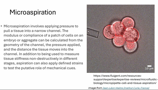
Microaspiration
Microaspiration involves applying pressure to pull a tissue into a narrow channel. The modulus or compliance of cells on an embryo or compliance of a patch of cells on an embryo or aggregate can be calculated from the geometry of the channel, the pressure applied, and the distance the tissue moves into the channel.
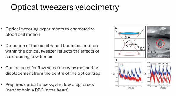
Optical tweezers velocimetry
optical tweezing experiments to characterize blood cell motion
detection of the constrained blood cell motion within the optical tweezer reflects the effects of surrounding flow forces
can be sued for flow velocimetry by measuring displacement from the centre of the optical trap
requires optical access, and low drag forces
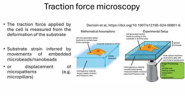
traction force microscopy
the traction force applied by the cell is measured from the deformation of the substrate
substrate strain inferred by movements of embedded microbeads/nanobeads or displacement of micropatterns
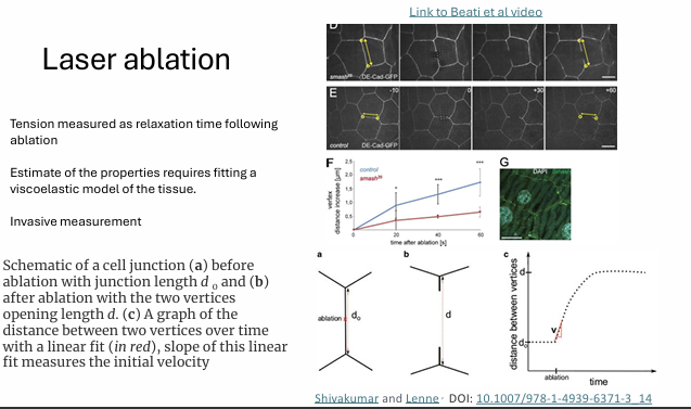
laser ablation
tension measured as relaxation time following ablation
estimate of the properties requires fitting a viscoelastic model of the tissue
invasive measurement
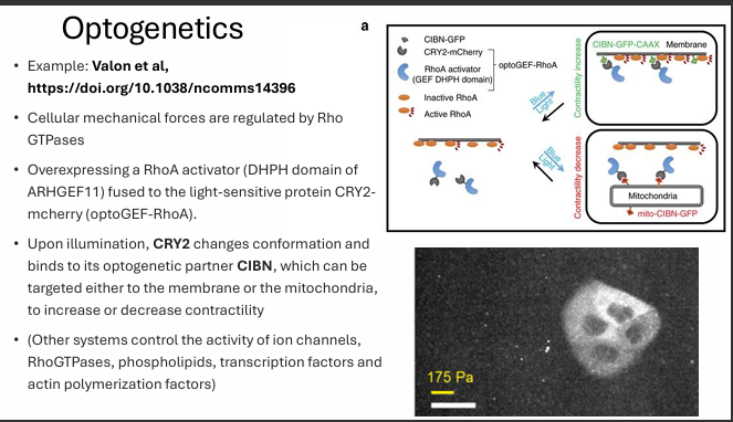
optogenetics - How is optogenetics used to control cell contractility through RhoA?
Optogenetics can control cell mechanics by using light-sensitive proteins to switch RhoA signaling on or off.
Example system
A RhoA activator (the DHPH domain of ARHGEF11) is fused to the light-sensitive protein CRY2 → optoGEF-RhoA
Light causes CRY2 to change conformation and bind its partner CIBN
What happens after illumination?
CIBN can be targeted to different places:
Membrane-targeted CIBN
→ recruits optoGEF-RhoA to the membrane
→ activates RhoA
→ increases actomyosin contractilityMitochondria-targeted CIBN
→ pulls optoGEF-RhoA away from the membrane
→ reduces RhoA signaling at the cortex
→ decreases contractility
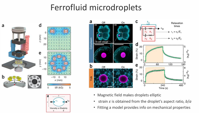
ferrofluid microdroplets
Magnetic field makes droplets elliptic
strain ɛ is obtained from the droplet′s aspect ratio, b/a
Fitting a model provides info on mechanical properties
Mechanical properties can be
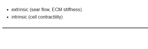
extrinsic or intrinsic
What is an important contribution to tissue and organ development
biomechanics of the microenvironment
Mechanotransduction contributes to…
cell orientation, migration and differentiation
Many new tools are available to…
measure the mechanical properties and perturb the micromechanical environment, and measure cellular response to mechanical cues
What is needed to fully understand the biomechanics of the microenvironment and causal effects with the cellular response
A combination of mechanical modelling and image analysis
biomechanics
mechanical processes that directly shape living organisms. Genes and environment are involved in directing material properties
compliance
the ability to deform under an applied force
compression
the object is under compression when it experiences a negative strain
elastic modulus
defines the elastic behaviour under an applied stress

force
an interaction with a magnitude and direction that changes the motion of an object or deforms it
mechanobiology
the feedbacks from mechanical processes that guide biology
tension
experiencing a positive strain

stiffness
the resistance to deformation under an applied force
strain
a dimensionless term to describe the deformation of an object caused by the force applied
stress
amount of force that is applied to a unit area
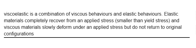
viscoelasticity
combination of viscous behaviours and elastic behaviours