clinical notes part one: pelvic limb
 Call Kai
 Learn
Learn Practice Test
 Spaced Repetition
 Match
Match Flashcards
 Knowt Play
1/59

There's no tags or description
Looks like no tags are added yet.
Name | Mastery | Learn | Test | Matching | Spaced | Call with Kai | Chat |
|---|
No analytics yet
Send a link to your students to track their progress
60 Terms
where does pelvic fracture usually fractured? how to solve if it is a minimal displacement?
car accidents, in fractures with minimal displacement, spontaneous recovery can occur in quiet setting without surgical interference.
since pelvis is box shaped, there are usually TWO fractures- if one site is fixed, the other usually assurmes to approximate position
what can the iliac crest/ dorsal spine be used for
bone marrow sample
can the ischatic tuberosity be palpated on dog?
yes,
what is the ischiatic tuberosity and tuber coxae common sites for
bc bone lies directly under the skin tuber coxae and ischiatic tuberosity are common sites for pressure sores in debilitated animals
what can happen to pelvic symphysis after trauma
it can be separated and is usually accompanied by sacroiliac luxation
abnormal shallowness of acetabulum is associated with
hip dysplasia
and can cause coxofemoral joint to become unstable and arthritic
shape of cat pelvis?
box shaped rather than flared
places for bone marrow samples
proximal end of femur, trochanteric fossa
greater trochanter is a useful landmark for
surgical approach to hip, detecting craniodorsal luxation of hip and placement of the intramedullary pin
Osteochondritis dissecans (OCD) can affect what?
the leisons may affect the lateral condyle of the femur
tibial tuberosity is an important landmark for
numerus clinical and surgical procedures
long bones in cat are longer/shorter?
longer than those of dog
what ligament of the pelvis is not present in a cat?
sacrotuberous ligament
popliteal LN is
superficially located and palpable
what muscle region is commonly used to give IM injections?
caudal muscles
iliopsoas injury can cause what? What dogs is it most common in? what symptoms do they exhibit?
hind limb lameness in dogs but can go undiagnosed due to self limiting nature
most common in agility dogs
exhibit decreased hind limb extension when jumping, decreased drive in hind limbs when weaving, and decreased weight bearing in the affected limb
how important is the QF? why? whats it innervated by? what can QF be used for in clinic?
very, its the primary extensor of the stifle joint and MUST be function for the animal to bear weight on the limb
innervated by femoral nerve
can be used for IM injections
fibular can also be referred to as
peroneal
same for fibular nerve.
traumatic disruption of the common calcaneus tendon results in?
animal walking with entire metatarsus on the ground
solus m in dog? what is it? location?
no its in cats
can see this muscle on lateral side of lateral head of gastrocnemius m
dont confuse it for the superficial digital flexor
well developed in humans and pigs
which joint is most commonly luxated?
coxofemoral, despite ligament of the femoral head, depth of acetabulum and surround muscles
ligament of the femoral head will be torn with luxation
what direction is patella luxation found?
medially
cranial cruciate ruptures require
surgical intervention at re-establishing proper alignment
diagnostic procedure for evaluating crutiate stability
tibial thrust- feel one femoral condyle and the tibial plateau with one finger and flex the hock
if tibia moves cranially with respect to the femoral condyles, this will indicate cranial cruciate instability
cranial drawer- use 2 hands, place thumb of one touching the fibular head caudally, and the first finger touches the tibial tuberosity, the thumb of the other hand palpates the lateral fabella and the first finger palpates the patella
if tibia moves cranially more than 2-3 mm, this indicates cranial cruciate ligament instability
why are the landmards around the femoral triangle important?
bc the femoral a. is commonly used for palpation of pulses and collection of arterial samples
what is the lateral saphenous vein good for? why? is it larger in dog or cat?
venipuncture, bc superficial location, size, lack of companion artery
larger in dog, in cat the medial saphenous vein is perferred
what artery is a common site to take peripheral pulse?
dorsal pedal running superficially over tarsus
can also be used to obtain arterial sample as well as catheter in critical patients
what results from damage to femoral nerve
inability to extend stifle joint and bear weight on limb
if damage occurs before saphenous nerve will give off, it will also cause loss of sensation on the medial side of hind limb
common fibular nerve is also called
peroneal nerve
common fibular nerve damage: distal to the level at which muscular branches are given off? proximal to where muscular branches are given off?
distal to the level at which muscular branches are given off: only cutaneous sensation will be lost (goes for TIBIAL TOO)
proximal to where muscular branches are given off: unable to flex tarsal or extend digital joint, as well as show sensory deficit
tibial nerve: proximal to where muscular branches are given off?
dog will be unable to flex digital joints and decreased ability to extend tarsal joints; sensory deficit
bc of thigh m. (contribute to common calcanean tendon), dog still has some ability to extend tarsal joints - limited
sciatic nerve damage: mid thigh? before muscular branches?
mid thigh: could occur bc of misplaced IM injection
will loose function of tibial and common fibular nerve, making patient unable to extend or flex digital joints or flex tarsal joints
could retain some extension of tarsal joints bc of caudal thigh muscles to common calcanean tendon
before muscular branches: loose ability to making patient unable to flex/extend tarsal and digital joint or flex stifle joint
if glute nerves are still intact, patient can still extend hip joint and bear weight on limb since femoral nerve is intact
happens with trauma/ surgical manipulation of the femoral head and neck

5 in cat
Gluteofemoralis m.
(5) Cd. vert to fascia lata “tail swisher”
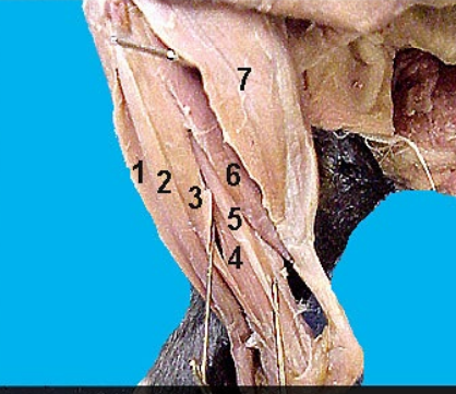
6?
Soleus m.
Fibula to tuber calcanei Extend tarsus
Hematopoiesis: occurs in?
occurs in the flat bones and the extremities of long bones.
when do you conduct Bone Marrow Aspirate/biopsy
when you see Non-regenerative anemia, Persistent neutropenia or thrombocytopenia, Neoplasia and/or infectious organisms.
areas to conduct Bone Marrow Aspirate/biopsy: small dog/cats, large, all small animals
small dog/cat: lateral aspect of wind of ilium ( for core biopsy), trochanter of proximal femur (for aspiration biopsies)
Large dog: dorsal approach to the iliac crest for aspiration and core biopsies
all small animals: proximal humerus is excellent site for both aspiration and core biopsies

ligaments of sacroiliac joint:

coxofemoral joint: movements, limited by what?
The head of the femur is spherical; all movements are possible: extension, flexion, abduction, adduction, and circumduction.
limited by ligament of the head of the femur, which extends from the acetabular fossa to the fovea capitis of the femoral head.
hip dysplasia: what is it
deformity of hip joint during growth period resulting in laxity of the joint
as laxity develops, the articular surfaces of the two bones lose contact with each other (subluxation) and this causes degenerative joint disease or osteoarthritis.
what causes the symptoms of hip dysplasia? can it be bilateral?
subluxation and the remodeling of the hip
yes it can affect both left and right hip

how to fix hip dysplasia
surgery
1. femoral head and neck excision (small patients)
triple pelvic osteotomy (young dogs that have minimal to no arthritic changes of the hips)
total hip replacement: large breed dogs
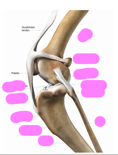
femoropatellar? patella, patellar ligament?

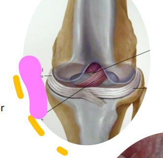
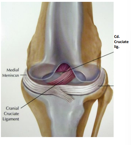
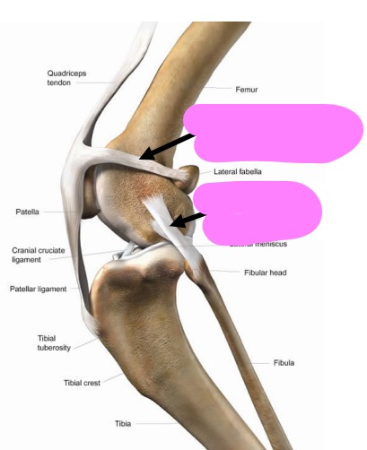


Cruciate ligaments limit the
sliding of the tibia forward and backward in relation to the femur.
Rupture of the cranial cruciate allows the tibia to be drawn forward on the femoral condyles, the cranial “drawer” sign; rupture of the caudal allows the tibia to be drawn caudally, the caudal “drawer” sign.
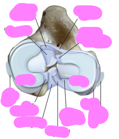
menisci are held in place by? A transverse ligament that extends between? A meniscofemoral ligament extends from the
The menisci are held in place by cranial and caudal meniscotibial ligaments that attach to the tibia.
A transverse ligament that extends between their cranial ends
. A meniscofemoral ligament extends from the caudal end of the lateral meniscus to the intercondylar surface of the lateral condyle.
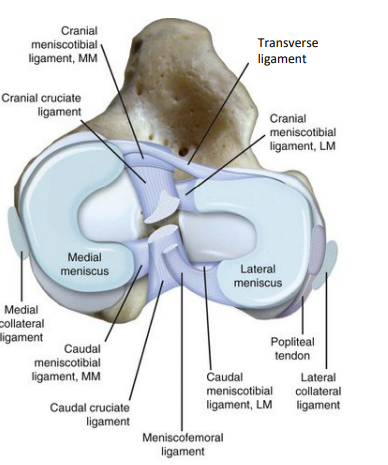
what does menisci do?
supply congruency in shape between articular surface and provide stability to joint
anchored to each other
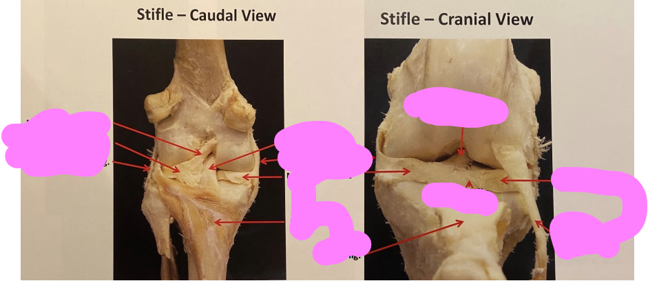

Patellar ligament:
Lateral femoropatellar ligament:
Medial femoropatellar ligament:
Lateral collateral ligament
Medial collateral ligament:
all connect to?

Cranial Cruciate Rupture
leading cause of lameness in the dog.

Cranial Cruciate Rupture/Cranial “drawer” sign
Anatomical Landmarks: Patella and lateral fabella (sesamoid) Tibial tuberosity (crest) and fibula:
With the femur held in place with the right hand, the examiner applies a firm cranially directed force to the tibia with the left hand. In a mature dog, the healthy, intact CrCL will not permit cranial tibial translation with the stifle held in extension or in flexion.
tibial thrust landmark
Tibial tuberosity and lateral fabella(sesamoid)
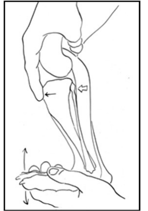
the right hand holds the stifle in a standing angle while the examiner's left hand flexes the hock to simulate weight-bearing.
this maneuver creates cranial tibial thrust by tensing the gastrocnemius muscle. In the CrCL-deficient stifle this cranially-directed shear force palpably shifts the tibia cranial relative to the femu
Cranial Cruciate Rupture surgery:
Tibial Plateau Leveling Operation (TPLO)
Tibial tuberosity Advancement (TTA)

Tibial tuberosity Advancement (TTA)