Development lecture 4
1/37
There's no tags or description
Looks like no tags are added yet.
Name | Mastery | Learn | Test | Matching | Spaced |
|---|
No study sessions yet.
38 Terms
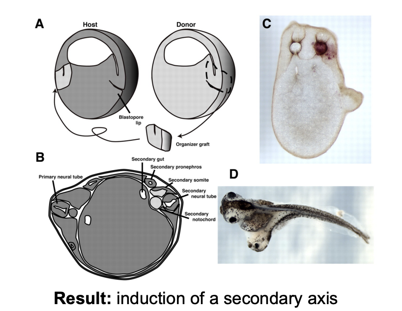
Organiser grafts showing inductin
Experiement: graft tissue from the dorsal side ontot hte ventral side
Result: Induction of a secondary axis
Where does the information come from?
From outside→ environmental factors (gravity, sperm entry point)
From inside→ maternal determinants put into egg cell
From outside→ cells signal to each other (induction)
after having been made different by maternal determinants
kidney
But how to find the machinery needed for induction
Need to find the different signalling pathways and regulatorys of gene expression
→ Genetic screen→ to find zygotic genome stuff
some definitions for the experiemnt
DTS→ dominant temperature sensitive
29 degrees→ kills flies or makes them infertile
easy way to get id of DTS carrying progeny
b→ balancer chromosome
lethal when homozygous and with clear markers
e.g beta-gal or GFP expression
or → curly wings for adults
How do this?
Treat a male with mutagen (EMS) (a/a)→ to form a* mutation sperm
Cross with a DTS/b female→ viable at 29 degrees
approx 5000 individual males outcrossed→ no. required for saturation of chromosome with mutations
i.e the mutant sperm from og fly→ mutant flies cross with more DTS/b female
Grow this F1 progeny→
b/b is embronicaly lethal
DTS/b and a*/DTS dead at 29 degrees
a*/b and a*/b mmale nad female surive!
Cross these a*/b x a*/b
Result:
a*/a*→ embryonic lethal (with pattern defect?? that can be studied)
a*/b→ heterozygous viable shock
b/b→ embryonic lethal
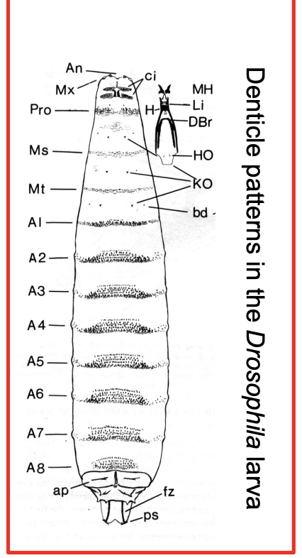
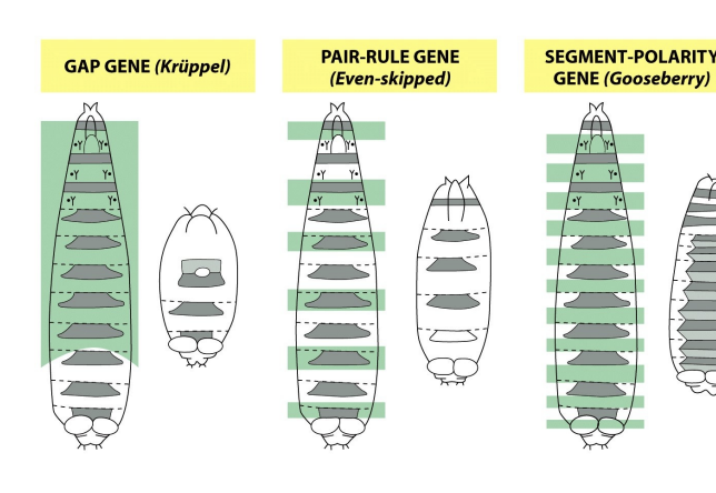
What does the a*/a* show?
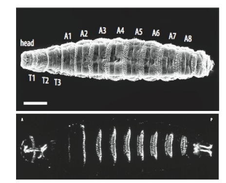
Denticle patterns that develop on the ventral of each segment of larvae
Used to look for aberrations in the normal pattern of development
→ THEREFORE→ find genes for each place in the larvae??
What ere the three patterns they found and their genes?
What are denticles used for?
feed
for larva to crawl on substrate in peristalsis
Therefore what did the mutants screens show?
mutations that disrupt this pattern
discovered gene beeded for normal development
and THEREFORE
→ Could show the gene encoded for:
elements of signalling pathways
and
regulatory networks controlling gene expression
Example of a mutant screen
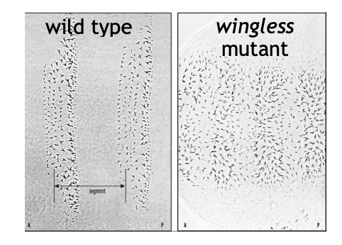
found a gene for wingless signalling pathway
→ found many factors required to build larvae
Why this was important in overal research of development
toolkit is largely conserved
same genes used to built a fly could build a fish
→ therfore→ studing fly found many basic mechanisms to contruct humans
why model organisms are really useful!
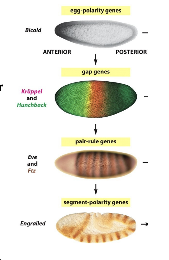
How did these mutatns interact with eachother
Formed a hierachy
→ showed that from intiail egg polarity→ gradual incrase in complexity to divide up the embryp into a large number of repeating units
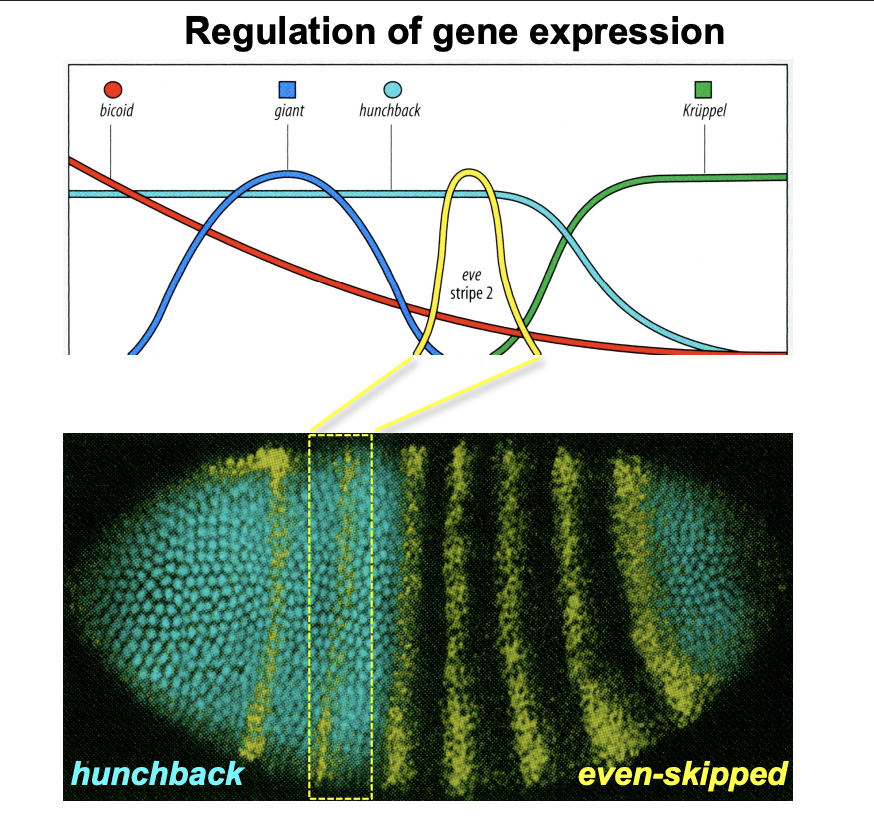
How is the gene expression regulated
Expression of eve stripe 2:
Respressor→ giant and Kruppel
Promotor→ bicoid and hunchback
Result→ get a peak of eve when there is low giant and kruppel but also high bicoid and hunchback
Overall: different cobinations of promotors and repressors→ to narrow down the effect of a specific region
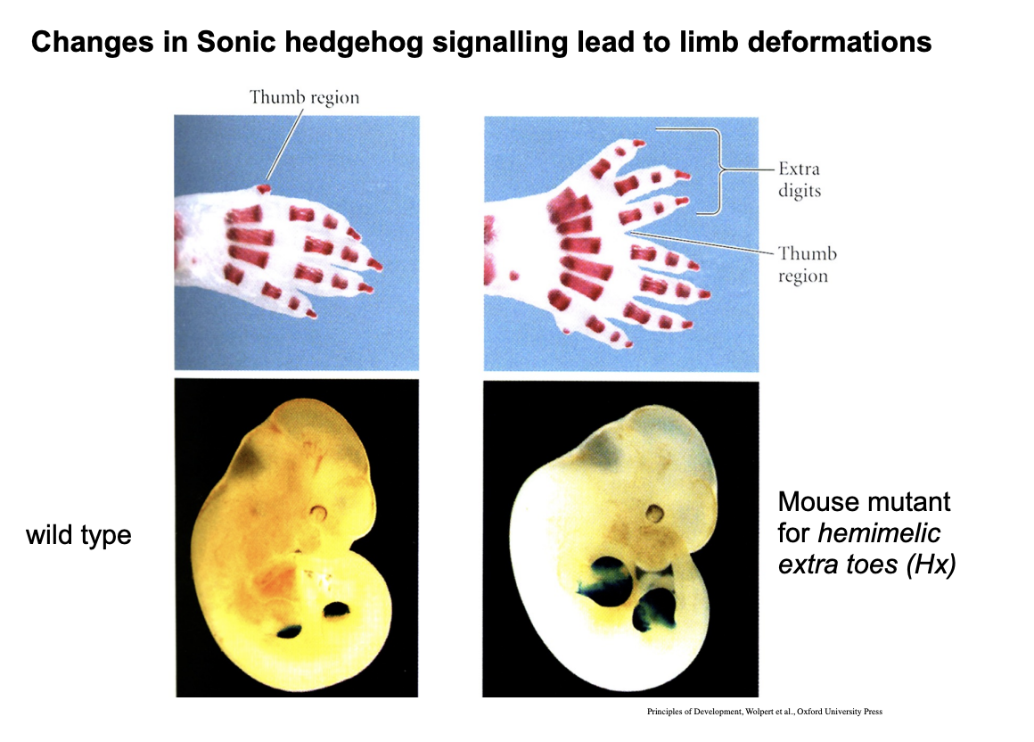
Example of conserved genes in Drosophila to vertebrates
Drosophila→ hegdeghog
mutant→ embryo has a prickly pattern on dentricles
encodes a diffusable signalling molcule needed for patterning the segment
Vertebrate→ sonic hedgehog
homolgous gene
key signalling molecules for vertebrate embryos
e.g central nervous system
limbs
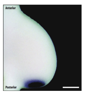
Sonic Hedgehog in developing limb of a chick embryo (or mouse/human)
In situ hybridisation dark staning→ shows SONIC HEDGHOG transitps
expressed by cells at the posterior margin of the limb bud
so Sonic hedgehog diffused anterioroly as a graded signal
→ This is critical for patterning of the limb in this axis!
So we know have information in the cytoplasm that regualted zygotic genes to lay out he ground plan for the embryo but now…
…We need additional mechanism which assign cells to particular pathways of differentiation bthe position that they have been assigned to!
The question to answer is
How do cells interpret positional information in the developing embryo?
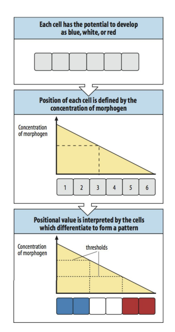
French flag Model
Organisms develop into groupd (fields), separated by boundaries (which is controlled by what is above)
Each field will be dedicated to making a part of the final strucutre e.g arm or leg→ they have gene unlcoked for specific parts
Fields generate information about the position→ A gradient of a diffusible substance established between the boundaries
Cells the assess their position by reference to the local level of the gradient→ differentiate accordingly
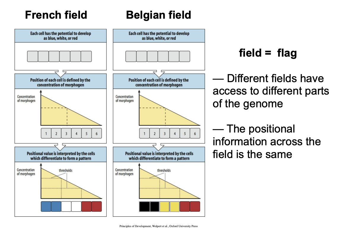
All fields generate the same information BUT cells of each field have access to a different part of the genetic programme!→ so the same gradient does not mean the same limb will be generated
Even though the positional information across the field is the same
Different fields have access to different parts of the genome
This is why→ in the diagram→ yess the positional information is the same→ so forms a flag of three colours in the same positions
BUT
Because the fields are different in each, the have access to different parts of the genome
→ OVERALL: make different colours→ different flags!
Overall two aspects of patterns which are working
Original field genetic assignments
Positional information
Example of evidence for this
The limb development of the chick
Why this is good evidence
→ the development of chick and mouse limbs are similar-. very ancient pathways!!
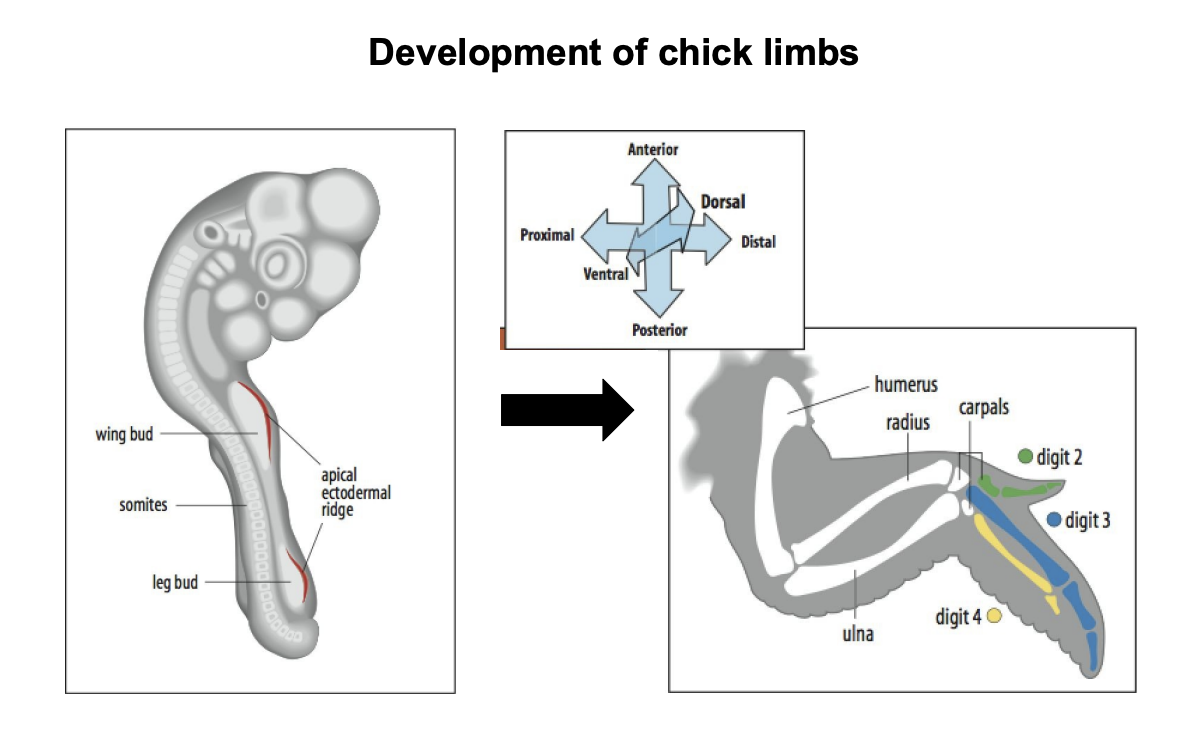
Axis of the chick limb
Proximal to Distal (closest to body→ away from the body)
Anterior→ Anterior
Dorsal→ Ventral
Testing French flag Model→ Prediction
Two fields of cells making different flags will have the same positional information
THEREFORE we predict
Cells from one group transplanted to the other
Will read new position
Differentiate appropriately
BUT
STILL following the programme to which they have previsouly been assigned
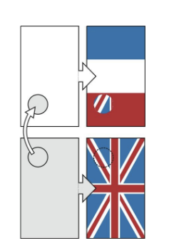
E.g explanation of this
Part of bottom unition flag put in top of french flag
Read its new position
STILL develops as a union flag
BUT→ as the top of the flag, instead of the bottom

Actual experimental evidence of this in chicks!
Experiment
Proximal leg
put into
Distal wing
Result:
Forms distal leg
→ Matches new position but develop as ind limb
Conclusion:
cells are assigned in groups to make different parts of the organism
psotional information in each group in the same→ must be universal cues but genetic code they have access to is different
Next question to ask
Hows is this positional information generated?
How are elements laid down in a proximal to distal sequence: the zones
Progress Zone→ region of dividing cells at tip of limb bud
Apical ectodermal ridge (AER)→ most distal layer of ectodermal cells
The most distal region!
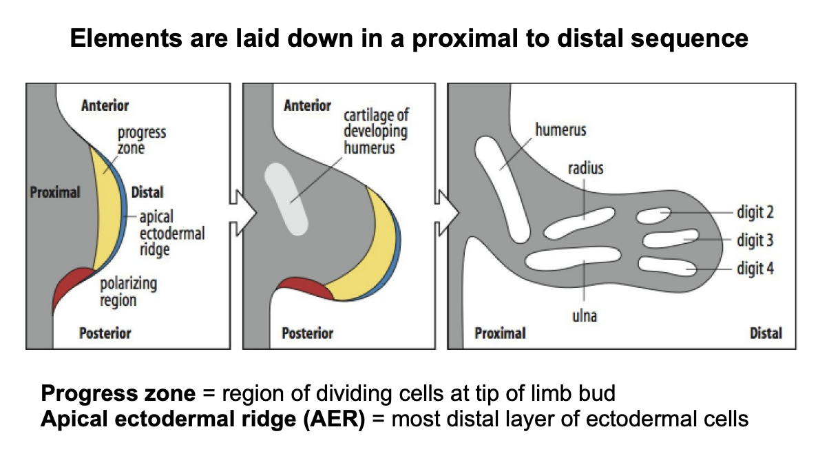
How do cells position in the porximo-disstal axis
Measuring the time they spend in a region of dividing cells at the tip of the limbs
→ progress zone
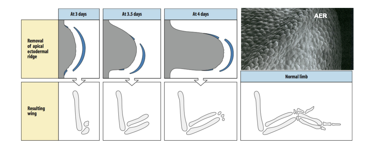
Evidence for this?
Removes the Apical ectodermal region at different time intervals
Results:
development of the limb stopped at different stages of development
Conclusion:
- once leaves the progress zone, stop dividing and differentiate, laying down elements of the pattern in a proximo-distal sequence
What happened if transplanted an AER from a mouse?
Recovered the limbs
→ shows conserved mechanism!
What in the Apical Ectodermal Region maintains proliferation?
Fibroblast Growth Factors (FGFs)
→ The more distal the part of the limb→ the most time it has spent in the AER
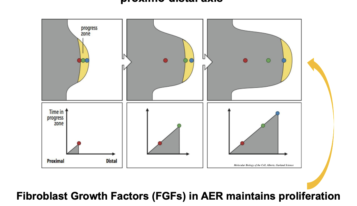
How we know the FGFs do this?
Even if AER is removed
the FGFs are sufficent to elicit cell division
How is Antero-posterior axis patterned?
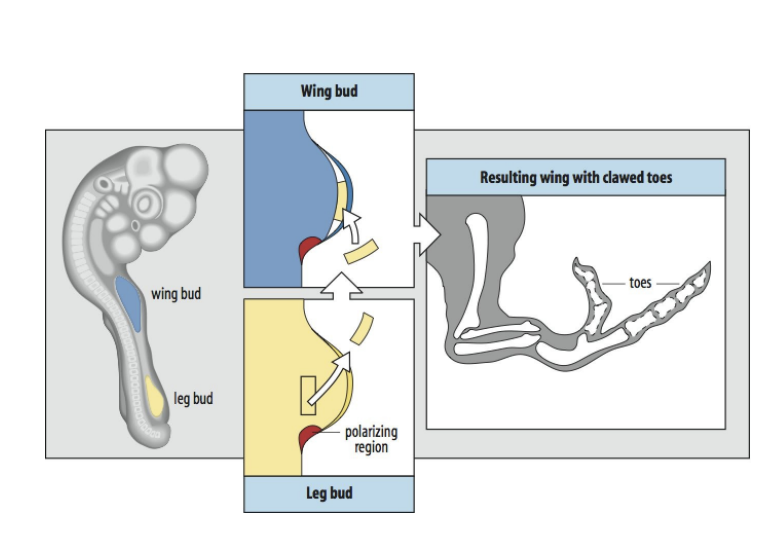
Layed down by measuring their position to the zone of polarising activity ZPA
This is the posterior region
What does the ZPA do to cause these patterned?
Generates a gradient of a substance
Diffuses across the limb
cells respond according to their level in the gradient
Experiemnts to show this
Transplanting the Zone of Polarising Activity ZPA
Add extra ZPA to the anterior margin
→ Cell differentiate to make mirror image duplication of the posterior digits
Add small amount to the anterior margin
→ Get a duplication of the small anterior limb but not symmetric→ does not reach high enough levesl to form another posterior type part of the limb
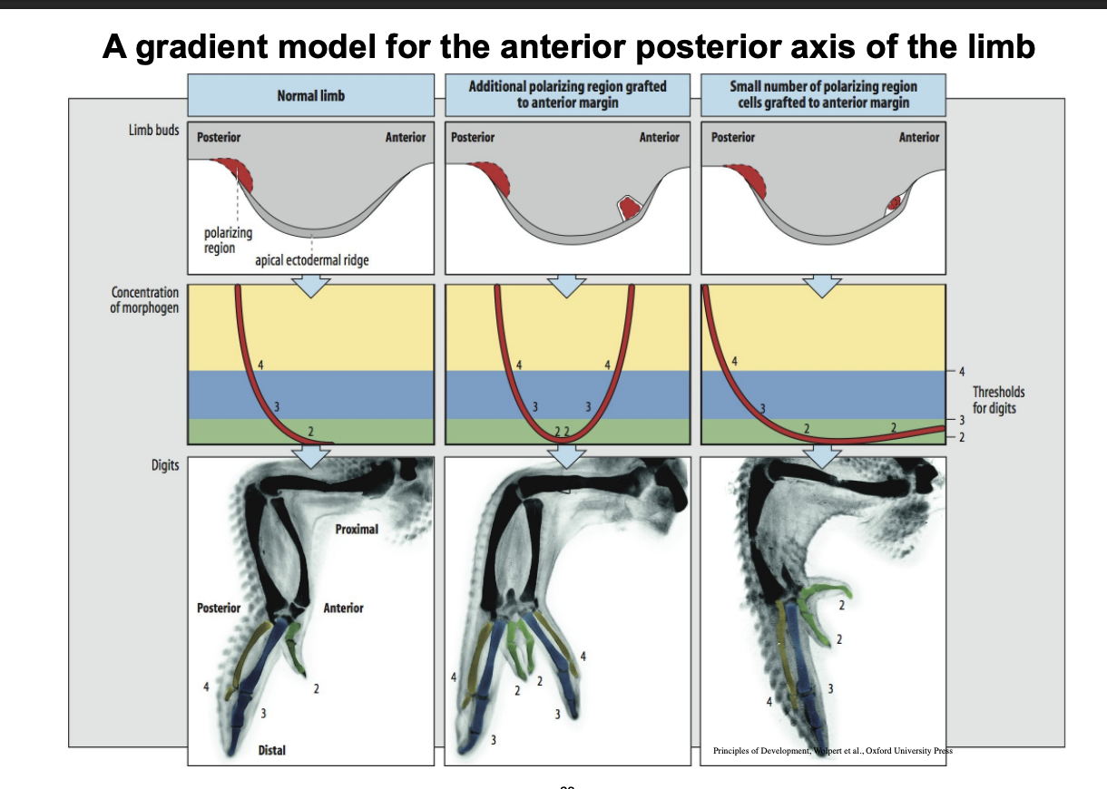
What we now know is the gradient signal
→ Sonic Hedgog synthesised by the cells in this region
However we now know the two signalling centres that organise limb bud development and what variable they are based on
Poximo-distal
FGFs from the AER
time based
Antero-posterior
Shh from ZPA
spatial based
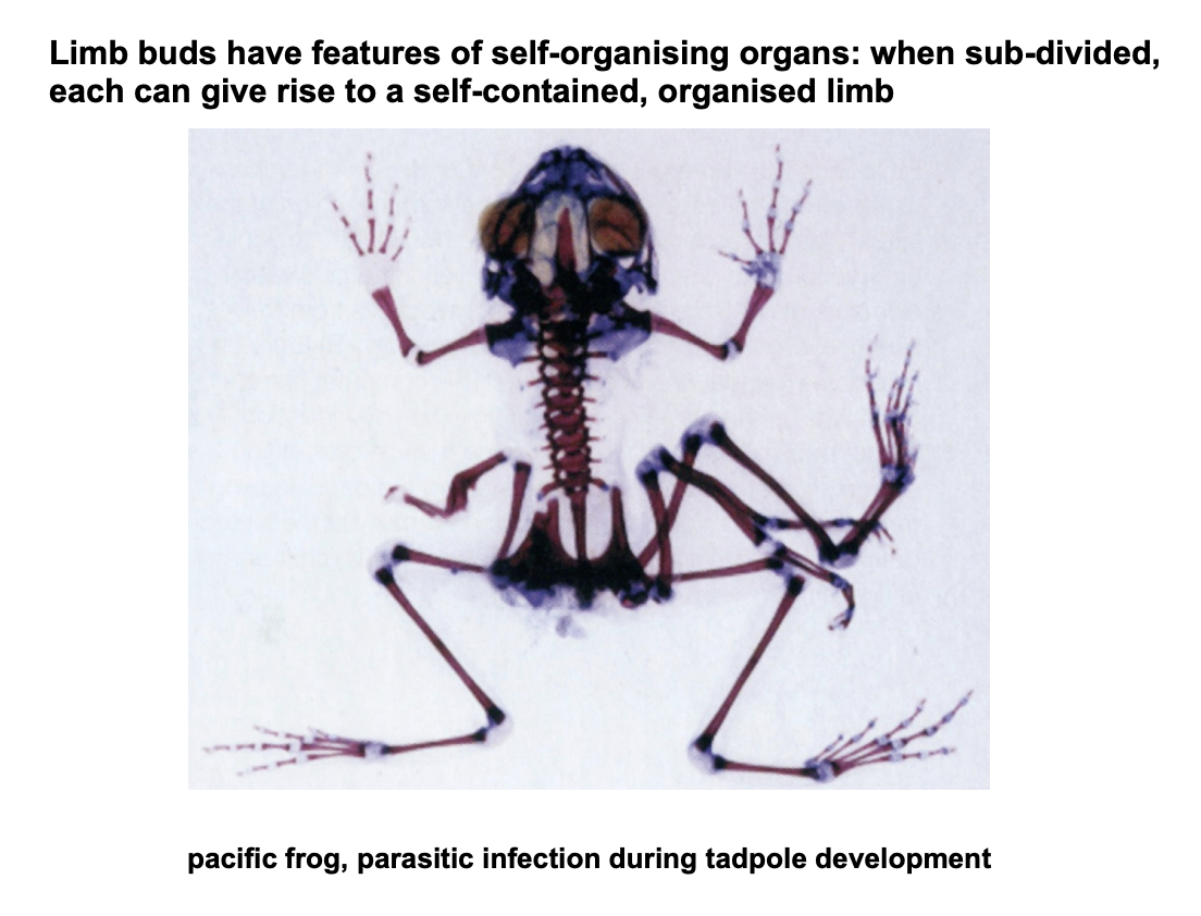
Parasite in frog→ example of limbs buds behaving like self-organised organs
Example of how one mutation/paraite
Can cause rise of self-contained organised limb
WHY?→ limb buds have features of self-organising organs