Class 6 Lecture
 Call Kai
 Learn
Learn Practice Test
 Spaced Repetition
 Match
Match Flashcards
 Knowt Play
1/14

There's no tags or description
Looks like no tags are added yet.
Name | Mastery | Learn | Test | Matching | Spaced | Call with Kai | Chat |
|---|
No analytics yet
Send a link to your students to track their progress
15 Terms

Protein folding is influenced by:
• Conformational degeneracy of the peptide chain.
• Hydrophobic effect
• Non-covalent interactions of atoms within the peptide backbone
• Non-covalent interactions of amino acid side chains
• Covalent disulfide bonds between cysteine side chains
favors U or F
Protein folding is influenced by:
• Conformational degeneracy of the peptide chain U
• Hydrophobic effect F
• Non-covalent interactions of atoms within the peptide backbone F
• Non-covalent interactions of amino acid side chains F
• Covalent disulfide bonds between cysteine side chains F
protein folding logistics
proteins are not stabilized by a lot (ex. 1-2 H-bonds)
proteins kind of go bad after a while (get oxidized)
ubiquitin destabilizes proteins and can’t do that if the protein is extremely stable
Non-covalent bonds/interactions
Chemical groups that are not close in a protein’s linear sequence can be close and interact in the folded state
Non-covalent bonds/interactions image helix

Intrahelical (within a single helix) and interhelical (between separate helices)
Cysteine
forms disulfide bonds

cell environment
outside cell: oxidizing, favors disulfide bonds
opposite inside
bacteria make proteins that digest the macromolecules of other bacteria (self-resistance)
insulin disulfide bonds
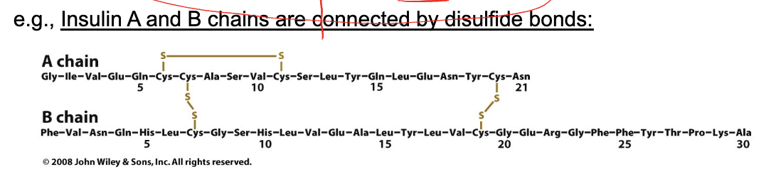
top first cystein bond with other cystein further away, one next to it bond below
Characteristics of misfolded proteins associated with disease

switch to beta-sheets → many copies form aggregates
Why do misfolded proteins have b- strands and not a-helices?
difference in alpha helix and beta strand interaction
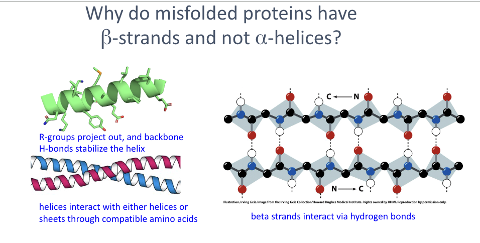
Why do misfolded proteins have b- strands and not a-helices? deepseek
β-strands dominate in disease-causing aggregates because:
β-strands H-bond between chains → form stable, self-templating intermolecular β-sheets (amyloid cross-β structure).
α-helices H-bond within the same chain → internally satisfied, so they don't aggregate or propagate.
Cross-β stacks are extremely stable — protease-resistant, heat-resistant, and act as a template for more misfolding.
Kinetics: when normal folding fails, exposed backbone readily forms intermolecular β-sheets; α-helical aggregates have no equivalent stable, repeating architecture.
One-liner: β-strands win in misfolding because their hydrogen bonds are intermolecular and self-propagating, whereas α-helix H-bonds are intramolecular and self-contained.
Transmissible Spongiform Encephalopathies (TSEs) – Misfolding of PrP
formation of fibrils that are long enough to break cells via mechanical stress
1-121: disordered
122-180 (end or protein): structured (alpha-helix)

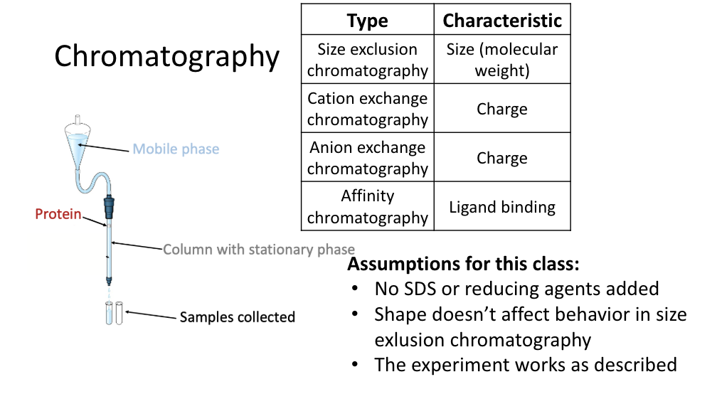
Experimental techniques for proteins
By charge/isoelectric point
ion exchange chromatography
isoelectric focusing
By molecular weight
Size exclusion chromatpgraphy (gel filtration)
SDS-PAGE
Both
2-dimensional gel electrophoresis
SDS-PAGE Assumptions for this class:
• SDS is added and proteins are unfolded
• Unaffected by charge of chemical groups of the protein
• Disulfide bonds are reduced
• Samples always start at the top of the gel represented in an image
• The experiment works as described
Chromatography