Family Medicine: Dermatology
 Call Kai
 Learn
Learn Practice Test
 Spaced Repetition
 Match
Match Flashcards
 Knowt Play
1/73

There's no tags or description
Looks like no tags are added yet.
Name | Mastery | Learn | Test | Matching | Spaced | Call with Kai | Chat |
|---|
No analytics yet
Send a link to your students to track their progress
74 Terms
Acne Vulgaris
Plugged, inflamed oil glands leading to open comedones, closed comedones, papules, pustules, nodules, or cysts that can scar
-Driven by excess oil, follicular plugging, Cutibacterium acnes, and inflammation
-Dx: clinical, mild has only comedones, moderate has papules and pustules, severe has numerous inflammatory lesions, and cystic has large nodulocystic lesions on face, jaw, and upper body
-Tx: topical retinoid, benzoyl peroxide, topical clindamycin, systemic antibiotic, isotretinoin
Topical Retinoid
What is the treatment of choice for Grade 1 acne?
-Comedones only
Topical Clindamycin
What should be added after topical retinoid and benzoyl peroxide in the treatment of Grade 2 acne?
-Papules and pustules
Doxycycline
What should be added to the treatment of Grade 3 acne?
-Numerous inflammatory lesions, high scarring risk
Isotretinoin
What should be added to the treatment of Grade 4 acne?
-Scarring/refractory
-Contraindicated in pregnancy
Contact Dermatitis
Well-demarcated erythema, erosions, and vesicles after being exposed to an allergen or irritant
-Dx: clinical, patch testing to identify the allergen
-Tx: avoid offending agent, topical steroids

Atopic Dermatitis
Itchy, dry, eczematous patches with lichenification that are classically seen in the flexor creases
-Dx: clinical, serum IgE may be elevated
-Tx: topical corticosteroids and emollients
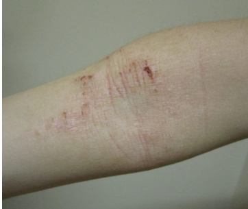
Seborrheic Dermatitis
Erythematous, yellowish, greasy scale caused by the yeast Malassezia
-Seen most often in the nasolabial folds, eyebrows, scalp, chest; worsened by stress, cold weather, and immunosuppression
-Dx: clinical
-Tx: Ketoconazole shampoo, topical antifungal + low-potency corticosteroid for the face

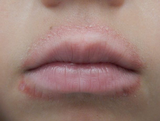
Perioral Dermatitis
Papulopustular plaques and scale around the mouth that spare the vermillion border
-Dx: clinical
-Tx: topical metronidazole
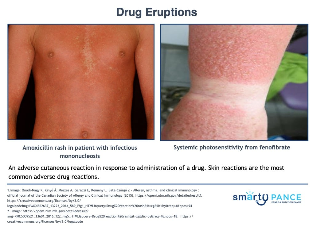
Drug Eruption
Adverse skin reaction to medicine, usually appearing within the prior 6 weeks
-Presentation: mild, morbilliform rash that clear after stopping the drug ranging to severe life-threatening reactions
-Dx: clinical with careful history
-Tx: withdraw the offending agent, antihistamines
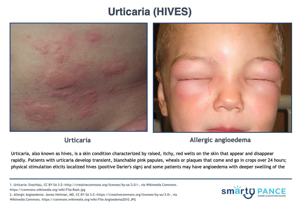
Urticaria
Transient, well-circumscribed, itchy, pink wheals that blanch with pressure and resolve within 24 hours
-Types: acute (IgE mediated), chronic (idiopathic), dermatographism, angioedema
-Dx: clinical with history, CBC, CMP, thyroid function, ANA
-Tx: H1 antihistamines, short course of steroids
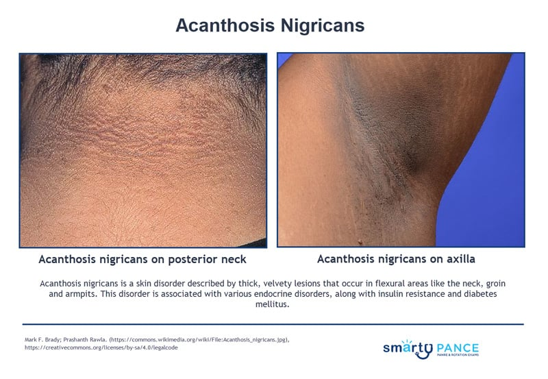
Acanthosis Nigricans
Dark, velvety patches in body folds and creases that are usually a sign of insulin resistance
-Most often occurs with obesity, PCOS, and type 2 diabetes
-Dx: clinical
-Tx: treat the underlying condition
Alopecia Areata
Well-demarcated, round patches of baldness from an autoimmune attack on hair follicles
-”exclamation point hairs” are characteristic
-Tx: intralesional corticosteroids, topical minoxidil, JAK inhibitors for severe disease
Tinea Capitis
Fungal scalp infection, causing broken hairs, scaling, and “black dots”
-Tx: oral griseofulvin or terbinafine
Telogen Effluvium
Diffuse shedding after a stressful event that occurred 6-16 weeks earlier
Androgenic Alopecia
Typical male/female pattern baldness, driven by dihydrotestosterone
-Tx: topical minoxidil
Bullous Pemphigoid
Chronic autoimmune blistering disease in which antibodies attack the hemidesmosomes that anchor the epidermis to the dermis, producing a deep, subepidermal split
-Presentation: large, tense bullae and crusts on the axillae, thighs, groin, and abdomen that are more tense, less fragile, and deeper than pemphigus vulgaris. Does not affect mucous membranes, negative Nikolsky sign
-Dx: skin biopsy, direct immunofluorescence shows linear IgG and C3 at basement membrane zone
-Tx: topical or systemic corticosteroids

Erythema Multiforme
Acute, immune-mediated rash with distinctive target lesions on the extremities, usually triggered by an infection (HSV is MC)
-Presentation: 3 zone “target” rash distributed symmetrically on extensor surfaces and palms
-Dx: clinical, skin biopsy if uncertain
-Tx: self-limited, treat underlying cause, valacyclovir suppressive treatment if recurrent and triggered by HSV

Hidradenitis Suppurativa
Chronic disease of follicular occlusion that produces recurrent inflammatory nodules, abscesses, sinus tracts, and scarring
-Presentation: pea to marble sized nodules that enlarge and drain pus where the skin rubs together like armpits, groin, buttocks, and under the breasts
-Dx: clinical, grade with Hurley staging
-Tx: intralesional clindamycin, oral doxycycline for moderate disease, consider adalimumab for moderate-to-severe

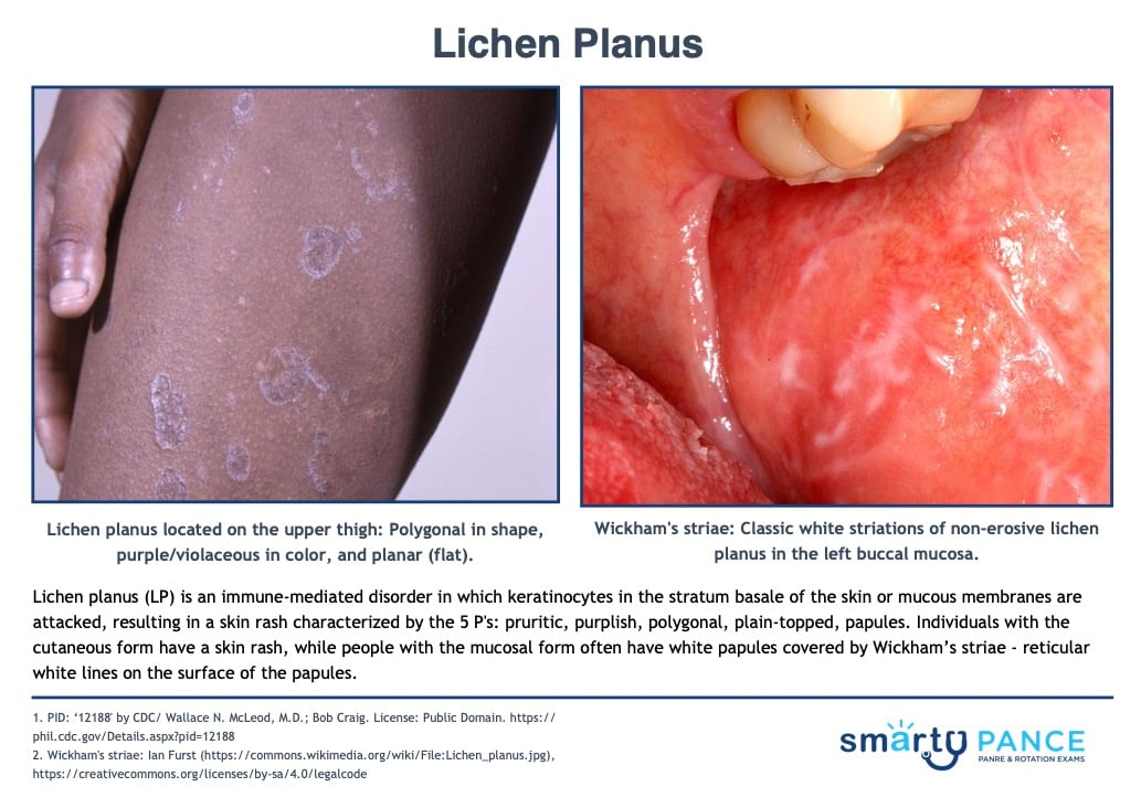
Lichen Planus
Chronic inflammatory skin condition characterized by purple, pruritic, polygonal, planar, papules, and plaques that appear on the wrists, ankles, lower back, genitalia, and mouth.
-Associated with hepatitis C infection
-Dx: clinical, confirm with skin biopsy
-Tx: high potency topical corticosteroids
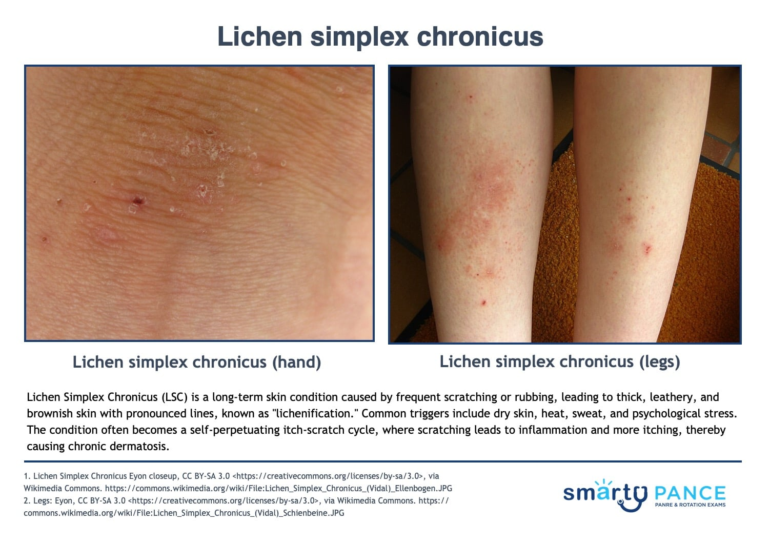
Lichen Simplex Chronicus
“The rash you scratch into existence”
-Presentation: thick, leathery, brownish skin in the posterior neck, wrists, ankles, genitals, or lower legs due to repeated scratching and rubbing
-Dx: clinical
-Tx: break the itch-scratch cycle, high-potency topical corticosteroids
Melasma
“Mask of pregnancy”
-Presentation: acquired hyperpigmentation driven by hormones plus the sun, leading to symmetric brown-gray patches on sun-exposed skin. Seen most often on the face, neck, and forearms
-Dx: clinical
-Tx: sun protection, topical hydroquinone, triple combination cream if refractory

Nummular Eczema
Coin-shaped, oozing, crusted plaques that are intensely itchy
-Common on the lower legs, buttocks, and trunk
-Dx: clinical, KOH to rule out tinea corporis
-Tx: high or ultra high potency topical corticosteroids
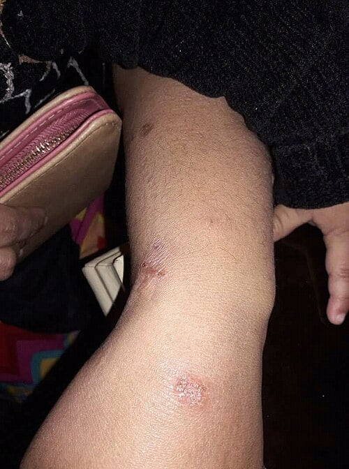
Pilonidal Disease
A pit or cyst that traps hair and debris, becoming infected in the natal cleft
-RF: young men, obesity, sedentary jobs, deep natal cleft
-Presentation: midline pit with a secondary abscess or sinus tract and foul-smelling drainage
-Dx: clinical
-Tx: I&D + abx if cellulitis
Pityriasis Rosea
Common, self-limited rash thought to follow reactivation of human herpesvirus 6 and 7
-Presentation: starts with a single “herald patch” and progresses to many smaller “daughter” lesions in a Christmas tree pattern over the course of 2 weeks, also seen along cleavage lines on the trunk. Spares the face, palms, and soles
-Dx: clinical, RPR to exclude secondary syphilis
-Tx: self-limited, oral antihistamines for itch

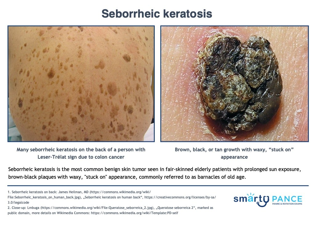
Seborrheic Keratosis
Benign skin growth in older adults, which is a harmless overgrowth of surface skin cells
-Presentation: brown, waxy, “stuck on” plaques with a well-defined, rough surface. Found on the face, trunk, and extremities. Can be paraneoplastic clue to an internal cancer
-Dx: clinical, shave or punch biopsy
-Tx: none needed
Tinea Versicolor
Scaly, oval hypo- or hyperpigmented macules that do not tan, which is due to the yeast Malassezia furfur
-Location: upper trunk, neck, and proximal arms; common in adolescents and young adults in warm/humid climates
-Dx: KOH prep shows “spaghetti and meatballs” appearance
-Tx: selenium sulfide 2.5%

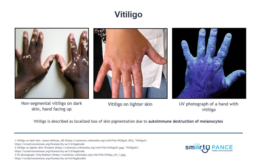
Vitiligo
Autoimmune condition in which the body destroys its own melanocytes, leaving depigmented patches of skin and hair
-Presentation: well-demarcated chalk-white patches with normal or hyperpigmented borders. Common on the face, hands, wrists, axillae and genitalia
-Common in Hashimoto thyroiditis, Graves disease, T1DM, Addison disease, alopecia areata
-Dx: Wood lamp
-Tx: sun protection, topical corticosteroids, Ruxolitinib cream
Abscess
Walled-off collection of pus in the skin, usually from Staph Aureus
-Presentation: painful, fluctuant, red nodule with surrounding cellulitis
-RF: skin trauma, injection drug use, diabetes, immunocompromise, MRSA contacts
-Dx: clinical, fluctuance is the key finding
-Tx: I&D is definitive + cephalexin / bactrim if MRSA coverage is needed
Cellulitis
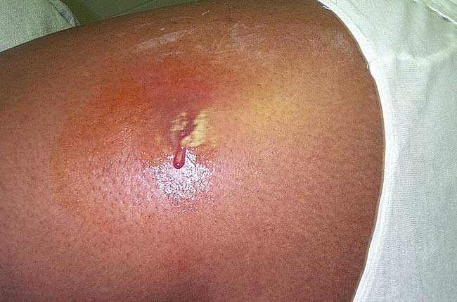
Deep bacterial infection of the dermis and subcutaneous tissue with pain, erythema, warmth, and swelling
-Presentation: erythematous, painful, warm swelling of the skin with flat borders and poor demarcation
-Dx: clinical
-Tx: Cephalexin if non-purulent, Bactrim/doxy if purulent

Folliculitis
Inflammation/infection of hair follicles, producing papules and pustules centered on the follicles
-Most often S. aureus, Pseudomonas for hot-tub folliculitis
-Dx: clinical
-Tx: topical mupirocin + benzoyl peroxide

Impetigo
Contagious, crusting skin lesions on the face from S. aureus
-Presentation: sores around the nose and mouth that rupture and ooze, drying into a yellow “honey-colored” crust
-Dx: clinical
-Tx: topical mupirocin, oral cephalexin if extensive

Paronychia
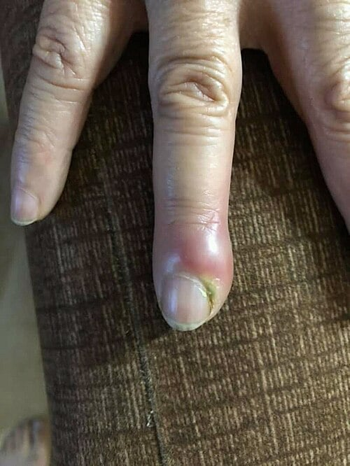
Infection of the nail fold, usually due to S. aureus
-Presentation: sudden pain, redness, swelling, and pus in a patient who bites their nails, sucks their fingers, has hangnails, or got a manicure
-Dx: clinical, culture pus
-Tx: warm soaks + topical mupirocin if without abscess, I&D if with abscess
Intertrigo
Cutaneous candidiasis infection of the skin folds, such as groin and under breasts, that presents as beefy red erythema with satellite pustules and maceration
-Tx: topical azoles +

Thrush
Oral candidiasis presenting as white plaques on the buccal mucosa that scrape off leaving a red base
-Seen in infants, elderly, immunocompromised, inhaled-steroid use
-Tx: nystatin swish-and-swallow for mild, fluconazole for moderate-severe
Diaper Rash
Cutaneous candidiasis presenting as satellite lesions beyond the main rash in a diaper distribution
-Tx: topical creams
Onychomycosis
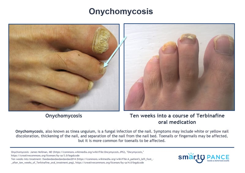
Fungal infection of the nail
-Presentation: thick, discolored, brittle, crumbling nails with debris under it
-Dx: KOH prep of nail scrapings show branching hyphae
-Tx: oral terbinafine
Tinea Capitis
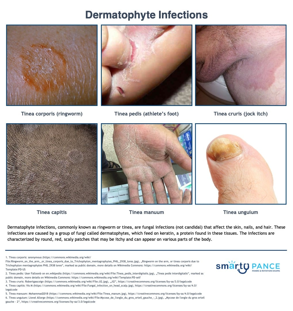
Tinea infection of the scalp, which is the MC fungal infection in children
-Presentation: broken hairs and scaling
-Tx: griseofulvin PO
Tinea Corporis
Tinea infection of the trunk/extremities
-Presentation: ring with central clearing and a scaly, advancing border
-Tx: topical antifungals

Tinea Pedis
Tinea infection of the feet/toes
-Presentation: itchy-scaling between the toes
-Tx: topical azoles
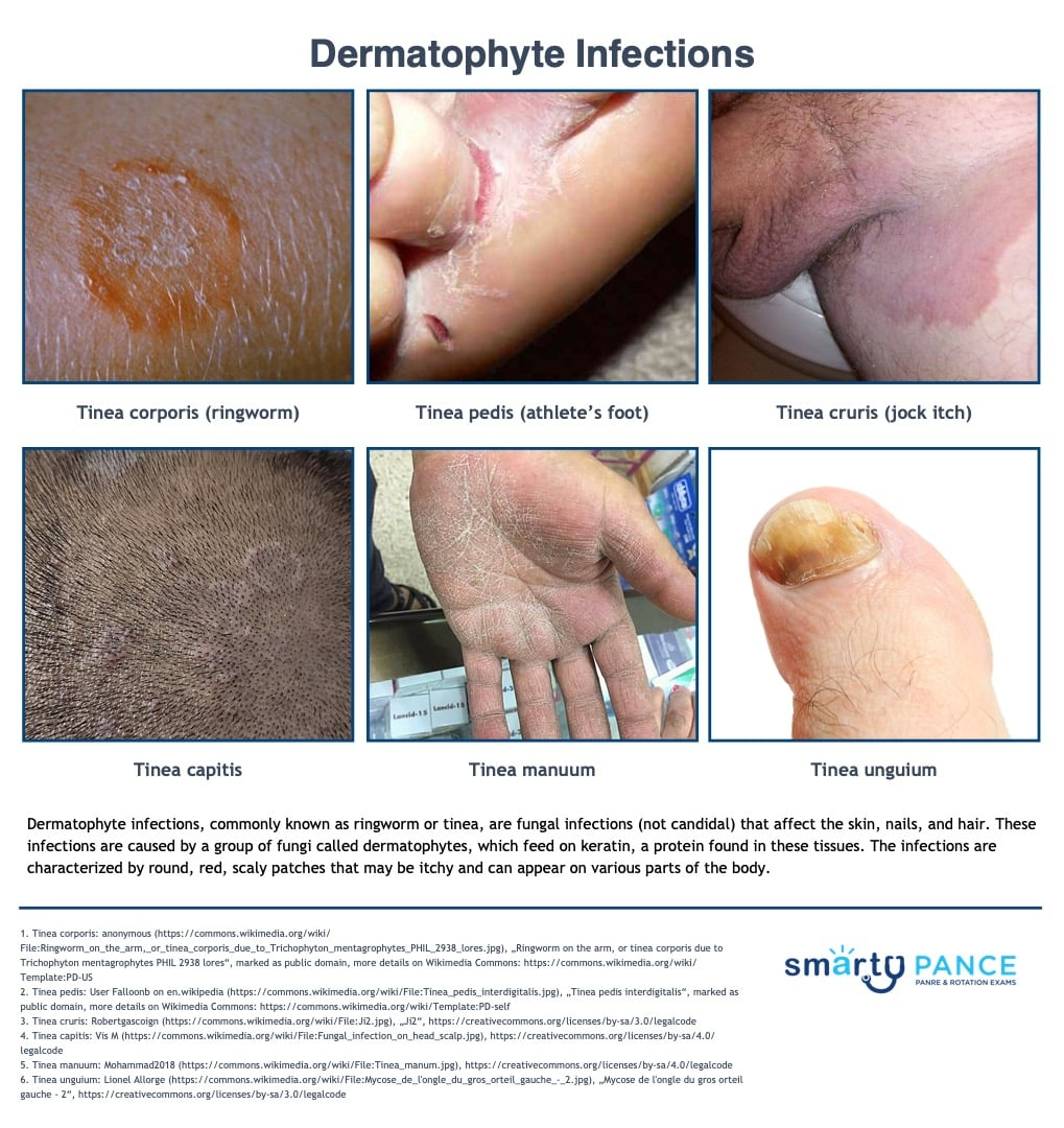
Hand, Foot, Mouth Disease (HFMD)
Very contagious infection due to Coxsackievirus A16, spread via fecal-oral and respiratory droplets
-Presentation: prodromal symptoms of low grade fever, malaise, and sore mouth for 1-2 days then develop painful oral ulcers, vesicles on the palms/soles/backs of hands and feet
-Dx: clinical
-Tx: supportive, self-limited
Shingles
Reactivation of latent varicella-zoster virus in a sensory ganglion, usually triggered by older age, immunosuppression, or stress
-Presentation: burning/itching/shooting pain along a dermatome for 1-5 days before a grouped vesicular rash develops. Can involve the eye (Zoster ophthalmicus)
-Dx: clinical, PCR if unsure
-Tx: antivirals within 72 hours of rash onset (valacyclovir), Shingrix vaccine if > 50

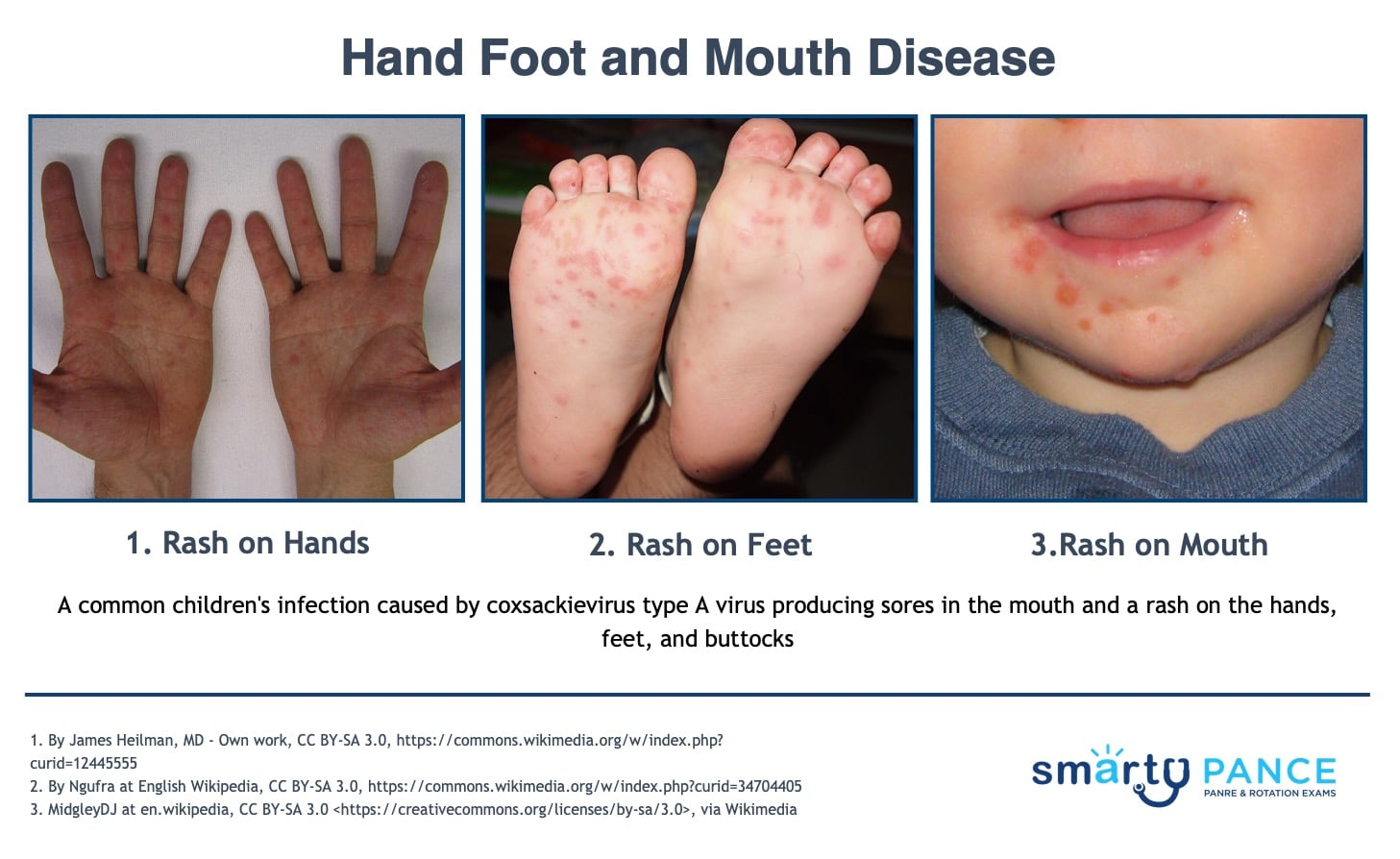
Measles (Rubeola)
Infection with the contagious, airborne measles morbillivirus
-Presentation: 3-5 days of high fever/cough/conjunctivitis/coryza, Koplik spots on the oral mucosa, then maculopapular rash that begins at the hairline and spreads down
-Dx: clinical, confirm with serum measles IgM or PCR, notify public health
-Tx: Vitamin A for hospitalized children < 2 years old, post-exposure prophylaxis with MMR within 72 hours
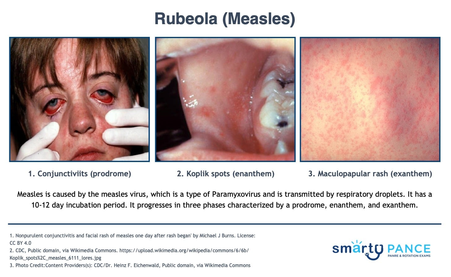
Molluscum Contagiosum
Common viral skin infection caused by a poxvirus
-Presentation: smooth, dome-shaped, waxy, flesh-colored papules with a central umbilication that spreads via skin-to-skin contact
-Dx: clinical
-Tx: often self limited (clears in 6-12 months) or use Cantharidin to blister / cryotherapy, Imiquimod if immunocompromised or on the genitals

Rubella
Infection due to the rubella virus, which is spread by respiratory droplets, and prevented by the MMR vaccine
-Presentation: prodromal symptoms of low-grade fever/malaise/LAD (posterior auricular/posterior cervical/suboccipital), pink-red maculopapular rash that starts on the face and spreads downward
-Dx: serum rubella IgM
-Tx: supportive, immune globulin for non-immune pregnant women exposed

Congenital Rubella
First-trimester rubella infection causing deafness, cataracts, cardiac defects, microcephaly, and “blueberry muffin” rash
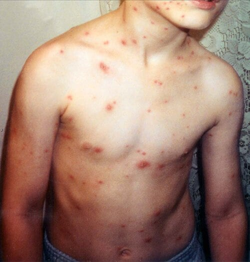
Varicella
Primary infection with the varicella-zoster virus, which is highly contagious by air and contact
-Presentation: two day prodrome of low grade fever and malaise, pruritic vesicular rash with lesions in different stages of development from the trunk to face and limbs
-Dx: clinical
-Tx: supportive, avoid aspirin in kids, immunocompromised/pregnant adults need acyclovir/valacyclovir
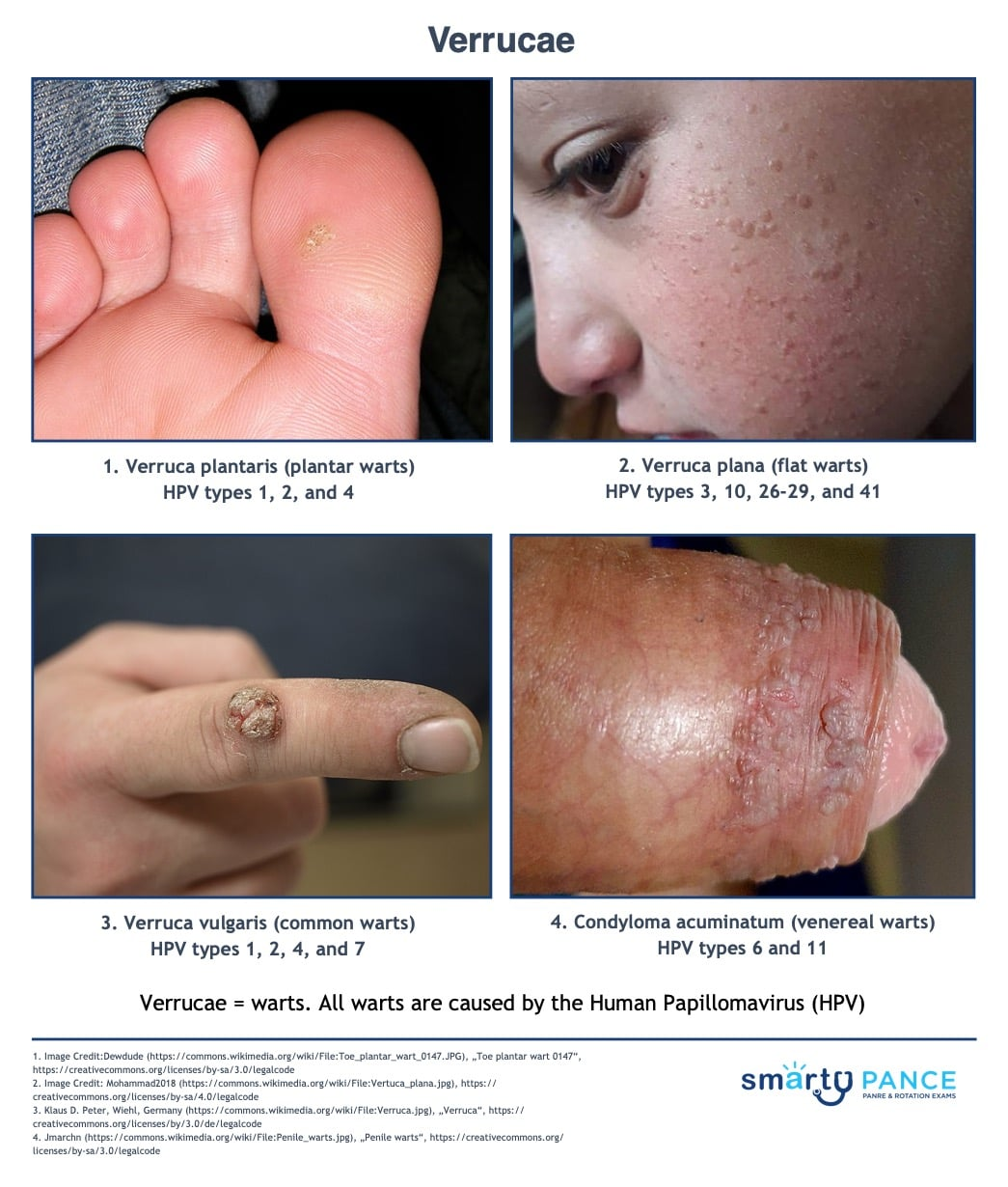
Verrucae
Warts caused by human papillomavirus (HPV)
-Can be seen on the fingers, elbows, knees, face, soles/heels, genitals, eyelids
-Dx: clinical
-Tx: salicylic acid, cryotherapy
Actinic Keratosis
Sun-damage growth that is a precursor to squamous cell carcinoma
-Presentation: flesh-colored, pink, or yellow-brown lesion with a rough sandpaper feel on sun-exposed skin like face, lips, ears, backs of hands, forearms, scalp, and neck
-Dx: clinical
-Tx: cryotherapy, imiquimod

Lipoma
Benign overgrowth of adipocytes
-Presentation: soft, doughy, mobile, painless subcutaneous nodule that is well-circumscribed with normal overlying skin. Common on the upper back, shoulders, neck, abdomen, and arms
-Dx: clinical
-Tx: observe if asx, surgical excision if symptomatic

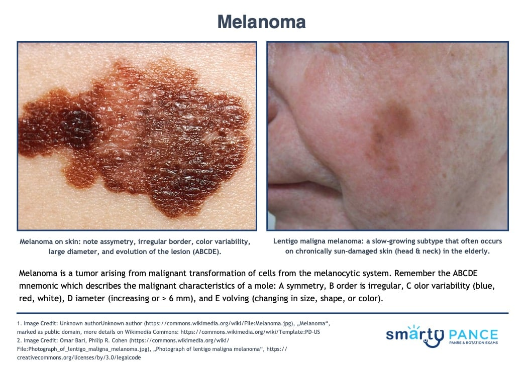
Melanoma
Malignant tumor of melanocytes, which is the deadliest skin cancer
-RF: UV exposure, fair skin, family history, many nevi
-Dx: ABCDEs, excisional biopsy is gold standard
-Tx: wide local excision, node biopsy if > 1mm thickness
Basal Cell Carcinoma
MC form of skin cancer, which arises from basal cells
-Presentation: pearly papule with raised, rolled borders, telangiectasias, and a central ulcer on a person with fair-skin
-Dx: shave or punch biopsy
-Tx: surgical excision

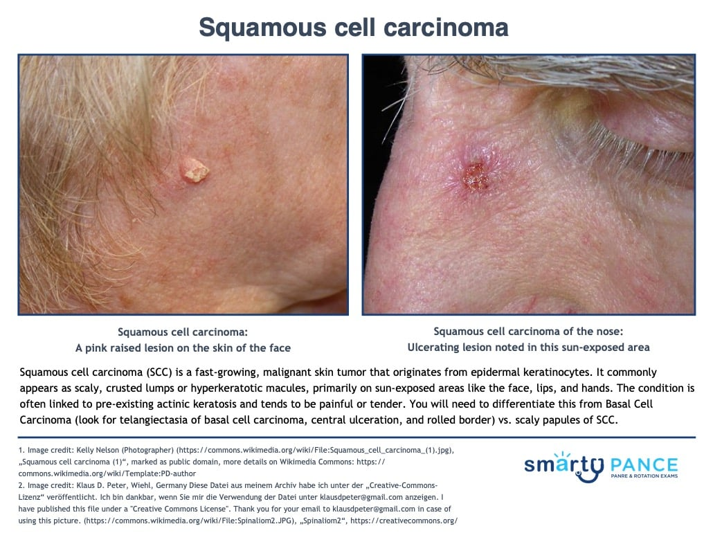
Squamous Cell Carcinoma
Cancer arising from keratinocytes
-Presentation: firm, scaly or ulcerated papule/nodule on sun-exposed skin that can arise from actinic keratosis, chronic wounds, scars, and mucosa
-Dx: shave or punch biopsy
-Tx: surgical excision with wide margins
Pediculosis
Infestation of parasitic lice, which can affect the head, body, or pubic area
-Dx: visualizing live lice or viable nits on hair shafts
-Tx: Permethrin 1% cream rinse for hair, permethrin for pubic lice

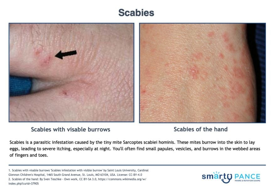
Scabies
Burrowing of Sarcoptes scabiei mites into the epidermis, which is spread by prolonged skin-to-skin contact
-Dx: clinical, skin scraping with mineral oil shows mites/eggs
-Presentation: intense itch especially at night, burrows especially in the finger webs and flexor wrists, and crusts
-Tx: permethrin 5% cream, repeat in one week
Keloid
Excessive scar tissue that grows beyond the original wound, due to dysregulated collagen production
-Presentation: firm, rubbery, raised scar that extends beyond the original wound margins. More common in darker skin tones
-Dx: clinical
-Tx: intralesional corticosteroid injection is first line, silicone gel sheets for prevention, cryotherapy

Epidermal Inclusion Cyst
Benign cyst lined by skin and filled with keratin
-Presentation: smooth, firm, well-circumscribed, mobile cyst with a central punctum. Contents are thick, white-yellow, foul-smelling keratin. Common on the face, scalp, neck, back, and genitalia
-Dx: clinical, US to confirm a cystic structure
-Tx: asx can be observed, elective removal, or I&D if infected

Lyme Disease
Caused by Borrelia burgdorferi, transmitted by the deer tick
-Presentation: starts with erythema migrans, facial nerve palsy, Lyme carditis, meningitis, and arthritis
-Dx: clinical diagnosis if early, serology if late
-Tx: doxycyline
Rocky Mountain Spotted Fever
Infection caused by Rickettsia rickettsii, transmitted by Dermacentor ticks
-Presentation: fever, maculopapular rash that begins on the wrists and ankles and spreads centrally. May become petechial
-Dx: clinical + serology
-Tx: Doxycycline
Abrasion
Superficial wound from friction/scraping that removes the epidermis and sometimes the superficial dermis without full-thickness loss
-Dx: clinical
-Tx: wound irrigation, debridement of devitalized tissue, moist wound healing, tetanus prophylaxis
Black Widow Bite
Spider bite with alpha-latrotoxin venom
-Presentation: causes pinprick pain then severe diffuse muscle cramps (rigid, board-like abdomen), diaphoresis, hypertension
-Dx: clinical
-Tx: opioids + benzos

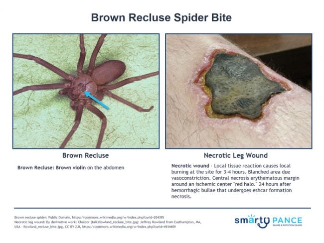
Brown Recluse Bite
Spider bite with sphingomyelinase D venom
-Presentation: initial painless bite, then a slowly expanding necrotic ulcer
-Tx: wound care, rest, ice, compression, elevation
Superficial
What type of burn is being described?
-Epidermis only
-Dry, red, painful. No blisters
-Heals in 3-5 days
Superficial partial thickness
What type of burn is being described?
-Blisters, moist, pink, and very painful
-Heals in 7-14 days
Deep partial thickness
What type of burn is being described?
-Pale, less painful
-Heals > 21 days with scarring
-May need grafting
Full-thickness
What type of burn is being described?
-Leathery, charred or white/waxy
-Painless
-Requires grafting
Stage 1
What stage of pressure ulcer is being described?
-Intact skin with non-blanchable erythema

Stage 2
What stage of pressure ulcer is being described?
-Partial thickness loss, shallow open ulcer or ruptured blister

Stage 3
What stage of pressure ulcer is being described?
-Full-thickness loss, fat visible, no bone/tendon/muscle exposed

Stage 4
What stage of pressure ulcer is being described?
-Full-thickness with exposed bone, tendon, or muscle

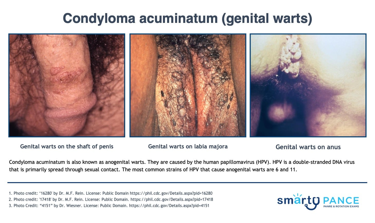
Condyloma Acuminatum
Soft, skin-colored, fleshy growths caused by human papillomavirus types 6 and 11
-Dx: clinical
-Tx: imiquimod if patient applied, TCA therapy in office
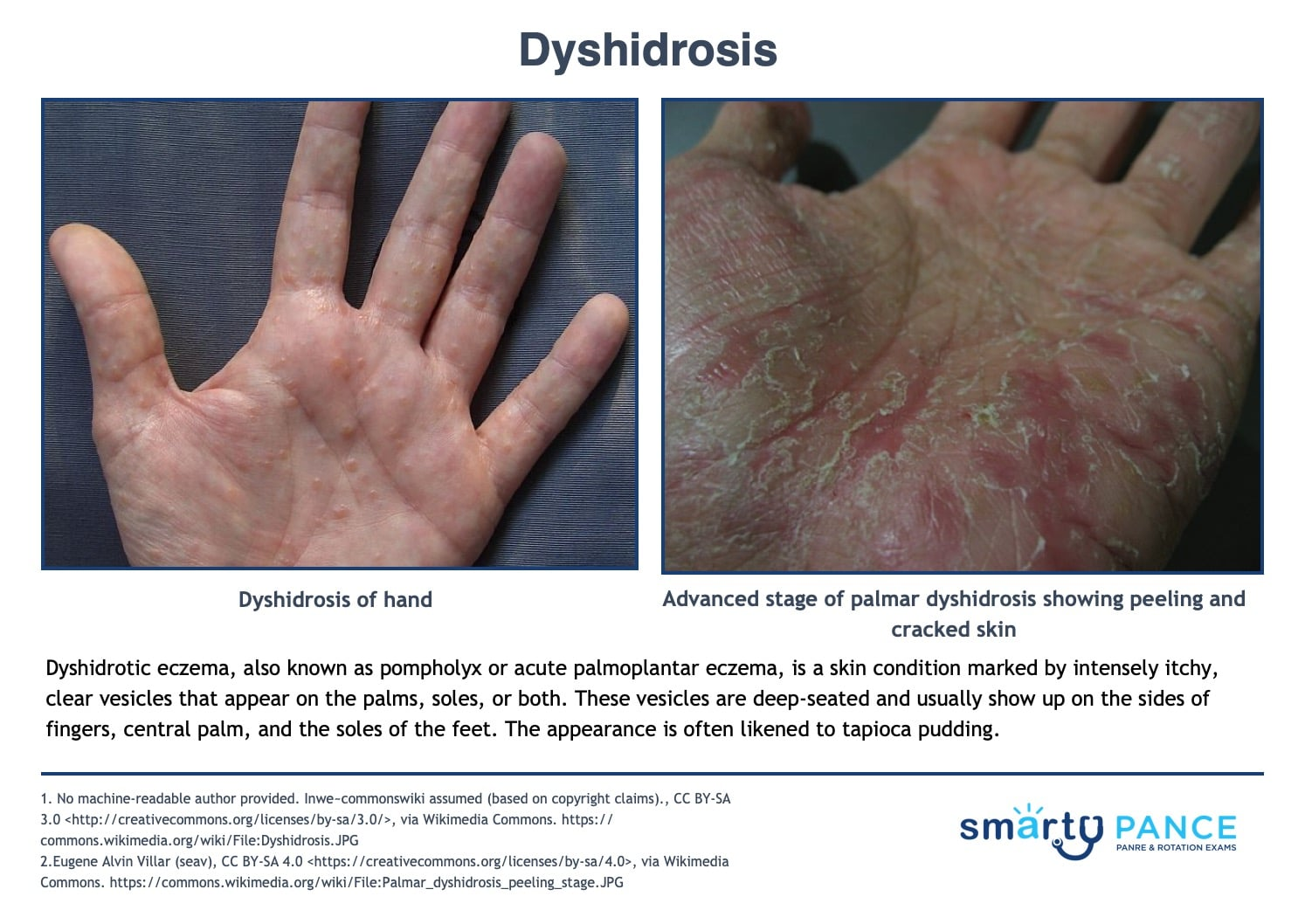
Dyshidrosis
Itchy vesicular eruption of clear, deep-seated vesicles without surrounding erythema on the lateral fingers, palms, and soles
-Described as “tapioca pudding” triggered by stress, sweating, contact allergens
-Dx: clinical
-Tx: high potency topical corticosteroids
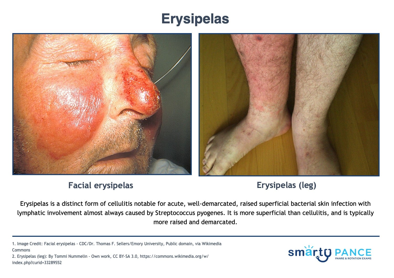
Erysipelas
Cellulitis with sharply demarcated, raised borders and lymphatic involvement. Almost always due to Group A Strep
-Presentation: looks like cellulitis but is well-demarcated and superficial. Redness, pain, fever, and chills
-Dx: clinical
-Tx: penicillin
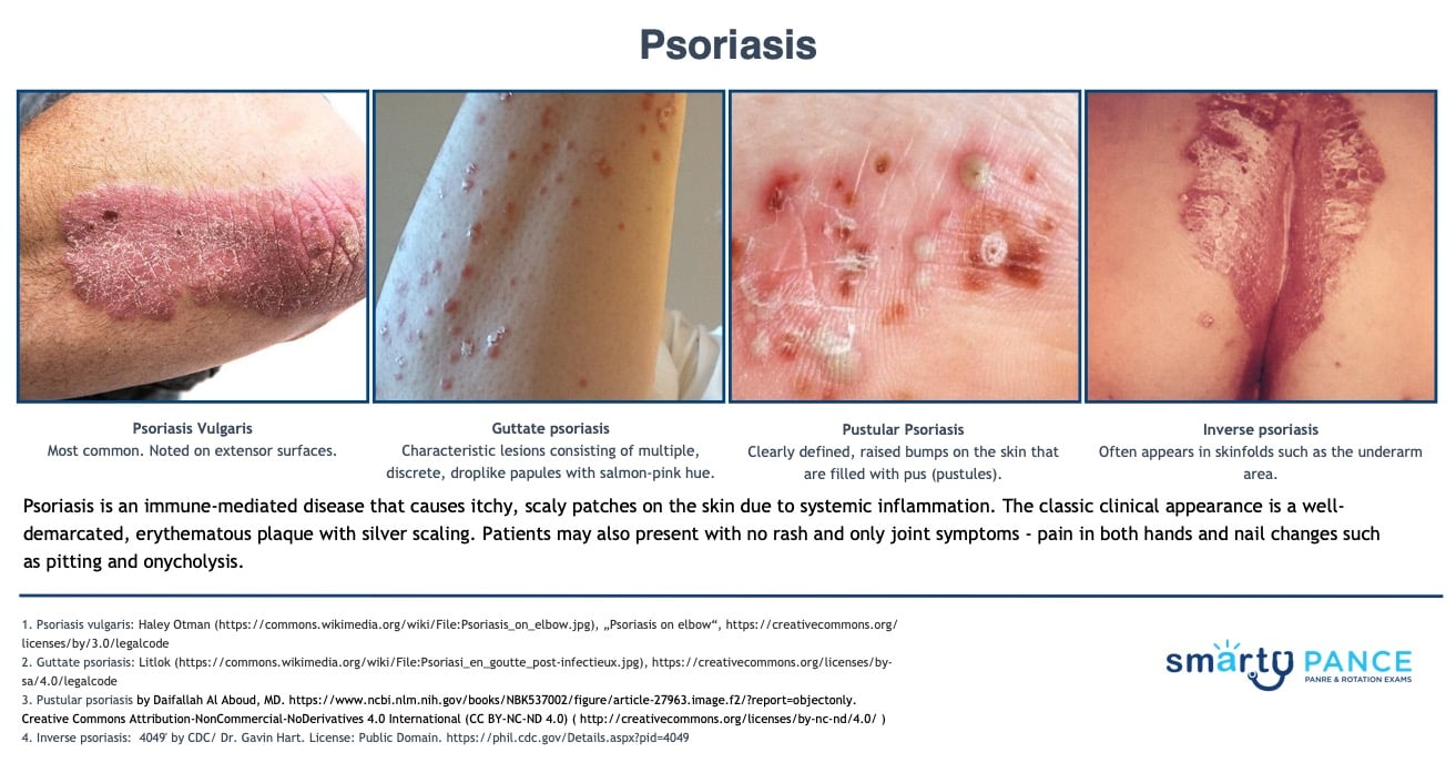
Psoriasis
Chronic autoimmune inflammatory skin disease with well-demarcated plaques covered by silver-white scale, classically on extensor surfaces
-Presentation: Auspitz sign (pinpoint bleeding when scale is removed), Koebner phenomenon, associated with psoriatic arthritis
-Dx: clinical
-Tx: topical corticosteroids