kaap309 exam 2
1/197
There's no tags or description
Looks like no tags are added yet.
Name | Mastery | Learn | Test | Matching | Spaced | Call with Kai |
|---|
No analytics yet
Send a link to your students to track their progress
198 Terms
define afferent nerves
associated with sensory. info coming TO spinal cord, CNS and brain (input)
define efferent nerves
associated with motor. spinal cord sends info DOWN to organs, muscles, etc
define somatic nerves
controls muscles, voluntary
define visceral nerves
controls organs, involuntary, automatic
describe sensory input
afferent, sensory receptors monitor changes throughout the body. when they detect the change that theyre specialized for, they send the signal to the brain
describe integration
processing of afferent info, interpretation, and decision on what should happen next
describe motor output
efferent, acts of organs, glands, muscles. coming from spinal cord to our target muscles
CNS vs PNS name for a group of cell bodies
CNS: nucleus
PNS: ganglia
CNS vs PNS name for a group of axons
CNS: tract
PNS: nerve
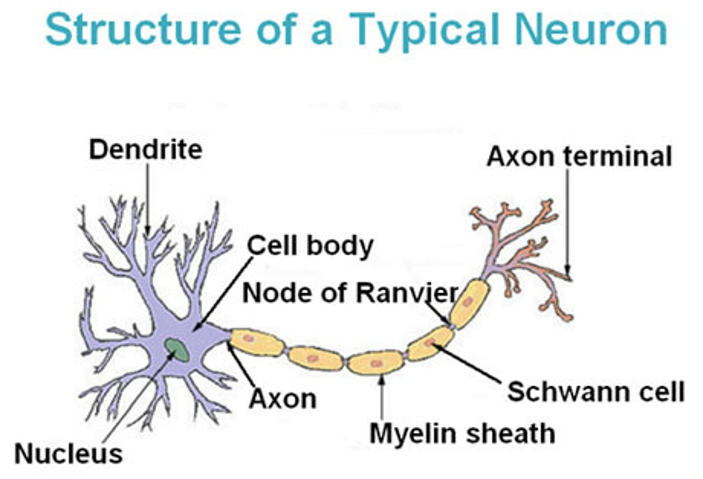
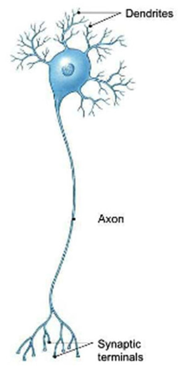
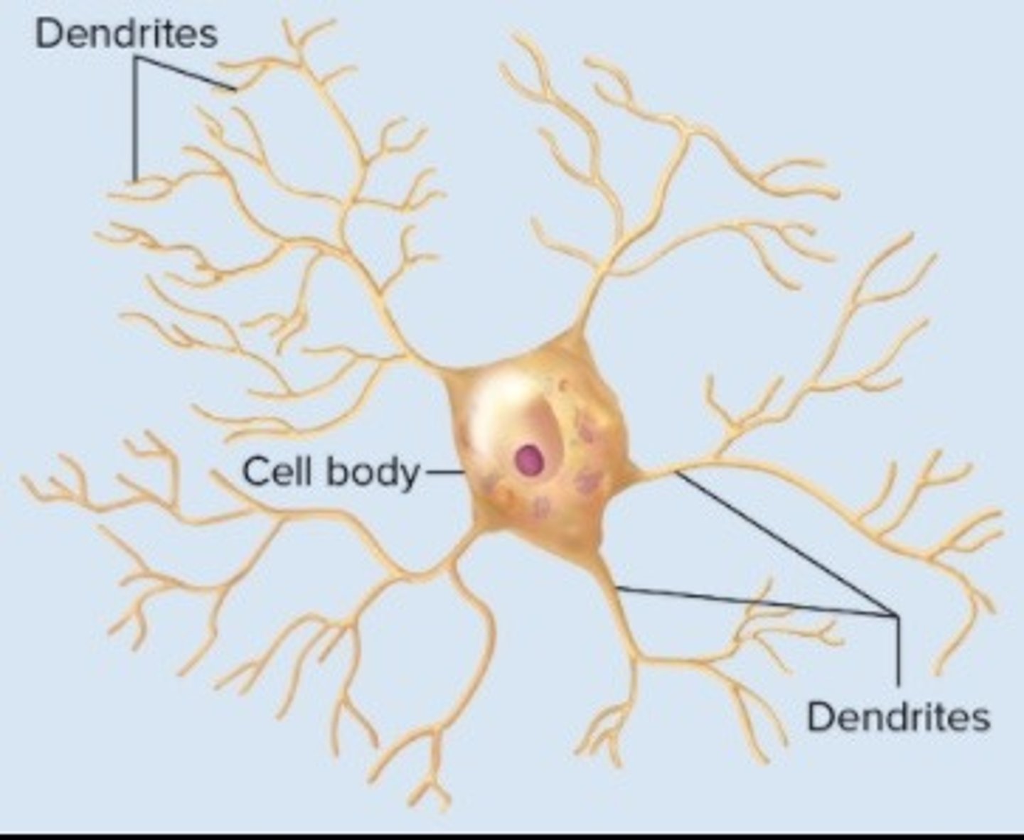
what are the 3 parts of a neuron
cell body: spherical, large nucleus, metabolic center of neuron
dendrite: extending off cell body, lots of variability, motor neurons typically contain more
axon: 1 axon, arises from cell body
what are the 4 structure types of neurons
multipolar, unipolar, bipolar, anaxonic
describe multipolar neurons
most common type, contains LOTS of dendrites.
most are interneurons that conduct impulses within the CNS
describe bipolar neurons
less common, found in retina of eye, receptors or nose, ear.
2 process extend from the cell body, one is a dendrite the other is an axon.
all bipolar neurons are sensory neurons located in special sene organs

describe unipolar neurons
cell body sits up and floats on its own, peripheral process is usually long, central process is short
starts bipolar in embryonic development
usually found in dorsal root ganglion areas of PNS

describe anaxonic neurons
do not contain axons nor deal with APs.
send signals between other neurons
found in networks within the brain, retina of eye, adrenal medulla
describe variations of structure of multipolar, bipolar, and unipolar neurons
mulitpolar: purkinje of the cerebellum, pyrimidal cell
bipolar: olfactory and reitnal cells
unipolar: dorsal root ganglion cell
describe the 3 types of neurons
sensory: afferent, pick up info from receptor in PNS and bring it to CNS
interneuron: only in CNS< relay info to appropriate motor neurons, 90% of neurons
motor: sneds info to muscles and organs
what are the 4 supporting cells of nervous tissue in CNS?
oligodendrocytes, ependyma,microglia, astrocytes
what are the 2 supoporting cells of enrovus tissue in the PNS?
satellite and schwann cells
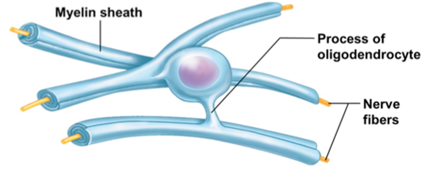
describe oligodendrocytes
found in CNS, branching cells, can help on multiple axons at once
F: wrap around axons of CNS neurons, creating an insulating layer known as the myelin sheath in brain and spinal cord
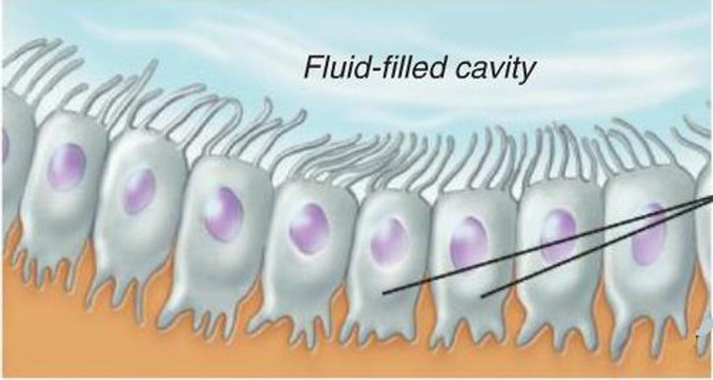
describe ependymal cells
found in CNS, wrapping garment, typically columnar or squamous, containing cilia
F: line cavities of brain and spinal cord, circulate CSF, create a fairly permeable barrier
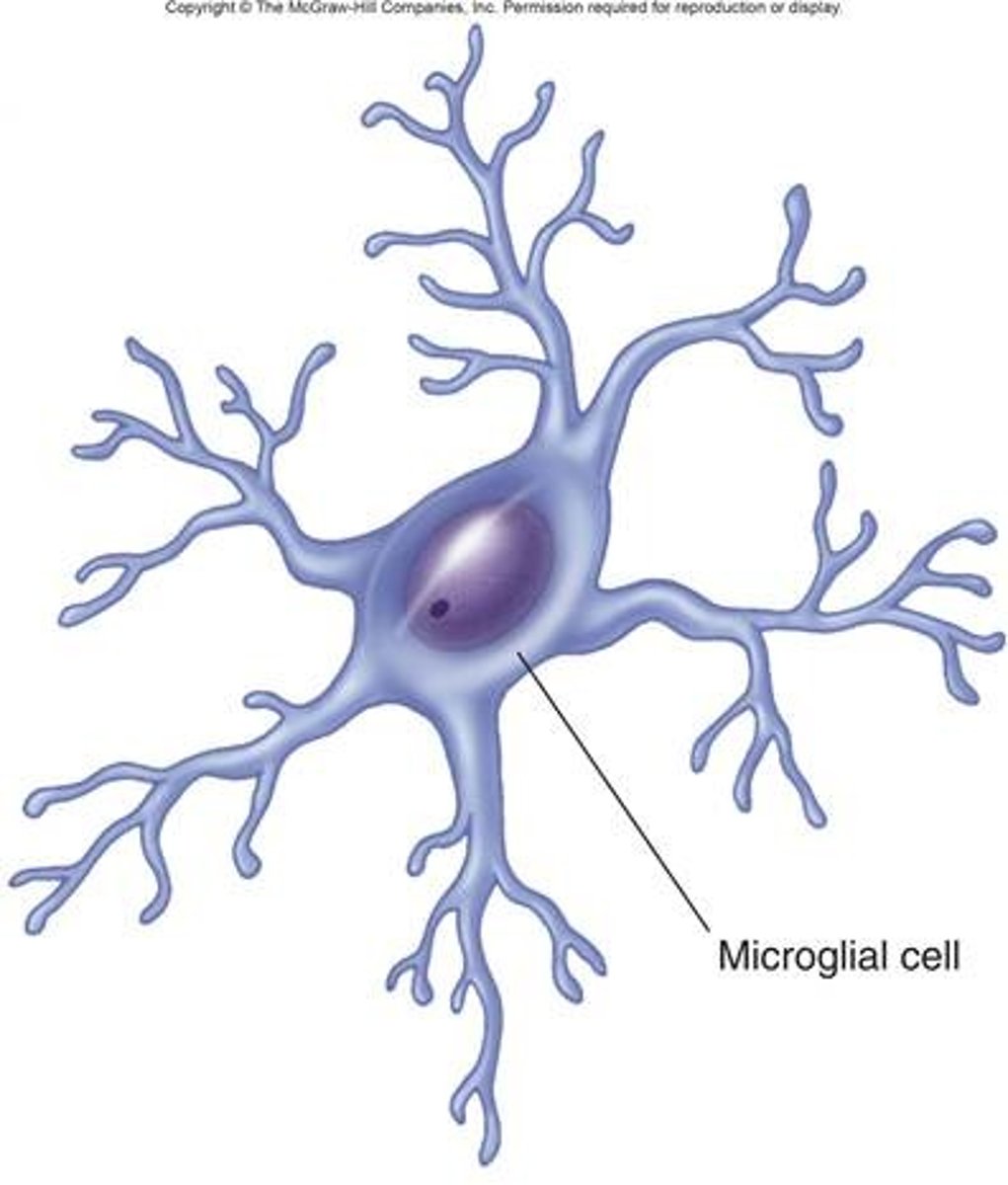
describe microglia
found all over CNS, small ovoid shape
F: monitor health of neurons(migrate to neurons in distress), transform to macrophages
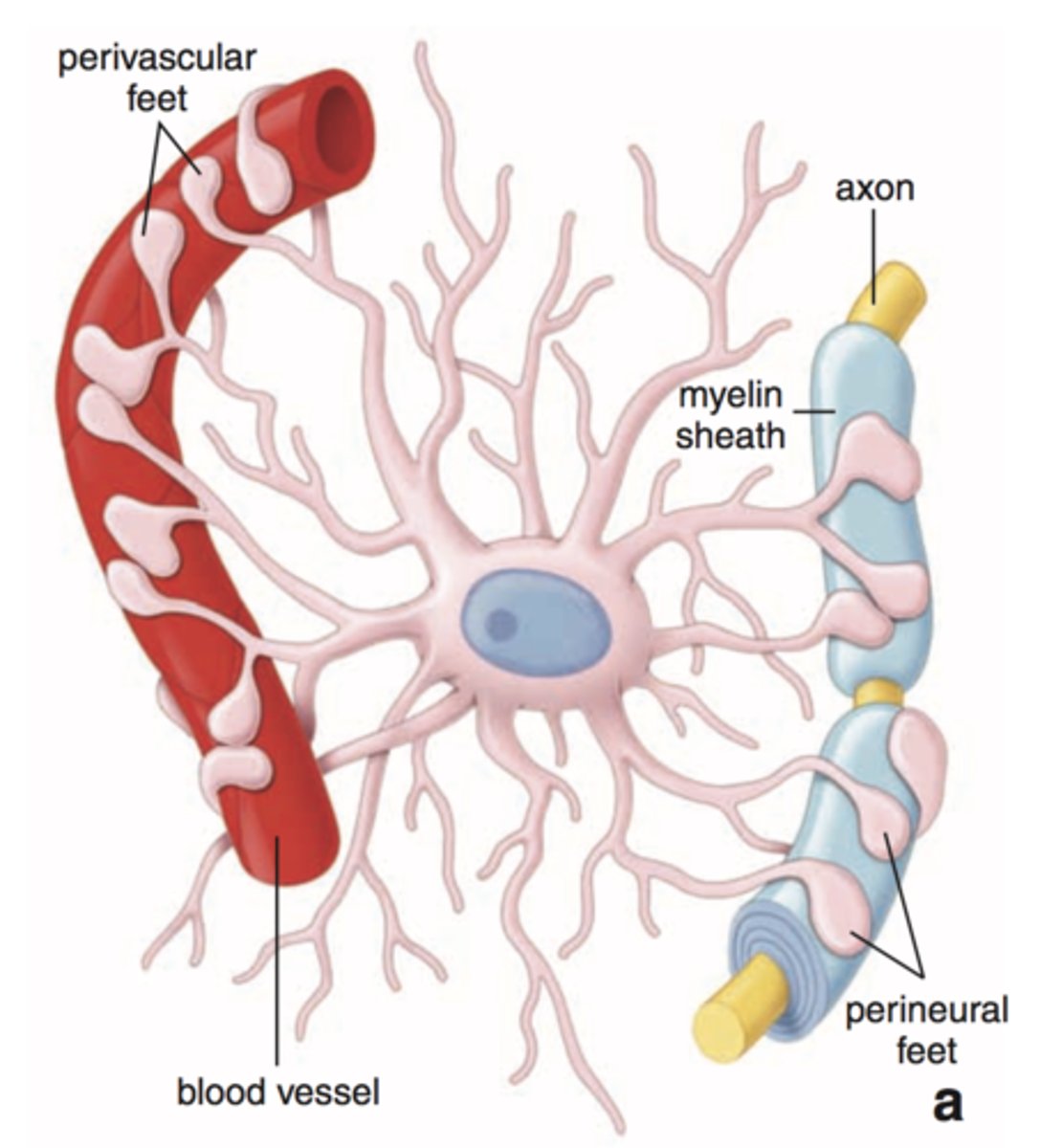
describe astrocytes
found in CNS, star cells, most abundant and versatile neuroglia
F: everything, exchanges between capillaries and neurons, control chemical environment (mop up leaked K+)
describe satellite and schwann cells
found in PNS
satellite = astrocytes of PNS
Schwann = oligodendrocytes of PNS, only covers one segment of an axon with myelin
describe myelin
fatty protein lipid, develops rapidly within the first 2 years of life
F: insulation and increase the speed of electrical impulse
describe the nodes and ranvier
gaps found about every 1mm on axon, first gap is known as initial segment
describe unmyelinated axons
many within both CNS and PNS but in PNS, axons are still within a schwann cell, just not wrapped around
describe 3 types(size) of nerve fibers
large diameter: sensory and motor fibers for skin, muscles, joints; thick myelin sheaths, fastest
intermediate diameter: lightly myelinated, ANS and pain
small diameter: non myelinated, ANS and pain, slowest
how can you change a resting membrane potential?
alter ion concentrations or change the permeability to an ion
what can changing the resting membrane potential produce?
graded potential: short distance
action potential: long distance
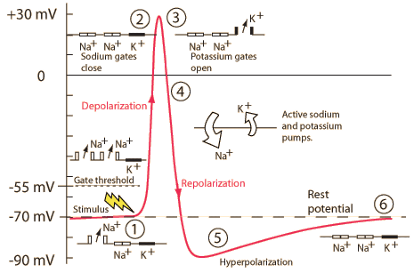
what is the resting membrane potential?
-70 mV
changing resting membrane potential: leaks/voltage-gated channels
Na+ leaks in while K+ leaks out, happens naturally, Na+/K+ pump compensates for this to maintain homeostasis, working constantly
changing resting membrane potential: ligand (chemcial) gated
ligand binds to a receptor, channel opens and Na+ floods into the neuron, shifts the membrane to depolarization
changing resting membrane potential: mechanically gated
ion channels that open under mechanical stress
what are the 4 types of local potentials?
-graded
-decremental: as we move away from stimulus, it will have less effect
-reversible: when stimulus ends, cell goes back to normal
-excitatory/inhibitory
what are the 3 types of action potentials?
- all or none
-nondecremental: once it starts, same level you have is going to continue all the way down
-irreversible
what are the 7 steps of action potentials?
1. A local current arrives at the axon hillock, depolarizing the area
2. A minimum threshold (critical voltage) of -50mV must be reached
3. Voltage gated channels open (Na+ fast, K+ slower). Na+ channels continue to open
4. At 0 mV the Na+ channels close, and final voltage peaks between +35-50mV
5. At the peak, K+ gates are open and K+ floods out of the cell
6. K+ channels stay open, resulting in more K+ leaving than Na+ entering. Voltage is now 1-2 mV more negative than RMP
7. Na+ begins to re-enter the cell, returning to normal RMP
describe the propagation of an action potential
they are sefl propagating away from the point of origin, continue as a consistent velocity. NOT the same as conduction
what influences impulse propagation?
axon diameter(larger=faster) and degree of myelination(present=faster)
continous vs saltatory propagation
myelinated: continuous, APs are triggered in nodes, only change seen in resting MP is seen in gaps
nonmyelinated: saltatory
what is a synapse and their 3 locations?
a junction that mediates info from one neuron to another
L: axosomatic, axodendritic, axoaxonal
what are neurotransmitters?
molecules synthesized by a neuron, released when a nerve signal reaches an axon terminal, have a specific effect on physiology
describe acetylcholine
L: neuromuscular junctions, most synapses of ANS, retina, aprts of brain
F:excites skeletal muscles, inhibits cardiac muscle
describe glutamate
L: cerebral cortex and brainstem
F: accounts for about 75% of all excitatory synaptic transmission in the brain, involved in learning and memory
describe aspartate
L: spinal cord
F: effects similar to glutamate
describe glycine
L: inhibitory neurons of the brain, spinal cord, retina
F: most common inhibitory neurotransmitter in the spinal cord
describe GABA
L: thalamus, hypothalamus, cerebellum, occipital lobe, retina
F: most common inhibitory neurotransmitter in the brain
describe norepinephrine
L: SNS, cerebral cortex, hypothalamus, brainstem, cerebellum, spinal cord
F: involved in dreaming, waking, mood; excites cardiac muscle; can excite or inhibit smooth muscle and glands depending on location
describe epinephrine
L: hypothalamus, thalamus, spinal cord, adrenal medulla
F: similar to norepinephrine
describe dopamine
L: hypothalamus, limbic system, cerebral cortex, retina, substantial nigra of midbrain
F: involved in elevation of mood and control of skeletal muscles
describe serotonin
L: hypothalamus, limbic system, cerebellum, retina, spinal cord, also secreted by platelets and intestinal cells
F: involved in sleepiness, alertness, thermoregulation, mood/behavior
describe histamine
L: hypothalamus, a potent vasodilator released by mast cells of CT and basophils
F: causes allergy symptoms
describe substance P
L: basal nuclei, midbrain, hypothalamus, cerebral cortex, small intestine, pain receptor neurons
F: mediates pain transmission
describe enkephalins
L: hypothalamus, limbic system, pituitary, pain pathways of spinal cord, nerve endings of DT
F: act as pain relievers by inhibiting substance P, inhibit intestinal motility, modulate immune responses
describe beta endorphin
L: digestive tract, spinal cord, parts of brain, also secreted as a hormone by the pituitary
F: surpasses pain, secretion rises sharply during labor and delivery
describe cholecystokinin
L: cerebral cortex and small intestine
F: suppresses appetite
what are the 4 primary regions of the brain?
cerebrum, diencephalon, brain stem, cerebellum
describe white matter
myelinated neurons, found mainly on internal of brain
describe gray matter
short, unmyelinated neurons, external aspect of brain but some clusters internally
describe the cerebral cortex
2 hemispheres, divided into 5 lobes. accounts for 83% of brain mass
gyri: "twisters"(elevated ridges)
sulci: "furrows"(shallow grooves)
describe the frontal lobe
motor areas(efferent somatic)
prefrontal cortex: controls personality, critical thinking, spatial memory
describe parietal lobe
sensory areas: where afferent neurons end up
describe the insula
found deep in the brain, controls taste and equilibrium
describe motor areas of the brain
primary motor cortex: pre central gyrus
premotor cortex: process thinking about movement patterns
brocas areas: deals with speech production
frontal eye field
describe sesnory areas
primary sensory cortex: post central gyrus
somatosensory association cortex
visceral sensory area
frontal eye field
what is the main purpose of basal nuclei (ganglia)?
primarily involved in control of movement
"who" is basal nuclei?
caudate nucleus, putamen, globus pallidus
who helps out basal nuclei?
subthalamic nuclei, substantia nigra
what neurotransmitters do basal nuclei primarily use?
GABA, dopamine, glutamate
what is parkinsons disease?
-results from a degeneration of the dopamine-releasing neurons from the substantia nigra
-basal nuclei become overactive, results in persistent tremors and slow initiation of movement
describe the diencephalon
central core, paired structures: thalamus, hypothalamus, epithalamus
describe the thalamus
bilateral egg shaped nuclei where info is sorted and relayed, accounts of 80% of diencephalon
describe the hypothalamus
vital for homeostasis
some functions include: body temp, thirst, sleep/wake cycles, control ANS, response to emotion, food intake
describe the epithalamus
visible as pineal gland, secretes melatonin
describe the midbrain
1.superior cerebellar peduncles
2. superipr colliculi: vision reflexes
3. inferior colliculi: auditory reflexes
4. substantia nigra
5. red nuclei: limb flexion pathways
describe the pons
composed of mainly tracts: longitudinal(brain to spinal cord) and transverse(pons to cerebellum)
describe the medulla
crucial for ANS, site of decussation(crossing), inferior cerebellar peduncle controls spatial awareness
important nuclei: cardiac and respiratory center, cochlear nuclei
describe the cerebellum
provides the precise timing and appropriate patterns of muscle contraction (subconscious)
primarily made of gray matter and ipsilateral
describe the 4 steps of cerebellar processing
1. motor cortex relays info to the cerebellum their intention for movement
2. cerebellum is simultaneously receiving info from the proprioceptors throughout the body
3. cerebellum calculates the best way to coordinate the force, direction, and extent of muscle contraction
4. cerebellum dispatches a blue print of movement to the cerebrum
what are meninges?
layers(from superficial to deep): dura mater, arachnoid mater, pia mater
what are the functions of meninges?
protect CNS, protect vasculature, house CSF, form partition in the skill
describe the dura mater
2 layers of fibrous CT: periosteal and meningeal
encloses the dural venous sinuses
describe the dural foldings of the dura mater
falx cerebri: separates right and left hemis of cerebellum, sits in longitudinal fissure
falx cerebelli
tentorium cerebelli: separates cerebrum from cerebellum
describe arachnoid mater
spider like, fine elastic CT
describe pia mater
gentle, direct contact with brain tissue
describe the 3 meningeal spaces
dura: 1. under epidural space, only found in some sections (real/potential) 2. above subdural space(potential)
arachnoid: subarachnoid, CSF flowing through it (real)
what does CSF do for us?
provides nutrients and reduces brain weight by 97%
where does CSF come from
choroid plexuses, about 500mL of CSF is formed daily and about 50mL is replaces every 8 hours
what is the pathway of CSF?
1. the choroid plexus of each ventricle produces CSF
2. CSF flows through the ventricles and into the subarachnoid space via the median and lateral apertures
3. CSF flows through subarachnoid space
4. CSF is absorbed into the dural venous sinuses
describe hydrocephalus
water on the brain, CSF is produced faster than it drains. treatment options include a shunt to reduce pressure
what is included in the limbic system
amygdala, cingulate gyrus(express emotion thru gestures), hypothalamus, hippocampus
what does the limbic system control
-why we react emotionally to things we understand to be happening(emotion> logic)
-aware of our emotions(logic>emotion)
describe language in regards to the brain
originally though to be entirely left sided
includes brocas area and wernickes area
what is the spinal nerve composed of?
ventral root and dorsal root
describe ventral and dorsal roots
ventral: motor, skeletal muscle
dorsal: sensory, receptors
describe spinal nerve rami path
afferent info enters through the dorsal rami, then travels to the spinal nerve, then to the dorsal root, then finally the spinal cord.
efferent info leaves the spinal cord and goes into the ventral root, then the spinal nerve, then exits through the ventral rami to go to muscles
how many pairs of spinal nerves are there
8 cervical
12 thoracic
5 lumbar
5 sacral
1 coccygeal
how many vetebrae each are there?
7 cervical, 12 thoracic, 5 lumbar (breakfast at 7, lunch at 12, dinner at 5)
how are spinal nerves named in regards to their location?
cervical region: spinal nerve is named for vertebrae below
thoracic and lumbar region: spinal nerve is named for vertebrae above
what are the 5 PNS stimulus receptors and what do they detect?
-mechanoreceptors: physical change(stretch, vibration, pressure)
-thermoreceptors: temp changes
-photoreceptors: changes in light
chemoreceptors: chemical changes
nociceptors: pain