Lecture 8 & 9. Pelvic Limb mm & Joints
 Call Kai
 Learn
Learn Practice Test
 Spaced Repetition
 Match
Match Flashcards
 Knowt Play
1/189

There's no tags or description
Looks like no tags are added yet.
Name | Mastery | Learn | Test | Matching | Spaced | Call with Kai | Chat |
|---|
No analytics yet
Send a link to your students to track their progress
190 Terms
sacroiliac joint- shift weight to pelvic limbs when standing, drive force of limbs into trunk while moving
name and significance of the indicated joint
cartilaginous synchondrosis (a semi mobile cartilaginous joint)
what kind of joint in the sacroiliac joint is indicated by the green arrow in the crainial area

synovial plane joint (a mobile synovial joint)
what kind of joint in the sacroiliac joint is indicated by the blue arrow in the caudal area
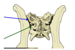
cartilaginous synchondrosis (a semi mobile cartilaginous joint)
what aspect of the sacroiliac joint is shown here
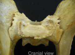
synovial plane joint (a mobile synovial joint)
what aspect of the sacroiliac joint is shown here
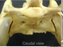
dorsal and ventral sacroiliac ligg.
what lig. is being indicated

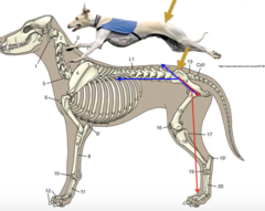
sacrotuberous ligg.
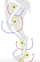
what is being indicated in both of these images

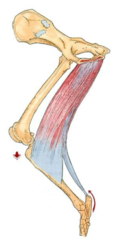
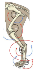
do you know those angles yet? (the tarsal joint angle is different than the carpal joint angle, remember that...)
(blue = extension, red = flex)
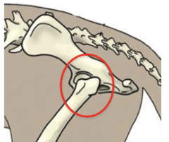
coxal/hip joint- ball and socket synovial joint
what name and kind of joint is this
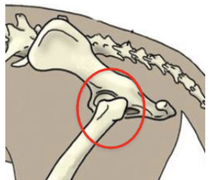
synchondrosis forms to "replace" the femoral head
in the event that you do an FHO on this joint, how does the patient heal
1. gluteal group
2. deep group
3. caudal group
4. medial group
primary muscles of the hip joint
1. extensor group
2. flexor group
primary muscles of the stifle joint
1. craniolateral (flexors or tarsals, extensors of digits)
2. caudal (extensors of tarsals, flexors of digits)
primary muscles of tarsal and digital joints
1. tensor fascia lata
2. superficial gluteal
3. middle gluteal
4. piriformis
5. deep gluteal
***gluteals an generally extensors***
what muscles are included in the lateral group of those affecting the hip joint
tensor fascia lata
1

tuber coxae
origin of 1 (tensor fascia lata)

fascia lata (very wide insertion across femur)
insertion of 1 (tensor fascia lata)

1. tense fascia lata
2. flex coxal joint
3. extend genual joint
action of 1 (tensor fascia lata)

superficial gluteal
2
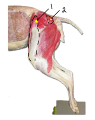
sacrum and 1st caudal vertebra
origin of 2 (superficial gluteal)
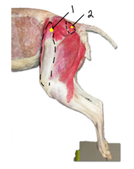
third trochanter (distal to lesser trochanter of femur)
insertion of 2 (superficial gluteal)
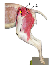
1. extend coxal joint
2. abduct pelvic limb
action of 2 (superficial gluteal)
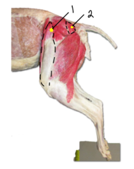
middle gluteal
what muscle is indicated
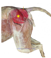
deep
where is the middle gluteal m. relative to the superficial gluteal m.

gluteal surface of ilium (wide origin area spanning much of the ilium wing)
origin of middle gluteal

greater trochanter
insertion of middle gluteal

1. extend coxal joint
2. abduct and rotate pelvic limb medially
action of middle gluteal

piriformis
2

sacrum and 1st caudal vertebra
origin of 2 (piriformis)
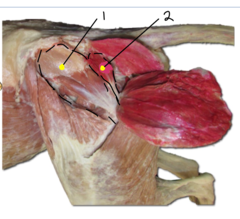
greater trochanter
insertion of 2 (piriformis)
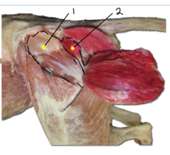
extend coxal joint
action of 2 (piriformis)
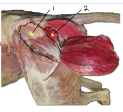
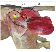
deep gluteal
1

body of ilium and ischiatic spine
origin of 1 (deep gluteal)
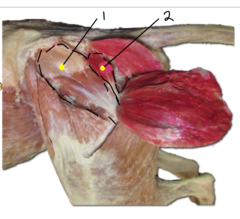
greater trochanter
insertion of 1 (deep gluteal)
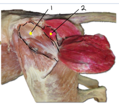
1. extend coxal joint
2. abduct and rotate pelvic limb medially
action of 1 (deep gluteal)
1. external obturator
2. internal obturator
3. gemelli
4. quadratus femoris
what muscles are included in the caudal group of those affecting the hip joint
external obturator
what muscle is being indicated

ventral aspect of os covae around obturator foramen
origin of external obturator
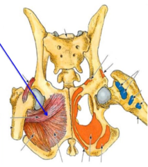
trochanteric fossa
insertion of external obturator
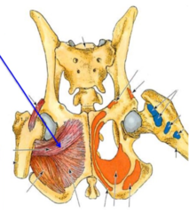
rotate limb laterally at the coxal joitn
action of external obturator
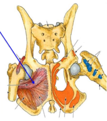
internal obturator
what muscle is being indicated

dorsal aspect of os coxae around obturator foramen
origin of internal obturator
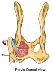
trochanteric fossa
insertion of internal obturator

rotate limb laterally at the coxal joint
action of internal obturator

gemelli
what muscle is being indicated

lateral aspect of ischium
origin of gemelli

trochanteric fossa
insertion of gemelli

rotate limb laterally at the coxal joint
action of gemelli
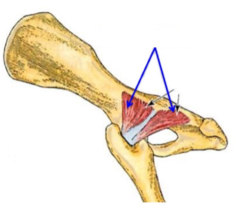
quadratus femoris
what muscle is being indicated
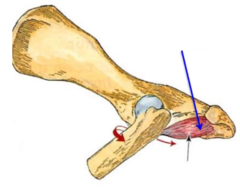
ventral aspect of ischium
origin of quadratus femoris
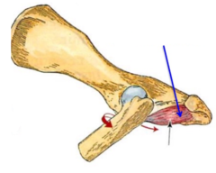
trochanteric fossa
insertion of quadratus femoris
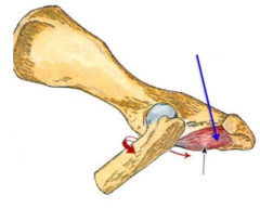
1. extend coxal joint
2. rotate limb laterally at the coxal joint
action of quadratus femoris

genual joint (stifle)
what is considered the largest synovial joint in the body
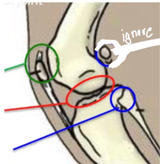
femoropatellar (hinge synovial)
what joint name and type is indicated by the green arrow in this image of the genual (stifle) joint
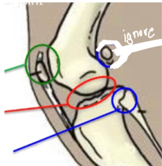
femorotibial (condylar synovial)
what joint name and type is indicated by the red arrow in this image of the genual (stifle) joint
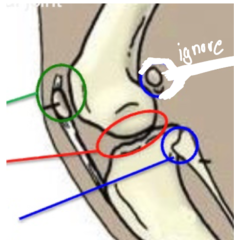
proximal tibiofibular (plane synovial)
what joint name and type is indicated by the blue arrow in this image of the genual (stifle) joint
1. lateral collateral lig
2. medial collateral lig
3. cranial cruciate lig
4. caudal cruciate lig
main ligaments in genual (stifle) joint
genual (stifle) joint capsule
what is indicated by this arrow

patellar ligament
1 indicates what aspect of the femoropatellar joint

quadriceps femoris tendon
what tendon ends up corresponding with the patellar lig upon entering the joint capsule***
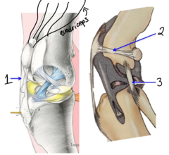
lateral femoropatellar lig- (there is a medial too)
2 indicates what aspect of the femoropatellar joint
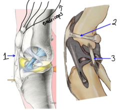
lateral fabellae
what structures are associated with the lateral femoropatellar lig

lateral collateral ligament/LCL (there is a medial too)
3 indicates what aspect of the femoropatellar joint

caudal cruciate lig.
1 is what aspect of the femorotibial joint

prevent the tibia from displacing caudally and prevent lateral rotation
what is the role of the caudal cruciate lig

cranial cruciate lig.
2 is what aspect of the femorotibial joint
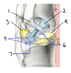
prevent the tibia from displacing cranially and prevent medial rotation (and hyperextension)
what is the role of the cranial cruciate lig
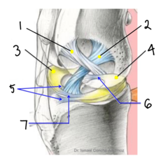
wherever the attach to the tibia is how they are named
how can we name the cranial vs caudal cruciate ligaments***

lateral meniscus
3 is what aspect of the femorotibial joint
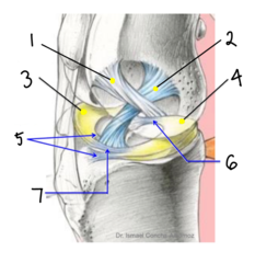
medial meniscus
4 is what aspect of the femorotibial joint
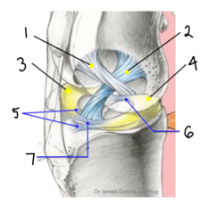
semi-moon shaped fibrocartilage that helps to fill the space left by the flat tibial condyles and strengthen the attachment between the tibia and femur
what is the purpose of the menisci
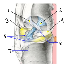
cranial tibial ligg. of menisci
5 (femorotibial joint)
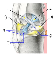
caudal tibial lig. of medial meniscus
6 (femorotibial joint)
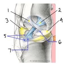
transverse lig.
7 (femorotibial joint)
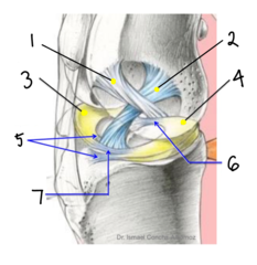
meniscofemoral lig
what lig on the caudal aspect of the stifle joint helps to attach the lateral meniscus to the femur
cranial drawer test (tibial thrust test)
how do we test for a cranial cruciate ligament rupture
capsule (extends proximal to patella to form sac-provides access for us to joint)
1 (genual joint capsule)
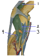
popliteal tendon
2 (genual joint capsule)
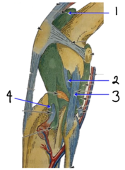
collateral ligg. (extracapsular)
3 (genual joint capsule)
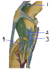
tendon sheat for long digital extensor tendon
4 (genual joint capsule)
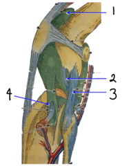
the tibial condyles are very flat so the joint has evolved to add extensive support to the area (and yet it still ruptures very commonly :)))
why does the genual/stifle joint have so much extra stuff added to stabilize it (its fr alot)...
done
just take a minute to study these angles and click "done"
1. biceps femoris
2. semitendinosus
3. semimembranosus
what are the caudal muscles of the thigh that act to extend the hip joint and flex the stifle joint
biceps femoris
what muscle is shown here
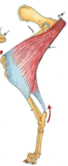
ischiatic tuberosity and sacrotuberous lig.
origin of indicated m (biceps femoris)
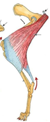
lateral aspect of fascia lata @ genual region and calcaneal tuber (via crural fascia),
insertion of indicated m (biceps femoris)

1. extend coxal joint
2. variable action on genual joint
3. extend tarsocrural joint
action of indicated m

semitendinosus
what muscle is shown here
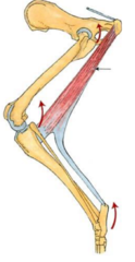
ischiatic tuberosity
origin of indicated m (semitendinosus)
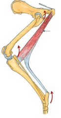
medial aspect of tibia and calcanean tuber (via crural fascia)
insertion of indicated m (semitendinosus)
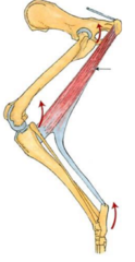
1. extend coxal joint
2. variable action on genual joint
3. extend tarsocrural joint
action of indicated m
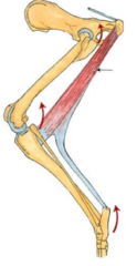
semimembranosus
what muscle is shown here
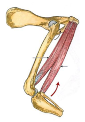
ischiatic tuberosity
origin of indicated m (semimembranosus)

medial aspect of distal femur and proximal tibia
insertion of indicated m (semimembranosus)
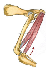
1. extend coxal joint
2. variable action on genual joint
action of indicated m.
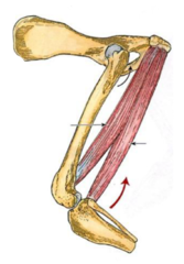
1. gracilis
2. pectineus
3. adductor
4. external obturator
5. sartorius
what are the medial muscles of the thigh that work to adduct the hip joint
gracilis
what muscle is shown here

pelvic symphysis
origin of indicated m. (gracilis)
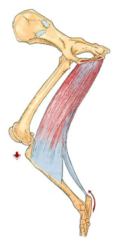
medial aspect of genual region and calcanean tuber (via crural fascia)
insertion of indicated m. (gracilis)
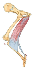
1. adduct pelvic limp
2. extend the tarsocrural joint
action of indicated m