Elbow Neural Entrapments & Injuries
 Call Kai
 Learn
Learn Practice Test
 Spaced Repetition
 Match
Match Flashcards
 Knowt Play
1/50

There's no tags or description
Looks like no tags are added yet.
Name | Mastery | Learn | Test | Matching | Spaced | Call with Kai | Chat |
|---|
No analytics yet
Send a link to your students to track their progress
51 Terms
radial nerve splits into
posterior interosseous nerve (motor) & superficial radial branch (sensation)
a posterior IO nerve problem will show
weakness
a superficial branch of radial nerve will show
pain/loss of sensation
median nerve branches into anterior interosseus nerve to innervate
deep flexors (FPL; OK symbol)
Neural “Entrapment” is neural compression leading to inflammation phase (2-3 wks). Prolonged compression or tension can result in
sensitization of neural structures
Distributions in axoplasmic flow
Distributions in blood supply
Neural “Entrapment” Difficult to diagnose, What is commonly used but usually only shows late signs of issue?
EMG studies (a (-) does not imply lack of neural problem)
Highly specific, Low sensitivity
Thorough clinical examination is essential
The consequences of prolonged neural compression starts w/ inflammation, then vascular changes, then nutritional disruption, then altered flow. What is seen to be lost first?
sensation, motor is lost last
intermittent ischemic nerve problem that does not typically lead to long term damage. This focal demyelination or ischemia is
Neuropraxia
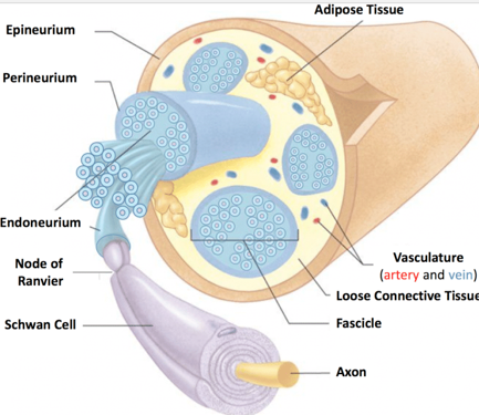
Long term damage to connective tissue in nerve:
Epineurium
Perineurium
Endoneurium
Axonotmesis
neurodynamic testing is better than EMG for determining
neural injury (not specific)
what is complete transection of nerve?
neurotmesis (complete loss of sensation & motor)
Axonotmesis may present with
loss of sensation, maybe some motor weakness
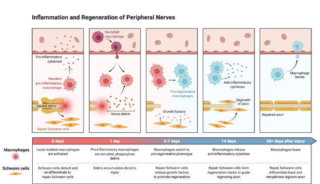
Wallerian Degeneration is retrograde degeneration of axon after nerve injury; 90% of axons damaged at site of injury, when does regeneration process begin?
1st 7 days after injury new axonal sprout growth will begin
Ulnar Nerve (C8-T1) innervates
Motor: FCU, FDP, Instrinics of hand
Sensation: Medial side of hand
Entrapment sites for the ulnar nerve
Arcade of Struthers
Cubital Tunnel (#1)
Flexor Carpi Ulnaris
ulnar nerve entrapment sensory changes are common (affects medial 1½ fingers); motor sxs include
Weakness/atrophy of FCU or medial ½ of FDP
Intrinsic of hand; palmar interosseous
Maybe weakness/atrophy of hypothenar eminence
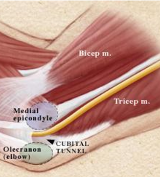
Cubital Tunnel (Funny Bone) has ulnar nerve pass posterior to medial epicondyle. Taut at
90° of flexion
On slack w/ full extension
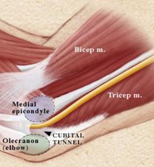
Cubital Tunnel Ulnar Nerve Entrapment causes include prolonged flexion of elbow, trauma to medial elbow, repeated trauma as in leaning on elbow, Inflammation of nerve; as well as a
cubitus valgus deformity (pitchers, Oly lifters), ↑ carrying angle
s/p misaligned healed fx may create tension along nerve
Recurrent dislocations of nerve out of cubital tunnel may be reason for neuritis
Entrapment may also occur between 2 heads of FCU (roof of cubital tunnel) (flexion & ulnar deviation can be MOI)
Cubital Tunnel Syndrome presentation sxs includes pain at the
medial elbow, Numbness & tingling into medial forearm + 4th & 5th digits
Muscle weakness in FCU or Instrinics
Muscle wasting or Claw deformity in severe cases
Cubital Tunnel Syndrome diagnostic tests
Elbow Flexion Test (3-5 mins, may not take that long)
Pressure on cubital tunnel
Tinel’s sign (hypersensitivity)
Neurodynamic test for ulnar nerve
Nerve conduction velocity tests
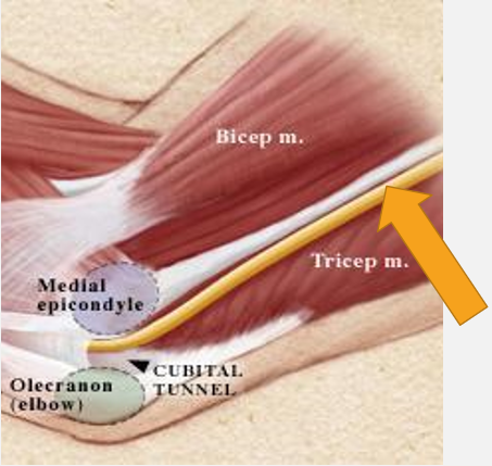
The Arcade of Struthers for ulnar nerve entrapment consists of a fibrous tunnel that is 8cm above the
medial epicondyle. Ulnar Nerve penetrates medial intermuscular septum
Palpation of region should reproduce sxs→ distally
if a pt has pain above medial epicondyle rule out what before going to arcade of struthers?
c-spine, TOS, cubital tunnel
Ulnar Nerve Interventions include Rest & avoid
repeated or excessive elbow flexion, trauma to area (resting elbow on desk, sleeping w/ elbow flexed)
Modalities for inflammation or desensitization: cortisone injections
ulnar nerve transposition surgery used as last resort (move ulnar nerve closer to median nerve). Moves nerve where?
anterior to medial epicondyle & stabilize it in CT (“anterior transposition”)
Excision of medial epicondyle
Divide tendinous insertion of flexor carpi ulnaris (arcuate lig.)
Median Nerve (C5-T1) innervates
Motor:
Anterior forearm
Thenar muscles
Lumbricals of lateral hand
Sensation: Palm & anterior 1,2,3rd & half of 4th digits
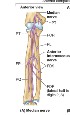
Common Entrapment Sites for median nerve
Ligament of struthers
Lacertus Fibrosus
Pronator Teres Syndrome
Flexor digitorum Superficialis
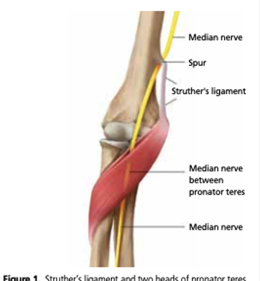
Ligament of Struthers is only present in 1% of population (Genetic). Testing will show all median nerve issues, includes sxs reproduced w/
resisted elbow flexion at 110-120°
Palpation of region
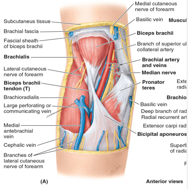
Lacertus Fibrosus is a fascial continuation of Biceps Tendon (bicipital aponeurosis), allows for bicep to be strong supinator. It is also an entrapment site for median nerve which can be compressed as it runs under its medial edge. What will test this site of entrapment?
Holding forearm in full pronation. have them supinate & adding elbow flexion
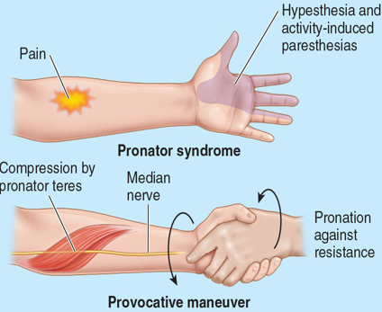
Pronator Syndrome (2nd most common entrapment) is compression of median nerve proximal to where the
AIN branch is given off between 2 heads of pronator teres
Pronator Syndrome presentation shows ↑ sxs w/ pronation. Motor involvement includes FCR, Palmaris longus, FDS. What are the 3 muscles affected by AIN syndrome?
FPL, FDP, PQ (OK sign)
Sensory: Lateral 3 1/2 fingers (median nerve)
(+) pinch grip & weak wrist flexion, finger flexion, sensory changes
Pronator Syndrome sxs can be elicited by
passive or active pronation
Pronator Syndrome testing
Resisted pronation while arm is in full supination
+Palpation of the pronator
Anterior Interosseous Nerve (AIN) entrapment will spare
lumbricals, superficial group of flexors
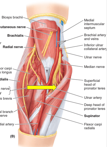
Anterior Interosseous Nerve (AIN) arises from median nerve at lower border of cubital fossa at Pronator Teres Muscle. It traverses distally along interosseous membrane between
FPL & FDP to reach pronator quadratus muscle. Ends by supplying sensory fibers to wrist
AIN Entrapment can occur between the
2 heads of pronator teres
Unclear etiology
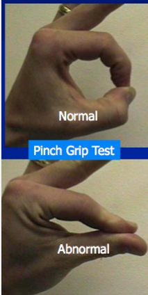
AIN Entrapment presentation muscle involvement
FPL: Lateral ½ of FDP, Index & middle finger
Pronator quadratus
Pinch deformity evident: (+) pinch grip test: can’t flex DIP, Difficulty picking things up
No cutaneous sensory function at wrist
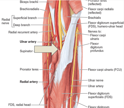
FDS entrapment of median nerve is concomitant w/ Pronator syndrome. resisted FDS performed w/
fingers in extension
Radial Nerve (C5-T1) innervates
Motor:
Posterior compartment of arm & forearm
Sensation:
Posterior arm, Dorsal Forearm, Dorsal of hand
radial nerve entrapment sites
Fibrous bands at radial head
Leash of Henry
The ECRB
Arcade of Frohse
Supinator
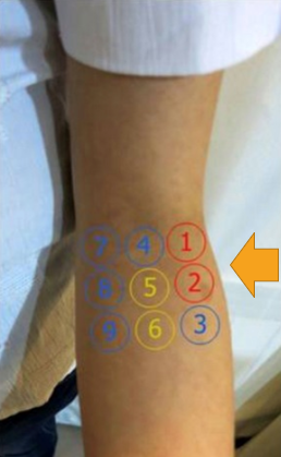
Radial Nerve (Tunnel) Syndrome sx presentation includes Diffuse lateral elbow pain, esp w/
gripping
Aching at night
Dorsal wrist pain
5 cm distal to lateral epicondyle: Irritation of this nerve may be concomitant w/ LE
Superficial Radial Nerve
PIO: motor weakness
OA or fibrous bands anterior to radiocapitellar jt or synovitis of radiocapitellar can impact what potential radial nerve compression site that may lead to RTS?
the lateral elbow jt in radial head
if a pt has a spiral groove fracture what muscles innervated are affected?
triceps & brachioradialis, ECRB (radial deviation & elbow extension affected)
Posterior Interosseous Nerve is a branch of radial nerve in forearm can become entrapped between
the 2 heads of the supinator
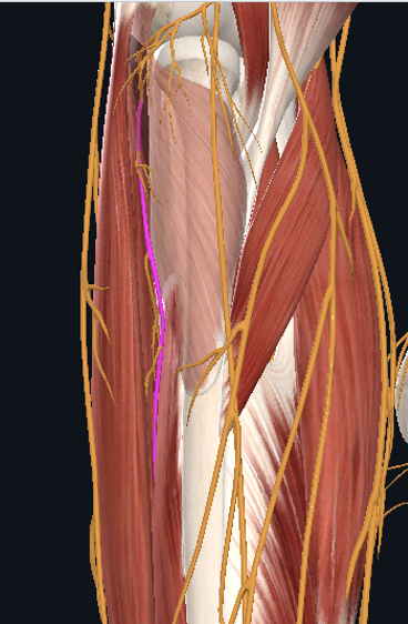
Posterior Interosseous Nerve MOI is result of trauma to the area
Presentation:
Motor: Extensor muscles of wrist
No sensory: but may have local pain
C6 radiculopathy, lateral epicondylalgia, radial tunnel could all be differential diagnosis for
lateral elbow pain
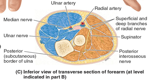
Acute Forearm Compartment Syndrome can be from
Supracondylar fx
Distal radial fx or direct trauma
IV line malfunction
Infections, Insect bites
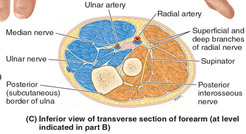
Forearm Compartment Syndrome presents w/ the 5 ps
Pain
Pallor
Passive stretch pain
Paranesthesia
Pulselessness
An arcade of anastomosing branches of recurrent radial artery at radial neck can impact what potential radial nerve compression site that may lead to (sensory) RTS?
the Leash of Henry
The leading/medial proximal edge of ECRB can impact what potential radial nerve compression site that may lead to (sensory) RTS?
fibrous edge of ECRB
The proximal edge of superficial layer of supinator muscle can impact what potential radial nerve compression site that may lead to (not sensory) RTS?
the Arcade of Frohse
The distal edge of supinator muscle can impact what potential radial nerve compression site that may lead to (not sensory) RTS?
distal edge of radial tunnel