Cardio Physiology 5 - Factors effecting cardiac output, Arteries
 Call Kai
 Learn
Learn Practice Test
 Spaced Repetition
 Match
Match Flashcards
 Knowt Play
1/69

There's no tags or description
Looks like no tags are added yet.
Name | Mastery | Learn | Test | Matching | Spaced | Call with Kai | Chat |
|---|
No analytics yet
Send a link to your students to track their progress
70 Terms
3 factors that effect stroke volume
End diastolic volume (preload, EDV)
Contractility of ventricular myocardium
Afterload
What is end diastolic volume
Volume of blood in the ventricles at the end of ventricular diastole (volume of blood in ventricles after filling is complete)
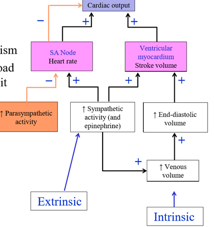
Intrinsic mechanism to alter stroke volume
Ventricles contract more forcefully when they have been stretched prior to contraction
Increased ventricle stretching causes increased stroke volume. What causes increased ventricular stretching?
Ventricles filled more fully with blood
How to fill ventricles with more blood
Increase venous return (blood flowing back to the heart through veins)
Effect of increased venous return
Increased end-diastolic volume, which increases stroke volume, which increases cardiac output
Why is end diastolic volume called the preload
Is the tension/load of ventricular myocardium before contraction
Extrinsic mechanism to increase filling of ventricles
Sympathetic stimulation of venous smooth muscle
Parasympathetic system effect on venous volume
No effect - system does not constrict/dilate veins
Frank Starling Mechanism
Independent mechanism that displays relationship between EDV and SV
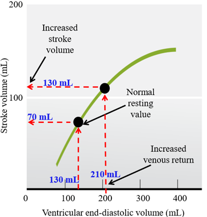
Relationship between EDV and SV
Increased EDV, increased SV
Why does increased EDV lead to increased SV
Ventricles fill with more blood, stretching out the sarcomeres, causing more forceful contraction
Frank-starling mechanism matches output of the two ventricles - what does this ensure?
Ensures the ventricles pump the same volume in blood, so blood does not accumulate in one circuit
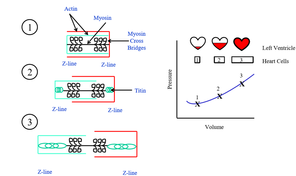
Mechanism of how increased blood volume impacts sarcomeres
More blood = more sarcomere stretch. The myosin cross bridges will connect to their correct actin. Titin protein is stretched out, and sarcomere is not crowded (high tension) = more forceful contraction
How to increase strength of contraction of ventricular myocardium
Increased sympathetic stimulation, allowing ventricles to eject more than the normal % of their blood volume
Effect of parasympathetic innervation on strength of ventricular myocardium contraction
No impact, ventricular myocardium does not receive parasympathetic innervation
How does sympathetic stimulation increase stroke volume
Stroke volume is greater at a given end-diastole volume with innervation = increase cardiac output
Ejection fraction
measurement of the volume percentage of left ventricular contents ejected with each contraction. = SV/EDV
Increased contractility on ejection fraction
Increased ejection fraction
Impact of sympathetic activity on rate of contraction/relaxation
Heart contracts and relaxes faster, giving more time for ventricles to fill (despite increase in heart rate)
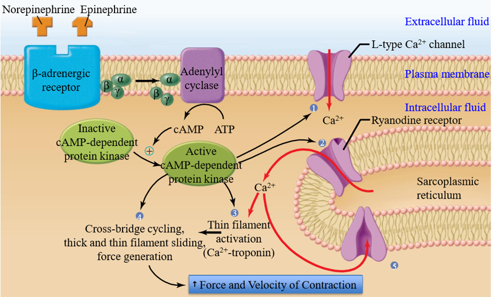
How is myocardial contractility regulated
G protein coupled mechanism
Sympathetic neurotransmitters for heart
Norepinephrine, epinephrine
Impact of sympathetic innervation on myocardial contractility regulation
Proteins involved in excitation-contraction coupling become phosphorylated, enhancing contractility
What proteins are phosphorylated leading to increased contractility
- L-type calcium channels (sarcolemmea)
- Ryanodine receptor in SR membrane
- Thin filament proteins (troponin)
- Thick filamens proteins associated with cross bridges
- Proteins that pump Ca2+ back into SR
Afterload
Tension against which ventricle must eject blood, related to arterial pressure
Effect of afterload on stroke volume
more ventricular afterload, lower stroke volume. Ventricle uses it's pressure to overcome the afterload
Endothelium continuity
Endothelium in blood vessels is continuous with the endocardium of the heart
Role of the endothelium in blood vessels
Provides smooth surface for blood to flow over
Why are pressures in the systemic circulation higher than the pulmonary circulation
Pressure needs to be generated to deliver blood to the entire body, and has to work against gravity (in legs)
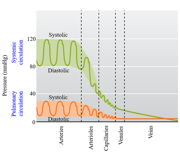
Why is the pulmonary vascular resistance much lower than systemic resistance
Larger vessels throughout the pulmonary system, and the arterioles have less smooth muscle
Artery wall composition
Smooth muscle, elastic fibers, connective tissue
Use of muscular wall in artery
Contraction and diameter change
Use of artery wall elasticity
Allows for passive changes in vessel diameter in response to changes in blood pressure (no contracting smooth muscle)
Vasoconstriction
Contraction of arterial smooth muscle, decreases artery diameter
Vasodilation
Relaxation of arterial smooth muscle, increases artery diameter
Types of arteries
elastic, muscular, arterioles
Elastic arteries
Contain elastic fibers and few muscle cells. Inside pulmonary trunk and aorta - tolerate pressure changes during cardiac cycle
Elastic artery stretch/contraction in cardiac cycle
Expand when stroke volume ejected
Recoil to original dimension in diastole
Muscular arteries
Contain smooth muscle cells, few elastic fibers. Makes most of the arterial system vessels.
Function of muscular arteries
Distribute blood throughout body
Arterioles
Smallest arteries in body
Role of arterioles
Regulate blood flow to organs by regulating blood flow to capillary beds. Determine mean arterial pressure (blood pressure)
Arterioles composition
Smooth muscle forms rings around endothelium - allows for constriction of arteriole
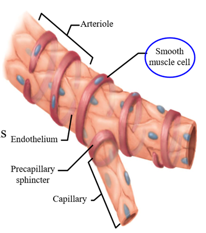
Use of arteriole's small diameter + constriction
Increases resistant to flow, allows less blood to reach organs
Role of arterioles in exercise
Muscular arterioles dilate, allowing more blood to flow to capillary beds to oxygenate muscles. GI arterioles contract to prevent blood from entering
Vasoconstriction of arterioles
Contracts arteriolar smooth muscle = decreased blood flow
Vasodilation of arterioles
Relaxes arteriolar smooth muscle = increased blood flow
Arteriole intrinsic tone
Partial contraction in absence of neural/hormonal stimuli
Increased arteriole tone =
Constriction
Decreased arteriole tone =
Dilation
Extrinsic factors that effect arteriole tone
External to the organ/tissue. Alter whole body needs. Includes nerves and hormones
Intrinsic factors that effect arteriole tone
Nerves and hormones that organs/tissues use to alter their own blood flow
Sympathetic innervation to arteriolar smooth muscle
Sympathetic nerve fibers release norepinephrine to cause vasoconstriction. Always discharge at basal level (but can be increased/decreased)
Importance of sympathetic innervation of arteriolar smooth muscle
Regulates blood pressure by influencing arteriolar resistance
Noncholinergic, noradrenergic neurons impact on arteriolar smooth muscle (No Ach, No NE)
Release nitric oxide, a vasodilator, onto smooth muscle
Role of epinephrine on arteriolar smooth muscle
Can cause vasoconstriction or vasodilation depending on which receptor it binds to
Examples of local controls effecting arteriolar resistance
Active hyperemia, reactive hyperemia, flow autoregulation
Active hyperemia
Local control acts to increase blood glow when metabolic activity of organ/tissue increases
Hyperemia
Excess of blood in vessels supplying organ or tissue
Steps of active hyperemia
1. Increased metabolic activity of organ
2. Decreased O2 (due to metabolism), Increased metabolites in organ interstitial fluid
3. Arteriolar dilation in organ
4. Increased blood flow to organ
How does the active hyperemia pathway get activated
Arteriolar smooth muscle is sensitive to local chemical changes in ECF surrounding arterioles - sensitive to O2, CO2, pH (changed by metabolism)
Where is active hyperemia most developed
Skeletal muscle, cardiac muscle, glands (where metabolic conditions are most altered)
Flow autoregulation
Locally mediated changes in arteriolar resistance do to organ/tissue experiencing change in blood pressure - occurs at constant metabolism
Steps of flow autoregulation at low pressure
1. Low arterial pressure in organ causes decreased blood flow to organ
2. Decreased oxygen, excess metabolites, decreased vessel wall stretch in organs
3. Arterioles dilate in organ
4. Restoration of blood flow to normal in organ
Steps of flow autoregulation at high pressure
1. High arterial pressure in organ causes increased blood flow to organ
2. Increased oxygen, decreased metabolites, increased vessel wall stretch in organ
3. Arterioles constrict in organ
4. Blood flow is constricted, restoration to normal flow
What mediates flow autoregulation
Changes in local chemical factors, and by myogenic response (direct smooth muscle response to stretch)
Are nerves/hormones involved in flow autoregulation
No - done by changes in chemical factors due to muscle wall response to stretch (myogenic response)
Reactive hyperemia
Form of flow autoregulation caused by occlusion (blockage) of blood flow. Allows for increased blood flow
Steps of reactive hyperemia
1. Occlusion of blood flow decreases O2 levels and increases metabolites
2. Arterioles dilate
3. Blood flow increases when occlusion is removed
Example of reactive hyperemia
When you remove a tightly tied string from finger, finger turns red due to blood rushing in (arterioles dilated)