Exam questions
 Call Kai
 Learn
Learn Practice Test
 Spaced Repetition
 Match
Match Flashcards
 Knowt Play
1/105

There's no tags or description
Looks like no tags are added yet.
Name | Mastery | Learn | Test | Matching | Spaced | Call with Kai | Chat |
|---|
No analytics yet
Send a link to your students to track their progress
106 Terms
List the 7 basics steps of image production in CT
x-ray tube directs x-ray beam towards pt
x-ray beam is attenuated
remnant photons are collected by detector
detector creates electrical radiation readings
radiation reading converted to digital signal
raw digital signal is processed into digital images
digital image sent to monitor
What components are located within the scanner gantry
x-ray tube
pre-patient collimator (determines beam width)
detectors (collects photons, converts to electrical signal)
post-pt/detector collimators (protects from scatter)
high-volt gens (produces variable kvp/ma)
analog to digital converter
Formula for Beam width
detector rows X slice thickness
What are the four photon interactions in CT?
Transmission
Coherent scattering
Photoelectric absorption
Compton scattering
Which photon interactions are NON-ionizing?
Transmission
Coherent scattering
Which interactions are IONIZING?
Photoelectric effect
Compton scattering
What is transmission?
X-rays pass through the body with no interaction.
What happens in coherent scatter? what is the alternate word?
Photon is absorbed and immediately re-emitted.
Rayleigh scattering
What happens in photoelectric absorption?
Photon is completely absorbed by an electron.
What happens in Compton scattering?
Photon interacts with an electron and loses part of its energy.
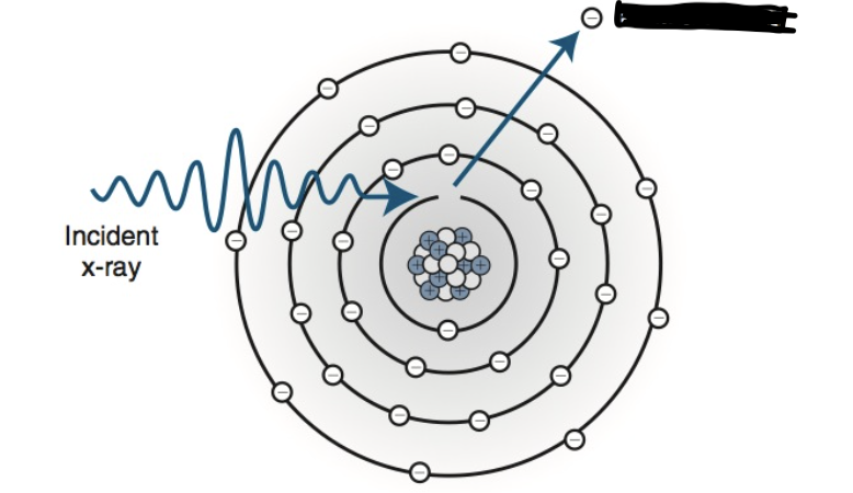
what photon interaction is this
Photoelectric absorption
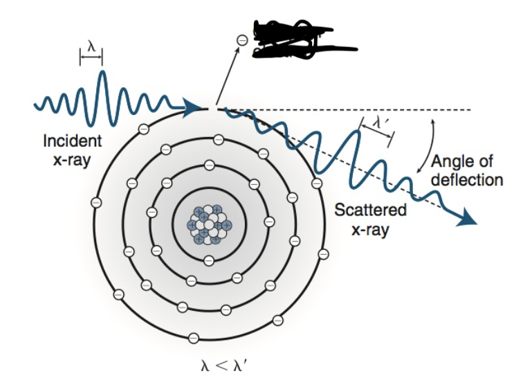
what photon interaction is this
Compton
what is overbeaming
The x-ray beam is widened so every detector gets equal x-ray exposure.
what is overranging
Extra radiation is used before and after the scan area so the computer has enough information to reconstruct accurate helical images.
abnormal levels of consciousness:
Alert: conscious have the ability to fully respond
lethargic: patient appears drowsy
but can be aroused.
obtunded: patient is in a more depressed level
of consciousness and may not easily be aroused
from a state of confusion.
Stupor (semicomatose): describes a state of near unresponsiveness
5. coma: patient is completely unresponsive to stimuli
Normal body temperature
97.7° to 99.5°F
Pulse rate for adults and children
Adults: 60 to 100 bpm
children: 70 to 120 bpm
Normal respiration rate for adults/children
Adults: 12 to 20 breaths per minute
Children: 20 to 30 breaths per minute.
Normal systolic and diastolic reading
systolic = 100-120
diastolic = 60-80
Normal pulse oxi range
90% to 100%
What is the first phase of heart contraction?
Contraction of the right and left atria.
What happens when the atria contract?
Blood is pushed into the ventricles.
What is the second phase of heart contraction?
Contraction of the ventricles.
What happens when the ventricles contract?
Blood is pumped into the pulmonary arteries and the aorta.
What does the P wave represent?
Electrical activity (depolarization) of the right and left atria.
What does the QRS complex represent?
Electrical activity (depolarization) of the ventricles.
What cardiac phase occurs during the QRS complex?
Ventricular systole (ventricular contraction).
What is the ST segment?
The period when the ventricles are contracting but no electrical activity is flowing.
Where is the ST segment located?
Between the QRS complex and the T wave.
What does the ST segment normally look like?
A straight, level line.
What does the T wave represent?
Ventricular repolarization. The ventricles reset electrically and prepare for the next contraction.
Which EKG waveform represents atrial depolarization?
P wave.
Which waveform represents ventricular depolarization?
QRS complex.
Which waveform represents ventricular repolarization?
T wave.
Which part of the EKG shows ventricular contraction without electrical activity?
ST segment.
Which CT exams commonly use EKG gating?
Cardiac CT and coronary CT angiography (CTA).
what drug can be administered to cause dilation of the coronary vessels, improving their visualization.
sublingual nitroglycerine
Cardiac CT images are typically reconstructed from data acquired during what phase
diastolic phase.
Which lab values evaluate kidney function?
BUN
Creatinine
GFR
Which lab values evaluate blood clotting?
PT
PTT
INR
Platelet count
What is the normal BUN range?
7–21 mg/dL
What is the normal creatinine range?
0.7–1.5 mg/dL
What is the normal GFR?
90–120 mL/min
What is the normal PT?
11–13.5 seconds
Why is a high PT a contraindication for interventional procedures?
increased risk of uncontrolled bleeding.
What is the normal PTT?
25–35 seconds
What is the normal platelet count?
150,000–400,000/µL
Pitch = 1
The table moves exactly the same distance as the beam width during one rotation.
What occurs when pitch = 1? how is image quality, dose and scan speed
No overlap and no gaps.
image quality = Moderate
dose = Moderate
scan speed = Moderate
What does a pitch greater than 1 mean?
The table moves farther than the beam width.
What occurs when pitch > 1? what happens to image quality, dose and scan speed?
Greater gaps no laps
image quality = decrease
dose = decrease
scan speed = increased
What does a pitch less than 1 mean?
The table moves less than the beam width.
What occurs when pitch < 1? what happens to image quality, dose and scan speed?
No gaps. Overlaps
image quality = Increases
dose = Increases
scan speed = Decreases
A technologist wants to lower patient dose. What pitch change should be made?
Increase pitch
A technologist wants faster acquisition. What pitch change should be made?
Increase pitch
A technologist wants maximum spatial resolution. What pitch change should be made?
Decrease pitch
What is the formula for pitch?
Pitch = Table Movement ÷ Beam Width
What is the formula for table movement?
Table Movement = Beam Width × Pitch
What is temporal resolution
The ability of a CT system to freeze motion and provide an image free of blurring.
WW & WL for displaying gray/white matter
WW 100, WL 35
WW & WL for displaying Hemorrhage/hematoma
WW 150, WL 75
WW & WL for displaying Acute ischemia
WW 25, WL 35
(variable, high-contrast windowing improves the CT visualization of acute stroke)
WW & WL for displaying Lung parenchyma
WW 1400, WL −450
WW & WL for displaying Mediastinum
WW 350, WL 40
What is the formula for calculating lower window limit?
WL - (WW ÷ 2)
What is the formula for calculating upper window limit?
WL + (WW ÷ 2)
Typical Abdomen Window?
WW 400
WL 40
Typical Bone Window?
WW 2500
WL 600
Typical Lung Window?
WW 1800
WL -600
WW increases. What happens?
Contrast decreases.
WW decreases. What happens?
Contrast increases.
WL increases. What happens?
Image becomes darker.
WL decreases. What happens?
Image becomes brighter.
what is An omental CT scan?
Scan of the abdomen focused on the omentum, a fatty layer of tissue inside the abdomen
What is the HU for air
-1000
What is the HU for lung
-700
What is the HU for fat
-50
What is the HU for CSF
+15
What is the HU for blood
+30 to +45
What is the HU for muscle
+40
What is the HU for bone
+1000
How does acquisition thickness affect spatial resolution?
Thinner acquisition thickness increases spatial resolution.
How should images be reconstructed to maximize spatial resolution?
Use the thinnest reconstruction slice thickness possible.
How does matrix size affect spatial resolution?
Larger matrix size increases spatial resolution.
Name three factors that affect image noise.
mA
kVp
Patient size
How does increasing mA affect contrast resolution?
It improves contrast resolution.
How does increasing kVp affect contrast resolution?
It improves contrast resolution.
How does thicker acquisition thickness affect contrast resolution?
It increases contrast resolution.
How does thicker reconstruction slice thickness affect contrast resolution?
It improves contrast resolution.
Which ROI measurement represents image noise?
Standard deviation.
Which reconstruction algorithm produces more image noise?
Bone (edge-enhancing) algorithm.
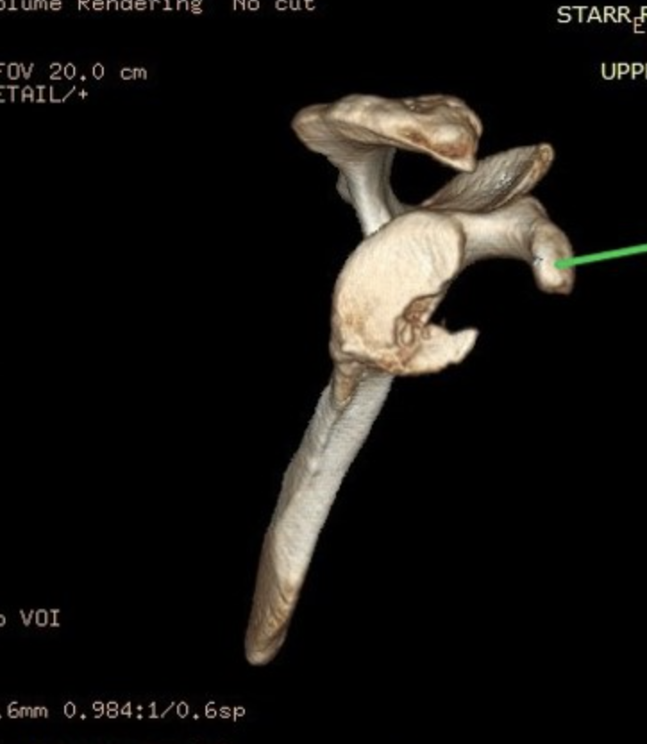
what is the green arrow pointing to
coracoid process
CT image quality is evaluated in terms of
spatial resolution and contrast resolution
the ______ data acquisition mode moves the bed through the CT gantry while the x-ray tube and detector array remain stationary
scout
list the phases of tissue enhancement after injection of contrast media
bolus, equilibrium, nonequilibrium
The _____ phase of liver enhancement is optimal for the evaluation of hypovascular lesions such as metastases that develop from primary tumors of the colon
portal venous
The falciform ligament extends from the liver to the
anterior abdominal wall and diaphragm