Periodontal Disease Pathogenesis
 Call Kai
 Learn
Learn Practice Test
 Spaced Repetition
 Match
Match Flashcards
 Knowt Play
1/50

Earn XP
Description and Tags
Lecture 3
Name | Mastery | Learn | Test | Matching | Spaced | Call with Kai | Chat |
|---|
No analytics yet
Send a link to your students to track their progress
51 Terms
What are the 4 components of the peridontium, which are the tissues that surround the tooth and hold it in place?
gingiva
periodontal ligament
cementum
alveolar bone
What is the most common disease of dogs and cats presented to small animal practitioners?
periodontal disease
What is the most common cause of oral infection and tooth loss?
periodontal disease
Is periodontal disease reversible?
No, once established it can only be managed and is chronically progressive
Describe healthy gingiva.
sharp, thin, and noninflamed margins
coral-pink if non-pigmented
firm and stippled
Describe the appearance of plague induced gingivitis.
marginal gingivitis with rounded edges
gingival margin — erythematous
may bleed upon probing
What colonizes on tooth surfaces within 24-36 hours after cleaning/brushing?
Bacterial plaque
_____ is an organic, transparent, adhesive biofilm consisting of glycoproteins, bacteria, and extracellular polysaccarhides.
Plaque
If not removed within a few days, plaque initiates what at the gingival margin?
inflammatory response
Normal oral flora is composed of primarily _____, _____ bacteria.
aerobic, gram positive
The accumulation of ______ and the subgingival location creates an anaerobic environment, causing an increase in anaerobic, gram negative, and spirochete bacteria.
dental calculus
Gingivitis is diagnosed when the inflammation is limited to the ____.
gingiva
Gingival inflammatory response to plaque consists of ____, _____, and _____.
vasculitis, edema, collagen loss
Advanced gingivitis leads to the potential development of _____.
periodontitis
True or False: Not all animals with gingivitis will ultimately develop periodontitis.
True
What are inflammatory factors (host’s immune response) in progression of gingivitis to periodontitis?
subgingival plaque bacteria
lipopolysaccharide from gram negative bacteria induce inflammatory mediator cytokines
activated neutrophils release proteolytic enzymes and reactive oxygen species
What is the generalized assessment of periodontal health evaluating gingivitis, plaque, and dental calculus?
gingivitis index
Evaluate gingivitis:
no inflammation
healthy
none
Evaluate gingivitis:
mild inflammation and color change
no bleeding on probing
GI1
Evaluate gingivitis:
moderate inflammation
bleeding on probing
GI2
Evaluate gingivitis:
severe inflammation
spontaneous bleeding on probing
GI3
List risk factors for periodontitis.
age (older)
breed (toy, brachycephalic, dolichocephalic)
persistent deciduous teeth
malocclusions
genetic predisposition
host’s immune response
non-abrasive diet
chewing habits
periodontal trauma
oral foreign bodies
Why are toy breeds at risk for periodontitis?
tooth crowding
Why are brachycephalic breeds at risk for periodontitis?
rotated and crowding of teeth
Why are dolichocephalic breeds at risk for periodontitis?
dry mouth
In periodontitis, the inflammatory factors associated with the ____ and ____ cause the destruction of the tissues surrounding the tooth.
anaerobic bacteria
host’s immune response
List clinical features of periodontal disease.
gingivitis
halitosis (bad breath)
gingival enlargement
gingival recession
periodontal pockets
abscessation
facial swelling
draining fistulae
oronasal fistula
osteomyelitis
osteolysis
mandibular fractures
systemic organ lesions from baceremia
opthalmic manifestations
oral pain
tooth mobility
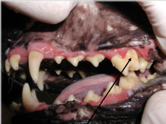
What is shown in this image?
gingival recession associated with periodontal disease
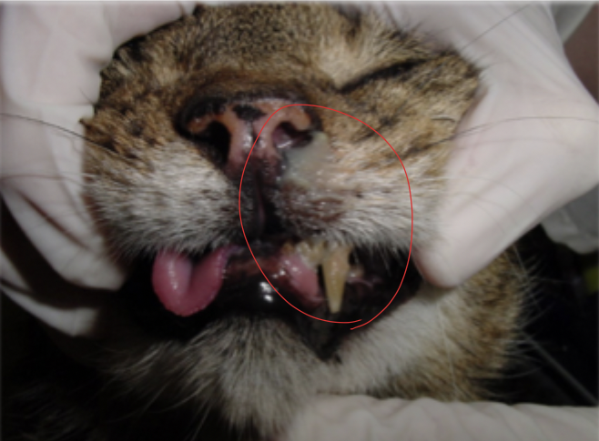
What is shown in this image?
oronasal fistula associated with periodontitis of maxillary canine tooth
Definitive diagnosis of periodontal disease stage requires ____, ____, and ____.
general anesthesia
dental radiography
periodontal probing
Does the degree of severity of periodontal disease relate to an individual tooth or the entire mouth?
individual tooth - a patient may have teeth that have different stages of periodontal disease
How should you write diagnosis of periodontal disease in the medical record from your physical exam?
“suspect grade of dental disease”
What refers to the destruction of the periodontium due to periodontitis?
attachment loss
The amount of attachment loss for a tooth is determined with _____, _____, and ____.
periodontal probing
measurement of gingival recession (if present)
radiographs
What is the distance (in mm) from the free gingival margin to the most apical point that the probe reaches when gently inserted into the gingival sulcus or pocket?
probing depth
What is defined, based on radiographs, as the distance from the alveolar margin to the cementoenamel junction relative to the length of the tooth?
attachment loss
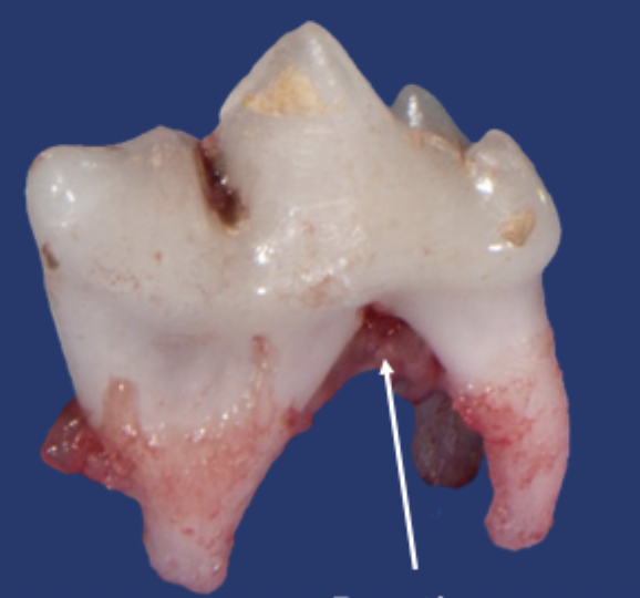
What is shown in this image?
tooth root furcation
Furcation Index:
probe extends less than halfway under the crown in any direction of a multi-rooted tooth with attachment loss
Stage 1 (F1)
Furcation Index:
probe extends greater than galfway under the crown of a multi-rooted tooth with attachment loss but not through and through
Stage 2 (F2)
Furcation Index:
probe extends under the crown of a multi-rooted tooth, through and through from one side of the furcation out the other
Stage 3 (F3)
Stages of Periodontal Disease:
clinically normal
gingival inflammation or periodontitis not clinically evident
Normal (PD 0)
Stages of Periodontal Disease:
gingivitis only without attachment loss
height and architecture of alveolar margin is normal
radiographic findings are normal
Stage 1 (PD 1)
Stages of Periodontal Disease:
early periodontitis or, at most, there is stage 1 furcation index
early radiographic signs of periodontitis
loss of periodontal attachment is less than 25%
Stage 2 (PD 2)
Stages of Periodontal Disease:
moderate periodontitis
25-50% of attachment loss
stage 2 furcation involvement
Stage 3 (PD 3)
Stages of Periodontal Disease:
advanced periodontal disease
greater than 50% attachment loss
stage 3 furcation
Stage 4 (PD 4)
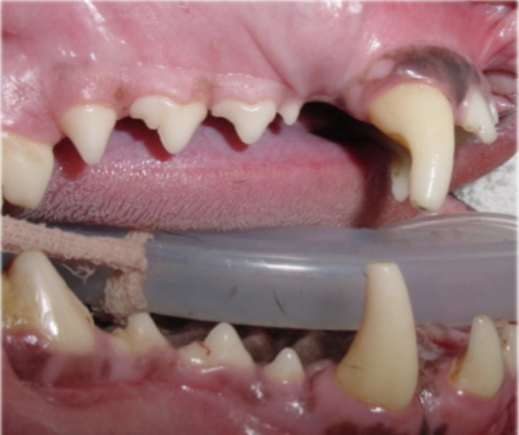
What is the stage of periodontal disease shown in this image?
Normal (PD 0)
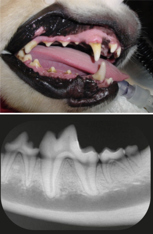
What is the stage of periodontal disease shown in this image?
Stage 1 (PD 1)
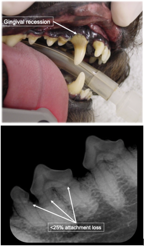
What is the stage of periodontal disease shown in this image?
Stage 2 (PD 2)
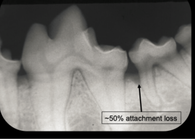
What is the stage of periodontal disease shown in this image?
Stage 3 (PD 3)
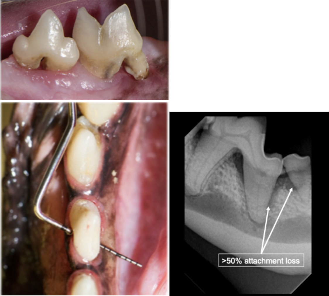
What is the stage of periodontal disease shown in this image?
Stage 4 (PD 4)
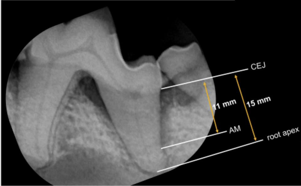
Calculate attachment loss based on the image.
11/15 = 0.733
Attachment loss = 73%