1750 Forearm, Elbow, Humerus IA
 Call Kai
 Learn
Learn Practice Test
 Spaced Repetition
 Match
Match Flashcards
 Knowt Play
1/59

There's no tags or description
Looks like no tags are added yet.
Name | Mastery | Learn | Test | Matching | Spaced | Call with Kai | Chat |
|---|
No analytics yet
Send a link to your students to track their progress
60 Terms
AP Projection; Forearm (CR & SS)
Perpendicular to the midpoint of the forearm
An AP projection of the forearm shows the elbow joint, the radius and ulna, and the proximal row of slightly distorted carpal bones
AP Projection; Forearm (Eval Criteria)
• Evidence of proper collimation
• Entire forearm, including wrist and distal humerus
• Slight superimposition of the radial head, neck, and tuberosity over the proximal ulna
• No elongation or foreshortening of the humeral epicondyles
• Partially open elbow joint if the shoulder was placed in the same plane as the forearm
• Open radioulnar space
• Similar image brightness and contrast of the proximal and distal forearm
• Soft tissue and bony trabecular detail
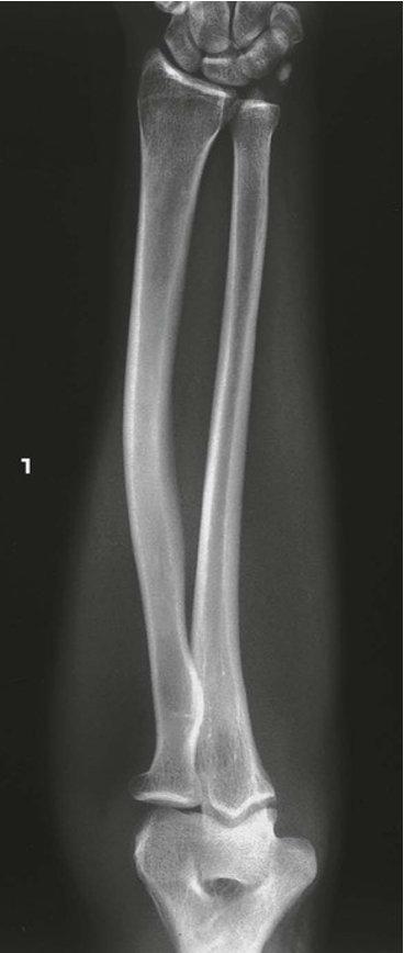
AP Projection; Forearm
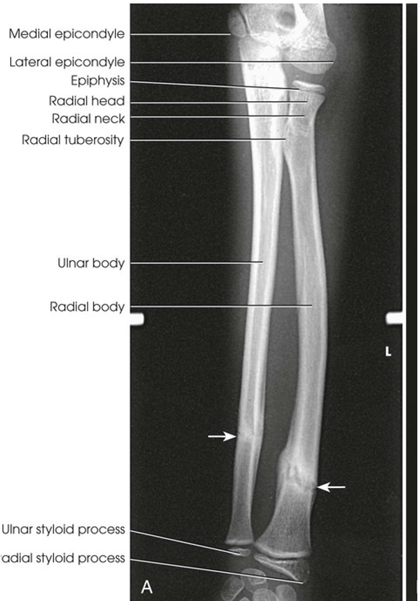
AP Projection; Forearm (Labeled)
Lateral Projection; Forearm; Lateromedial (CR & SS)
Perpendicular to the midpoint of the forearm
The lateral projection shows the bones of the forearm, the elbow joint, and the proximal row of carpal bones
Lateral Projection; Forearm; Lateromedial (Eval Criteria)
• Evidence of proper collimation
• Entire forearm, including wrist and distal humerus in a true lateral position:
• Superimposition of the radius and ulna at their distal end
• Superimposition of the radial head over the coronoid process
• Radial tuberosity facing anteriorly
• Superimposed humeral epicondyles
• Elbow flexed 90 degrees
• Soft tissue and bony trabecular detail along the entire length of the radial and ulnar bodies
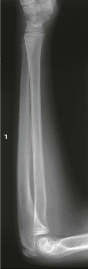
Lateral Projection; Forearm; Lateromedial
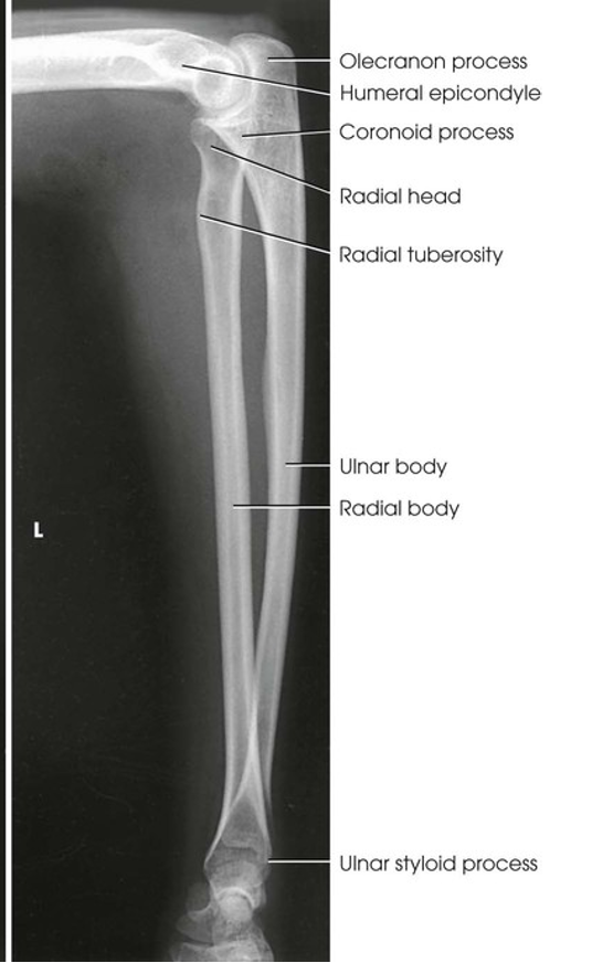
Lateral Projection; Forearm; Lateromedial (Labeled)
AP Projection; Elbow (CR & SS)
Perpendicular to the elbow joint
An AP projection of the elbow joint, distal arm, and proximal forearm is presented
AP Projection; Elbow (Eval Criteria)
• Evidence of proper collimation
• Radial head, neck, and tuberosity slightly superimposed over the proximal ulna (~.25”)
• Elbow joint open and centered to the central ray
• No rotation of humeral epicondyles (coronoid and olecranon fossae approximately equidistant to epicondyles)
• Soft tissue and bony trabecular detail
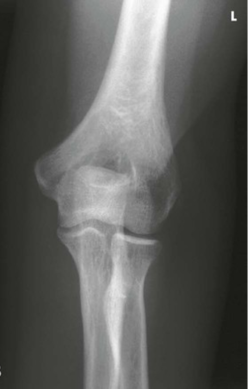
AP Projection; Elbow
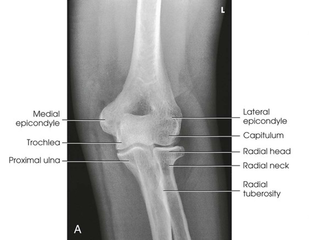
AP Projection; Elbow (Labeled)
AP Oblique Projection; Elbow; Medial Rotation (CR & SS)
Perpendicular to the elbow joint
The image shows an oblique projection of the elbow with the coronoid process projected free of superimposition
AP Oblique Projection; Elbow; Medial Rotation (Eval Criteria)
• Evidence of proper collimation
• Elbow joint open and centered to the central ray
• 45-degree medial rotation of elbow:
• Coronoid process in profile
• Elongated medial humeral epicondyle
• Ulna superimposed by the radial head and neck
• Trochlea
• Olecranon process within the olecranon fossa
• Soft tissue and bony trabecular detail
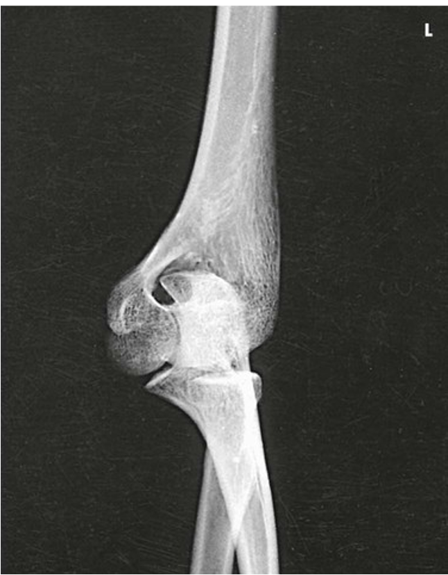
AP Oblique Projection; Elbow; Medial Rotation
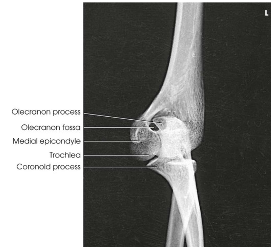
AP Oblique Projection; Elbow; Medial Rotation (Labeled)
AP Oblique Projection; Elbow; Lateral Rotation (CR & SS)
Perpendicular to the elbow joint
The image shows an oblique projection of the elbow with the radial head and neck projected free of superimposition of the ulna
AP Oblique Projection; Elbow; Lateral Rotation (Eval criteria)
• Evidence of proper collimation
• Elbow joint open and centered to the central ray
• 45-degree lateral rotation of elbow:
• Radial head, neck, and tuberosity projected free of the ulna
• Elongated lateral humeral epicondyle
• Capitulum
• Soft tissue and bony trabecular detail
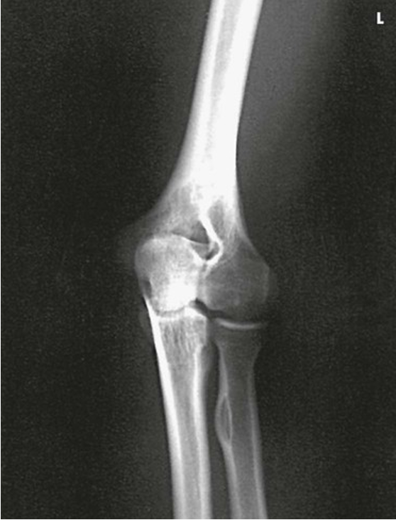
AP Oblique Projection; Elbow; Lateral Rotation
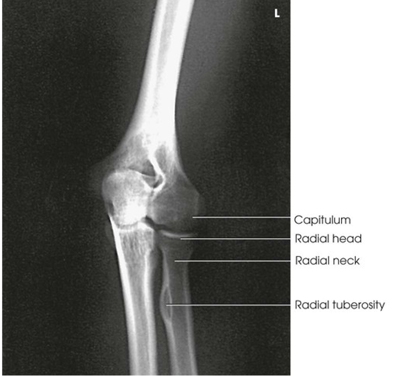
AP Oblique Projection; Elbow; Lateral Rotation (Labeled)
Lateral Projection; Elbow; Lateromedial (CR & SS)
Perpendicular to the elbow joint, regardless of its location on the IR
The lateral projection shows the elbow joint, distal arm, and proximal forearm
Lateral Projection; Elbow; Lateromedial (Eval Criteria)
• Evidence of proper collimation
• Elbow joint open and centered to the central ray
• Elbow in a true lateral position:
• Superimposed humeral epicondyles
• Radial tuberosity facing anteriorly
• Radial head partially superimposing the coronoid process
• Olecranon process in profile
• Elbow flexed 90 degrees
• Bony trabecular detail and any elevated fat pads in the soft tissue at the anterior and posterior distal humerus and the anterior proximal forearm
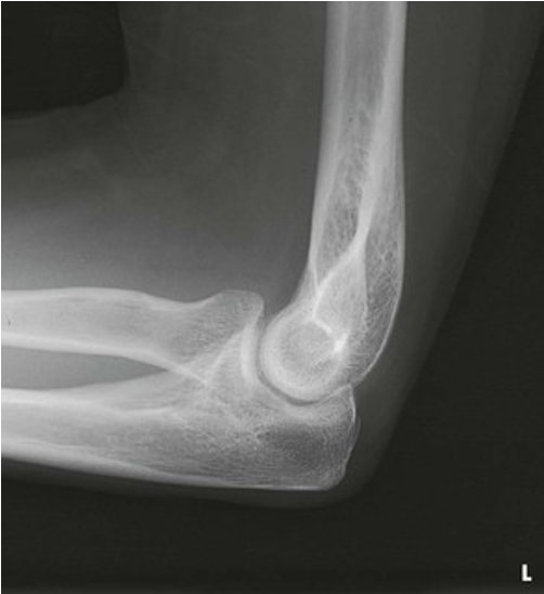
Lateral Projection; Elbow; Lateromedial
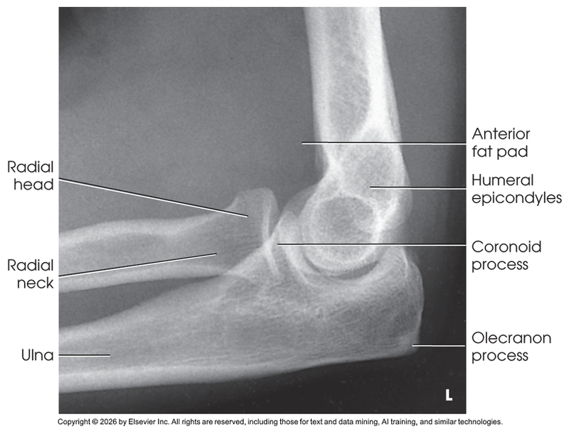
Lateral Projection; Elbow; Lateromedial (Labeled)
Distal Humerus; Elbow; AP Projection; Partial Flexion (CR & SS)
Perpendicular to the humerus, traversing the elbow joint (depending on the degree of flexion, angle the central ray distally into the joint)
This projection shows the distal humerus when the elbow cannot be fully extended
Distal Humerus; Elbow; AP Projection; Partial Flexion (Eval Criteria)
• Evidence of proper collimation
• Distal humerus without rotation or distortion
• Proximal radius superimposed over the ulna
• Closed elbow joint
• Greatly foreshortened proximal forearm
• Trabecular detail on the distal humerus
• Soft tissue and bony trabecular detail
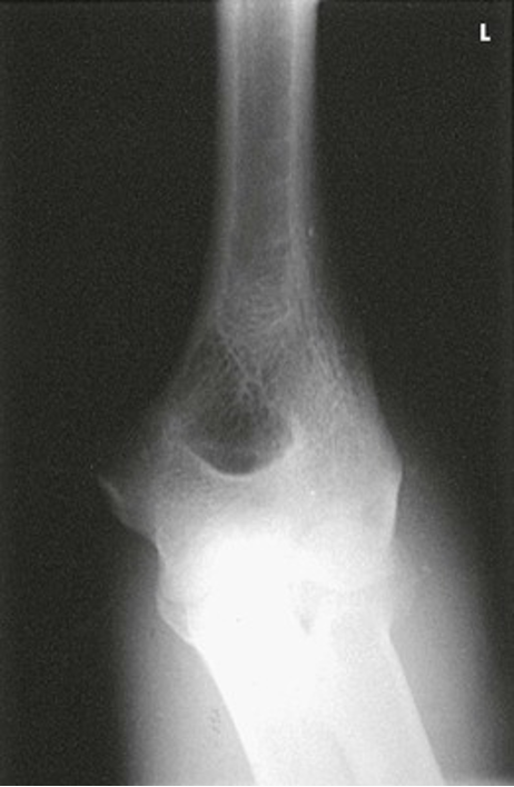
Distal Humerus; Elbow; AP Projection; Partial Flexion
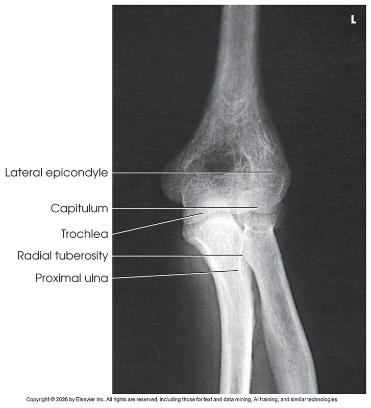
Distal Humerus; Elbow; AP Projection; Partial Flexion (Labeled)
Proximal Forearm; Elbow; AP Projection; Partial Flexion (CR & SS)
Perpendicular to the elbow joint and long axis of the forearm (Adjust the IR so that the central ray passes to its midpoint)
This projection shows the proximal forearm when the elbow cannot be fully extended
Proximal Forearm; Elbow; AP Projection; Partial Flexion (Eval Criteria)
• Evidence of proper collimation
• Proximal radius and ulna without rotation or distortion
• Radial head, neck, and tuberosity slightly superimposed over the proximal ulna
• Partially open elbow joint
• Foreshortened distal humerus
• Soft tissue and bony trabecular detail
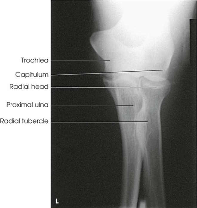
Proximal Forearm; Elbow; AP Projection; Partial Flexion
Lateral Projection; Elbow; Radial Head; Four-position Series; Lateromedial (CR & SS)
Perpendicular to the elbow joint
The radial head is projection in varying degrees of rotation
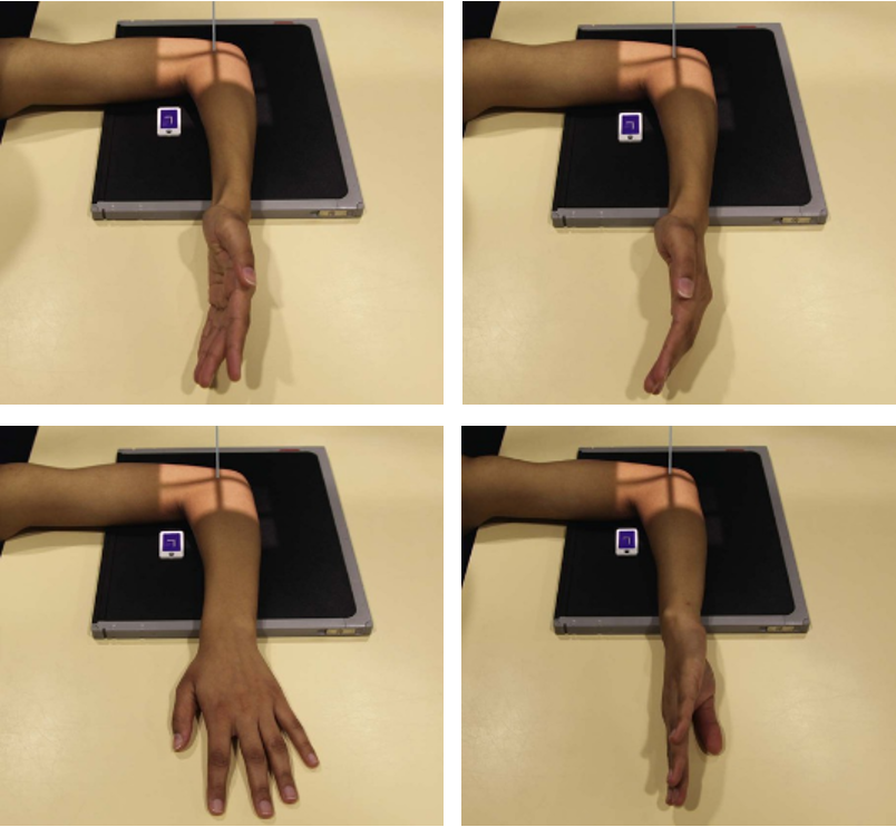
POSITIONING: Lateral Projection; Elbow; Radial Head; Four-Position Series; Lateromedial
Lateral Projection; Elbow; Radial Head; Four-position Series; Lateromedial (Eval Criteria)
• Evidence of proper collimation
• Radial tuberosity facing anteriorly for the first and second images and posteriorly for the third and fourth images
• Elbow flexed 90 degrees
• Radial head partially superimposing the coronoid process but seen in all images
• Soft tissue and bony trabecular detail
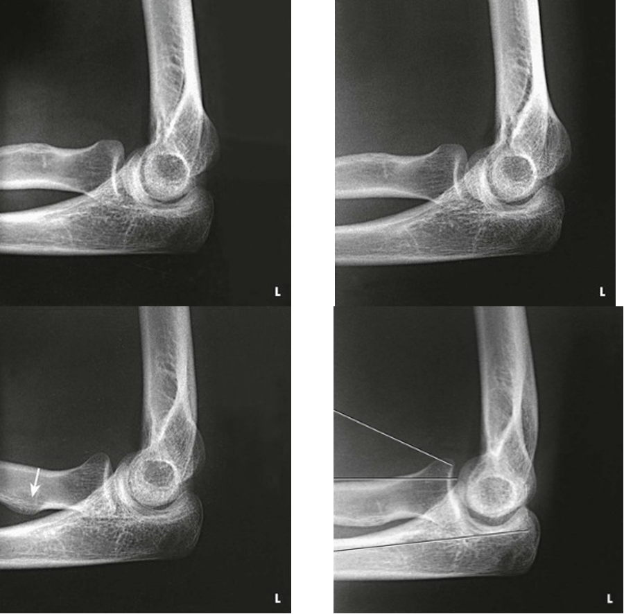
Lateral Projection; Elbow; Radial Head; Four-Position Series; Lateromedial
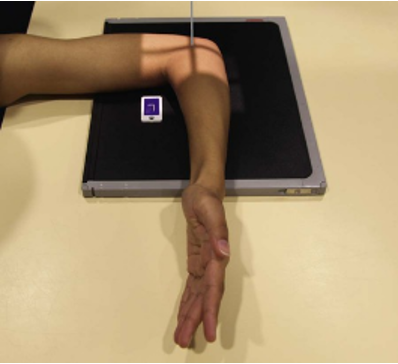
What position would the radial tuberosity face for this position?
Anterior
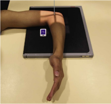
What position would the radial tuberosity face for this position?
Anterior
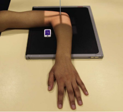
What position would the radial tuberosity face for this position?
Posterior
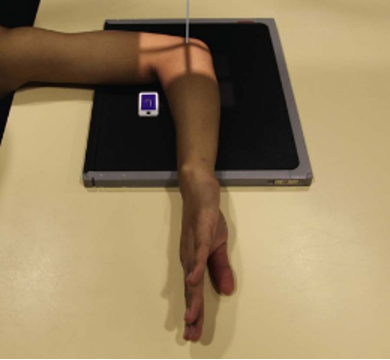
What position would the radial tuberosity face for this position?
Posterior
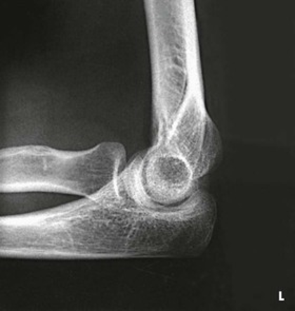
What position would the hand be in for this radiograph
Supinated
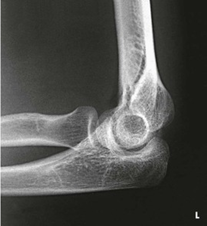
What position would the hand be in for this radiograph
Lateral
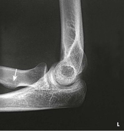
What position would the hand be in for this radiograph?
Pronated
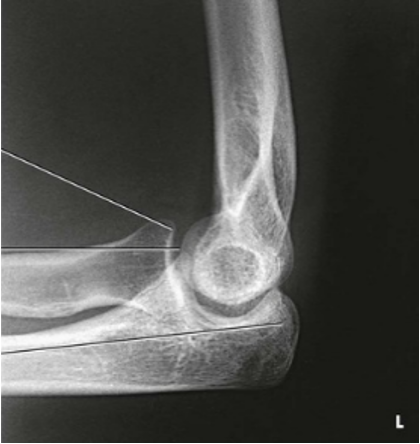
What position would the hand be in for this radiograph?
Internally rotated
AP Projection; Humerus; Upright (CR & SS)
Perpendicular to the midportion of the humerus and the center of the IR
The AP projection shows the entire length of the humerus. The accuracy of the position is shown by the epicondyles
AP Projection; Humerus; Upright (Eval Criteria)
• Evidence of proper collimation
• Elbow and shoulder joints
• Maximal visibility of epicondyles without rotation
• Humeral head and greater tubercle in profile
• Outline of the lesser tubercle, located between the humeral head and the greater tubercle
• Beam divergence resulting in distortion of the elbow joint
• Similar image brightness and contrast of the proximal and distal humerus
• Soft tissue and bony trabecular detail
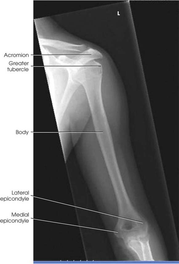
AP Projection; Humerus; Upright
Lateral Projection; Humerus; Upright (CR & SS)
Perpendicular to the midportion of the humerus and the center of the IR
The lateral projection shows the entire length of the humerus. A true lateral image is confirmed by superimposed epicondyles
Lateral Projection; Humerus; Upright (Eval Criteria)
• Evidence of proper collimation
• Elbow and shoulder joints
• Superimposed epicondyles
• Lesser tubercle in profile on medial aspect
• Greater tubercle superimposed over the humeral head
• Beam divergence resulting in distortion of the elbow joint
• Similar image brightness and contrast of the proximal and distal humerus
• Soft tissue and bony trabecular detail
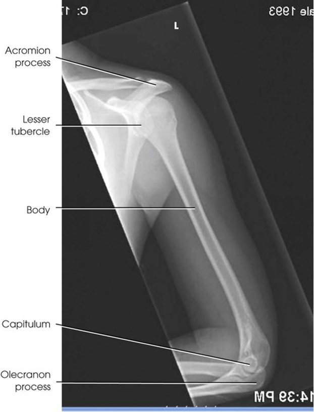
Lateral Projection; Humerus; Upright
Axiolateral Projection; Elbow; Radial Head and Coronoid Process; Coyle Method; Lateral (SS)
The resulting projections show an open elbow joint between the radial head and capitulum or between the coronoid process and trochlea, with the area of interest in profile. These projections are used to show the pathologic processes or trauma in the area of the radial head and coronoid process. The value of the projections is evident in the trauma images,
Axiolateral Projection; Elbow; Radial Head and Coronoid Process; Coyle Method; Lateral (CR Radial Head)
(Seated Position) Directed towards the should 45 degrees toward the radial head
(Supine Trauma Position) The horizontal CR is directed cephalad at an angle of 45 degrees to the radial head, entering the joint at mid-elbow
Axiolateral Projection; Elbow; Radial Head and Coronoid Process; Coyle Method; Lateral (CR Coronoid Process)
(Seated Position) Directed away from the shoulder 45 degrees to the coronoid process
(Supine Trauma Position) The horizontal CR is directed caudad at an angle 45 degrees to the coronoid process, entering the joint at mid-elbow
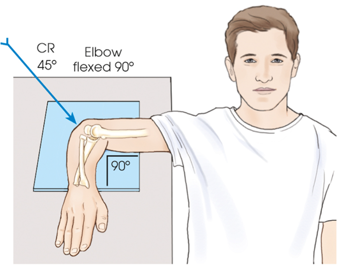
POSITIONING (Radial Head): Axiolateral Projection; Elbow; Radial Head and Coronoid Process; Coyle Method; Lateral
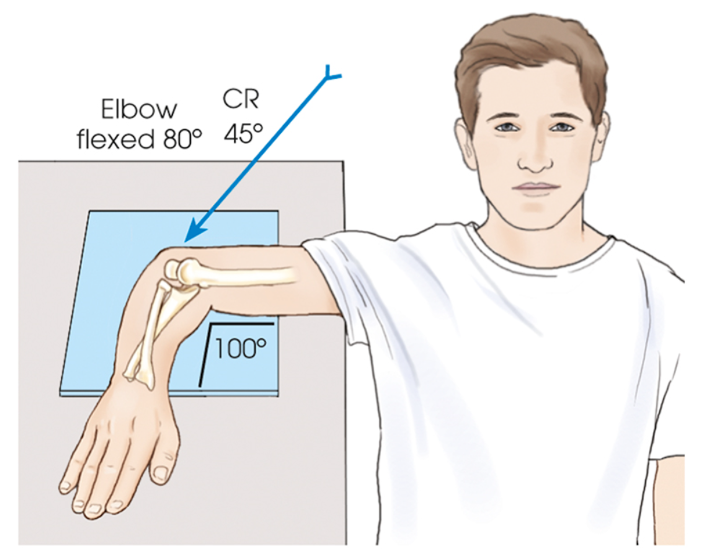
POSITIONING (Coronoid Process): Axiolateral Projection; Elbow; Radial Head and Coronoid Process; Coyle Method; Lateral
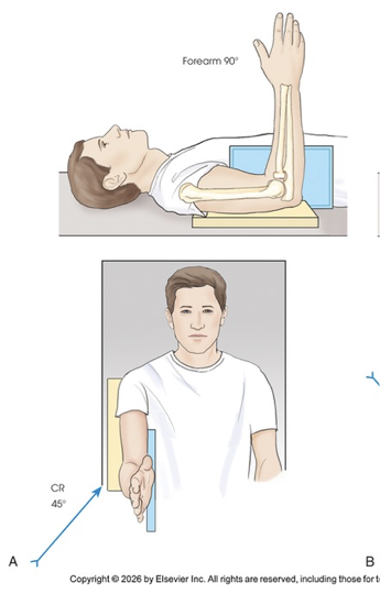
POSITIONING (Radial Head Trauma Position): Axiolateral Projection; Elbow; Radial Head and Coronoid Process; Coyle Method; Lateral
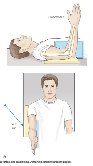
POSITIONING (Coronoid Process Trauma Position): Axiolateral Projection; Elbow; Radial Head and Coronoid Process; Coyle Method; Lateral
Axiolateral Projection; Elbow; Radial Head and Coronoid Process; Coyle Method; Lateral (Eval Criteria Radial Head)
The side marker placed clear of anatomy of interest
Open joint space between radial head and capitulum
Radial head, neck and tuberosity in profile and free from superimposition with the exception of a small portion of the coronoid process
Humeral epicondyles distorted, owing to CR angulation
Radial tuberosity facing posteriorly
Elbow flexed 90 degrees
Bony trabecular detail and surrounding soft tissues
Axiolateral Projection; Elbow; Radial Head and Coronoid Process; Coyle Method; Lateral (Eval Criteria Coronoid Process)
The side marker placed clear of anatomy of interest
Open joint space between coronoid process and trochlea
Coronoid process in profile and elongated
Radial head and neck superimposed by ulna
Elbow flexed 80 degrees
Bony trabecular detail and surrounding soft tissues
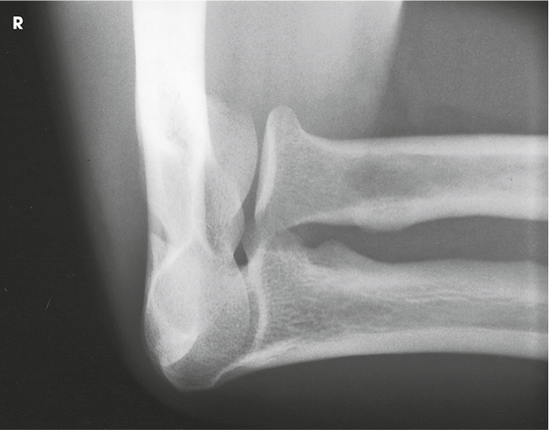
Axiolateral Projection; Elbow; Radial Head and Coronoid Process; Coyle Method; Lateral (Radial head and capitulum)
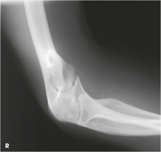
Axiolateral Projection; Elbow; Radial Head and Coronoid Process; Coyle Method; Lateral (Coronoid process and trochlea)