A&P - EXAM 1
 Call Kai
 Learn
Learn Practice Test
 Spaced Repetition
 Match
Match Flashcards
 Knowt Play
1/65

There's no tags or description
Looks like no tags are added yet.
Name | Mastery | Learn | Test | Matching | Spaced | Call with Kai | Chat |
|---|
No analytics yet
Send a link to your students to track their progress
66 Terms
Describe and give example of each level of organization from the simplest to most complex
Chemical - atoms, molecules
Cellular - single cells
Tissues - groups of cells
Organs - two or more tissue types
Organ System - organs that work together
Organismal - all organ systems combine to make the whole organism
Compare and contrast the fields and subfields of A&P and give an example of each
Anatomy - studies structure of body parts; asks where is it/ what is it (example: structure of the heart)
Physiology - studies the function of body parts; asks how does it work (example: how the heart pumps blood)
Integumentary System (structures and function)
Hair, skin, nails
Encloses internal body structures; site of many sensory receptors
Skeletal System
Cartilage, bone, joints
Supports the body
Enables movement (with muscular system)
Muscular System
Skeletal muscles, tendons
Enables movement (with skeletal system)
Helps maintain body temperature
Nervous System
Brain, spinal cord, peripheral nerves
Detects and processes sensory information
Activates bodily responses
Endocrine System
Pituitary gland, adrenal gland, thyroid gland, pancreas, testes
Secretes hormones
Regulates bodily processes
Cardiovascular System
Heart, blood vessels
Delivers O2 and nutrients to the tissues
Equalizes temperature in the body
Lymphatic System
Thymus, lymph nodes, spleen, lymphatic vessels
Returns fluid to the blood
Defends against pathogens
Respiratory System
Nasal passage, trachea, lungs
Removes CO2 from the body
Delivers O2 to the blood
Digestive System
Stomach, liver, gallbladder, large intestine, small intestine
Processes food for use by the body
Removes waste from undigested food
Urinary System
Kidneys, urinary bladder
Controls water balance in the body
Removes wastes from blood and excretes them
Male Reproductive System
Epididymis, testes
Produces sex hormones and gametes
Delivers gametes to the female
Female Reproductive System
Mammary glands, ovaries, uterus
Produces sex hormones and gametes
Supports embryo/fetus until birth
Produces milk for infant
Define and explain terms relating to homeostasis and give physiological examples for each
The maintenance of relatively stable internal conditions despite continuous changes in the environment
Maintained by contributions of all organ systems
The body is constantly monitored and regulated to maintain
Variables are factors that change: blood sugar, body temperature, blood volume, etc.
Three components involved: Sensor/Receptor, Control Center, and Effector
Negative Feedback System (with example)
Reverses/opposes the original change and moves the variable back towards its normal range
Change → body fights the change → back toward normal
Stimulus: Body temperature gets too high
Sensor: Nerve cells in skin and brain
Control Center: temperature regulatory center in brain
Effector: sweat glands throughout the body
Response: increased heat loss (temperature goes down)
Most used feedback mechanism in the body
Response reduces or shuts off original stimulus
Positive Feedback System
Reinforces/amplifies the original change, pushing the process farther in the same direction until a specific endpoint stops it
Change → body increases the change → increases more → endpoint
Stimulus: Childbirth; baby’s head stretches the cervix
Sensor: stretch receptors in the cervix detect the stretching
Control center: hypothalamus receives the signal and causes the posterior pituitary to release oxytocin
Effector: oxytocin travels through the blood to uterine smooth muscle
Response: uterine muscles contract more strongly, pushing the baby farther against the cervix; more stretching
Describe the structure of the plasma membrane and its components
Phospholipid bilayer (hydrophilic head, hydrophobic tail), containing many different molecular components, including proteins and cholesterol, and even attached carbohydrate groups
Describe the function of the plasma membrane and its components
Mechanical barrier - separates two of the body’s fluid compartments
Selective permeability - determines the manner in which substances enter or exit the cell
Electrochemical gradient - generates and helps to maintain the electrochemical gradient required for muscle and neuron function
Communication - allows cell-to-cell recognition and interaction
Cell signaling - plasma membrane proteins interact with specific chemical messengers and relay messages to the cell interior
Compare and contrast the types of transport across plasma membranes
Passive Transport - no energy required; high → low concentration
Down the concentration/electrochemical gradient
Simple diffusion (non-polar substances like O2 and CO2 pass through the cell membrane), facilitated diffusion, osmosis (movement of solvent, like water)
Active Transport - energy (ATP) required; low → high concentration
Can move against the gradient
Pumps, co-transport, vesicular transport
Explain how osmosis and tonicity result in the movement of water across the plasma membrane
Osmosis - the passive movement of water across a selectively permeable membrane
Water moves toward the side with less water and a higher solute concentration (water follows solute)
Tonicity - describes how a solution affects cell volume because of water movement
Hypotonic - water into cell → cell swells
Isotonic - no net movement → cell stays the same size (water moves in both directions)
Hypertonic - water out of the cell → cell shrinks
Example: hypotonic = higher solute inside → water goes in → cell swells
Hypertonic = higher solute solute inside → water goes out → cell shrinks
Describe acids
Acids - substance that increases the concentration of H+ in a solution
Acid → more H+ → less pH = acidic
Ex: HCl → H+ + Cl-
Describe bases
Bases - decreases the concentration of H+, often by accepting H+ or by releasing OH- that can combine with H+
Base → less H+ → more pH
Ex: H+ + OH- → H2O
Describe buffers
Buffers - resists sudden changes in pH
Ex: If H+ is too high → buffer can release H+ → prevents pH from rising dramatically
Describe how the pH scale is used
pH indicates how acidic or basic a solution is based on its hydrogen ion concentration
0-6.9 = acidic → H+ higher
7 = neutral
7.1-14 = basic/alkaline → H+ lower
What is the anatomical position?
Body standing upright
Facing forward
Arms at sides
Palms facing forward
Feet pointing forward
Person’s POV of right and left
Superior
Toward the head/above
Ex: chest is superior to the abdomen
Inferior
Toward the feet/below
The knee is inferior to the hip
Cranial
Toward the head
The chest is cranial to the pelvis
Caudal
Toward the lower end of the body
The abdomen is caudal to the chest
Anterior / ventral
Toward the front
The sternum is anterior to the heart
Posterior / dorsal
Toward the back
The spine is posterior to the heart
Medial
Toward the body’s midline
The nose is medial to the eyes
Lateral
Away from the body’s midline
The arms are lateral to the chest
Proximal
Closer to the trunk/point of attachment
The elbow is proximal to the wrist
Distal
Farther from the trunk/point of attachment
The fingers are distal to the elbow
Saggital plane
Divides the body into right and left portions
Frontal (coronal) plane
Divides the body into anterior (front) and posterior (back) portions
Transverse plane
Divides the body into superior (upper) and inferior (lower) portions
Oblique plane
Cuts through the body at an angle (diagonal cut)
What is a body cavity?
The space within the body that contains and protects internal organs
Dorsal body cavity
Located along the posterior (back) side of the body
Contains:
Cranial cavity → inside the skull = brain
Vertebral cavity → within the vertebral column = spinal cord
Ventral body cavity
Larger cavity along the anterior portion of the body
Divided into:
Thoracic cavity → the chest cavity located superior to the diaphragm
Subdivisions:
Right and left pleural cavities → contain the lungs
Mediastinum → central region between the lungs
Pericardial cavity → located within the mediastinum and associated with the heart
Abdominopelvic cavity → located inferior to the diaphragm
Divided into:
Abdominal cavity → superior portion
Pelvic cavity → inferior portion
Structures and functions of epithelial tissue
Sheets of cells that cover surfaces or cavities
Forms boundaries, protects, secretes absorbs, and filters
Skin surface; lining of the GI tracts and hollow organs
Structures and functions of connective tissue
Tissue that supports and connects other tissues
Supports, protects, and binds
Bones, tendons, fat/soft padding
Structures and functions of muscle tissue
Cells specialized to contract
Contracts to cause movement
Skeletal muscle, cardiac muscle, smooth muscle
Structures and functions of nervous tissue
Specialized cells involved in communication
Internal communication
Brain, spinal cord, nerves
Describe the structures and locations of specific types of epithelial tissues and classify them using microscopic images
A sheet of cells that covers body surfaces or cavities
Two main forms:
Covering and lining epithelia → found on external and internal surfaces (ex: skin)
Glandular epithelia → secretory tissues in glands (ex: salivary glands)
COVER, LINE, SECRETE
5 characteristics of epithelial tissues
Five characteristics:
Polarity
Apical surface - exposed to the surface or cavity
Some apical surfaces have microvilli
Basal surface - lower/attached surface
Attaches to the basal lamina
Located next to the underlying connective tissue
Specialized contacts
Epithelial tissues fit closely together to form continuous sheets
Tight junctions (seal) and gap junctions (communicate)
Supported by connective tissues
All epithelial sheets are supported by connective tissue
Consists of collagen fibers
Reinforces the epithelial sheet
Resists stretching and tearing
Defines the epithelial boundary
Avascular, but innervated
No blood vessels in epithelial tissues
Epithelia supplied by nerve fibers
Regeneration
High regenerative capacities
Stimulated by loss of polarity and broken lateral contacts
Due to friction, hostile substances, or damage
Requires adequate nutrients and cell division
What are the 6 major functions of epithelial tissue?
Protection
Absoprtion
Filtration
Excretion
Secretion
Sensory Reception
Describe cell junctions and the different types of junctions
Most cells are bound together to form tissues and organs
Types of Cell Junctions
Tight Junctions
Gap Junctions
Anchoring Junctions
Describe the structure and function of tight junctions
Integral proteins on adjacent cells fuse to form an impermeable junction that encircles the whole cell
Prevents fluids and most molecules from moving in between cells
Describe the structure and function of gap junctions
Transmembrane proteins (connexons) form tunnels that allow small molecules to pass from cell to cell
Main function: communication
Used to spread ions, simple sugars, or other molecules between cells
Allows electrical signals to be passed quickly from one cell to the next cell
Used in cardiac and smooth muscle cells
What are the specific types of epithelial tissue?
Simple Squamous
Simple Cuboidal
Simple Columnar
Stratified squamous
Pseudostratified columnar
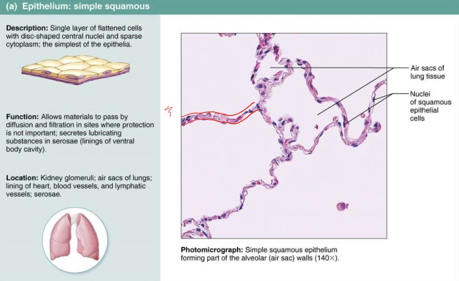
Describe the structures, functions, and locations of simple squamous epithelium
Single layer of flattened cells with disc-shaped central nuclei and sparse cytoplasm; the simplest of the epithelia
Function: allows materials to pass by diffusion and filtration in sites where protection is not important
Secretes lubricating substances in serosae (linings of ventral body cavity)
Location: kidney glomeruli; air sacs of lungs; lining of heart, blood vessels, and lymphatic vessels; serosae
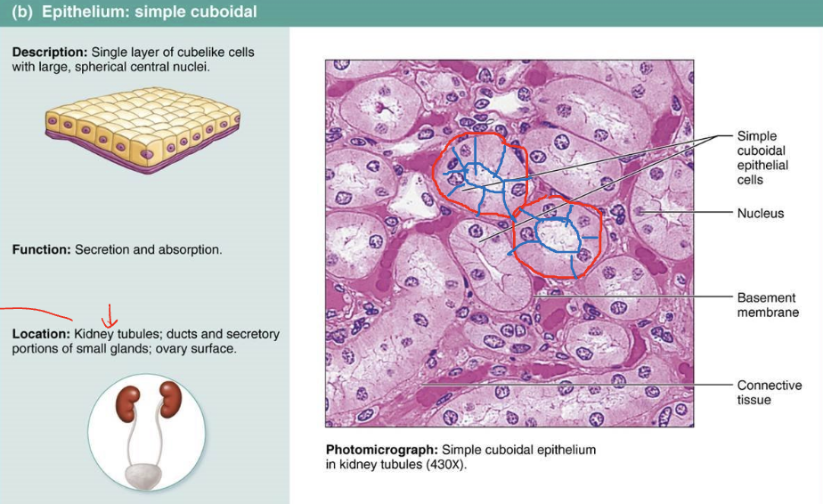
Describe the structures, functions, and locations of simple cuboidal epithelium
Single layer of cube-like cells with large, spherical central nuclei
Function: secretion and absorption
Location: kidney tubules, ducts and secretory portions of small glands; ovary surface
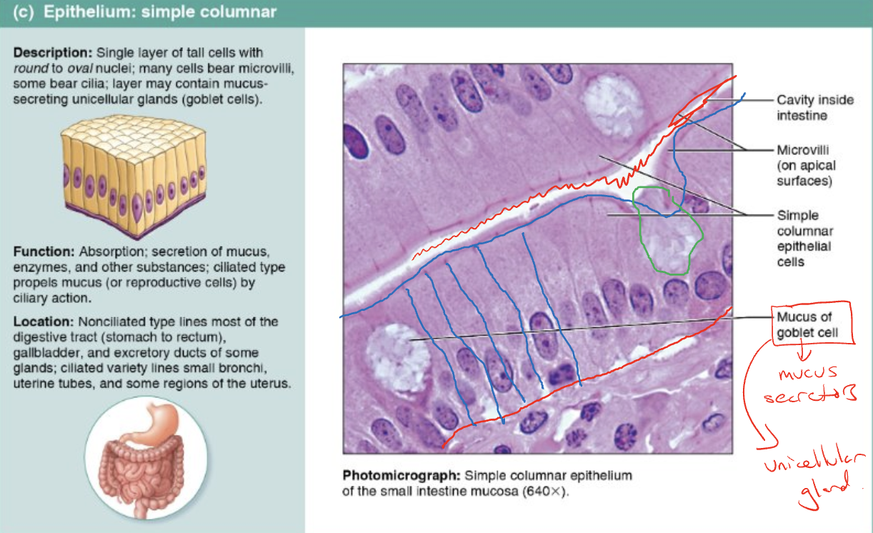
Describe the structures, functions, and locations of simple columnar epithelium
Single layer of tall cells with round to oval nuclei; many cells bear microvilli, while others bear cilia; layer may contain mucus-secreting unicellular glands (goblet cells)
Function: absorption; secretion of mucus, enzymes and other substances
Ciliated type propels mucus by ciliary action
Location: nonciliated type lines most of the GI tract (stomach to rectum), gallbladder, and excretory ducts of some glands; ciliated variety lines small bronchi, uterine tubes, some regions of the uterus
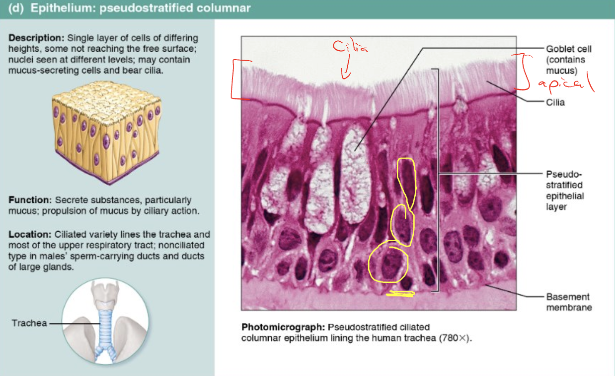
Describe the structures, functions, and locations of pseudostratified columnar epithelium
Single layer of cells of differing heights, not reaching the free surface
Nuclei are seen at different levels; may contain mucus-secreting cells and bear cilia
Function: secrete substances, particularly mucus; propulsion of mucus by ciliary action
Location: ciliated variety lines the trachea and most of the upper respiratory tract; nonciliated types in males’ sperm-carrying ducts and ducts of large glands
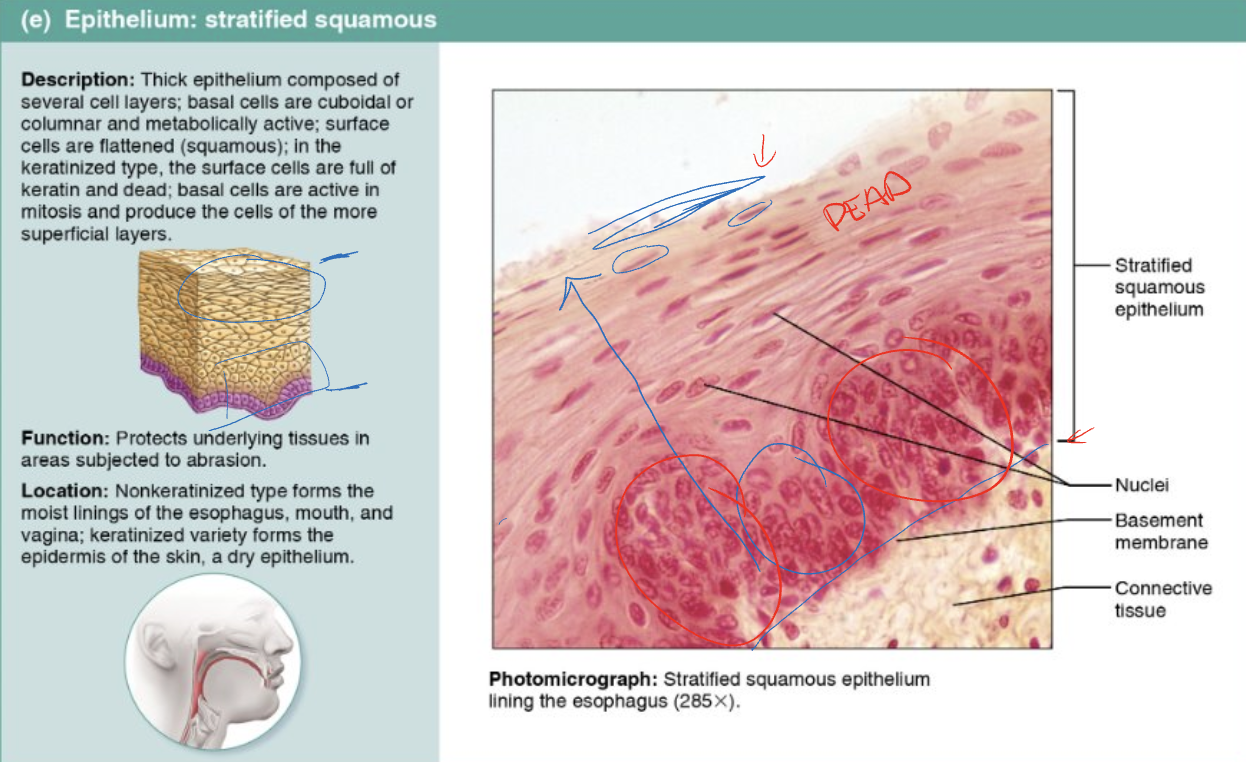
Describe the structures, functions, and locations of stratified squamous
Thick epithelium composed of several cell layers; basal cells are cuboidal or columnar and metabolically active; surface cells are flattened (squamous)
Function: protects underlying tissues in areas subjected to abrasion
Location: non-keratinized type forms the moist linings of the esophagus, mouth, and vagina; keratinized variety forms the epidermis of the skin, a dry epithelium
Describe the structures, functions, and locations of transitional
Resembles both stratified squamous and stratified cuboidal; basal cells are cuboidal or columnar, and surface cells are dome-shaped or squamous-like, depending on the degree of organ stretch
Function: stretches readily, permits stored urine to distend the urinary organ
Location: lines the ureters, bladder, and part of the urethra
Describe glands (glandular epithelia) and the different classifications
Gland - one or more cells that make and secrete an aqueous fluid called a secretion
Classified by:
Site of product release
Endocrine - internally secreting (ex: hormones)
Exocrine - externally secreting (ex: sweat)
Relative number of cells forming the gland
Unicellular (ex: goblet cells) or multicellular (ex: salivary)
Describe the structure and functions of endocrine glands
Ductless glands
Secretions are released into the surrounding interstitial fluid, which is picked up by circulatory system
Secrete hormones: messenger chemicals that travel through lymph or blood to their specific target organs
Describe the structure and functions of exocrine glands
Secretions are released onto body surfaces, such as the skin, or into body cavities
Secrete products into ducts
Ex: mucus, sweat, oil, salivary glands
Can be unicellular or multicellular
Describe the structure of unicellular exocrine glands
The only important unicellular glands are mucous cells and goblet cells
Found in epithelial linings of intestinal and respiratory tracts
All produce mucin, a sugar-protein that can dissolve in water to form mucus, a slimy protective, lubricating coating
Describe the structure of multicellular exocrine glands
Composed of a duct and a secretory unit
Classified by:
Structure
Mode of secretion
Describe the modes of glandular secretion in multicellular exocrine glands
In merocrine secretion, the cell remains intact (most sweat glands)
In apocrine secretion, the apical portion of the cell is released (sweat glands in armpits and genitalia)
In holocrine secretion, the cell is destroyed as it releases its product and the cell itself becomes part of the secretion (sebaceous glands of the skin)