C3.2: Defense Against Disease | IB Biology HL
1/42
Earn XP
Description and Tags
Name | Mastery | Learn | Test | Matching | Spaced | Call with Kai |
|---|
No analytics yet
Send a link to your students to track their progress
43 Terms
Define pathogen.
C3.2.1: Pathogens as the cause of infectious diseases.
An organism or virus that causes disease
List major pathogen types.
C3.2.1: Pathogens as the cause of infectious diseases.
Viruses
Bacteria
Fungi
Protists
Describe the importance of observation in addressing childbed fever.
C3.2.1: Pathogens as the cause of infectious diseases.
Dr. Ignaz Semmelweis noticed that infection after childbirth was a large issue in the 19th-century, but the likelihood of infection was less when doctors washed their hands.
He separated a group of doctors into 2 groups: Clinic 1 doctors came from dirty areas (like from dissections), and Clinic 2 doctors washed their hands prior to childbirth. He noticed that Clinic 2 had far fewer deaths and infections. This observation demonstrated the importance of hand washing to avoid infections like childbed fever.
Describe the importance of observation in addressing the Cholera outbreak in London in the 19th-century.
C3.2.1: Pathogens as the cause of infectious diseases.
At the time, the leading theory for sickness was the Miasma Theory (that dirty air caused sickness).
However, Dr. John Snow observed that cholera-affected symptoms included diarrhea and vomiting. Since these were digestive system issues, not respiratory system issues, he was convinced that dirty water caused cholera, not the air.
He also observed that those who drank water from the same pump were all affected. His observations eventually led to the understanding that cholera is caused by a pathogen.
This was later confirmed by Koch and Louis Pasteur. They disproved Miasma and led to the development of germ theory.
Define “primary defense.”
C3.2.2: Skin and mucous membranes as a primary defense.
The first line of defense that an organism (plant or animal) uses to protect itself from disease-causing microorganisms.
Outline the role of skin as a physical and chemical barrier to pathogens.
C3.2.2: Skin and mucous membranes as a primary defense.
Skin acts as a physical barrier to keep pathogens out of the body.
Skin also functions as a chemical barrier. The sebaceous glands release lactic acid and fatty acids, which creates an acidic environment. As a result, the acidic environment stops harmful bacteria from growing. The skin also produces lysozyme enzymes to break down bacterial cells walls and kill bacteria. In addition, the helpful bacteria in skin protects against harmful bacteria.
Outline the role of the mucous membranes as a barrier to pathogens.
C3.2.2: Skin and mucous membranes as a primary defense.
The mucous membranes are the soft areas of skin covered in mucus. It is in places like the nose and trachea.
The membranes aren’t a strong physical barrier, but they produce lysozymes. The mucus traps pathogens, and the lysozymes kill bacteria by damaging their cell walls.
Outline the structure and function of lymphocyte cells.
C3.2.6: Lymphocytes as cells in the adaptive immune system that cooperate to produce antibodies.
Lymphocytes are a type of leukocyte that are part of the lymphatic system. They produce antibodies to fight pathogens.
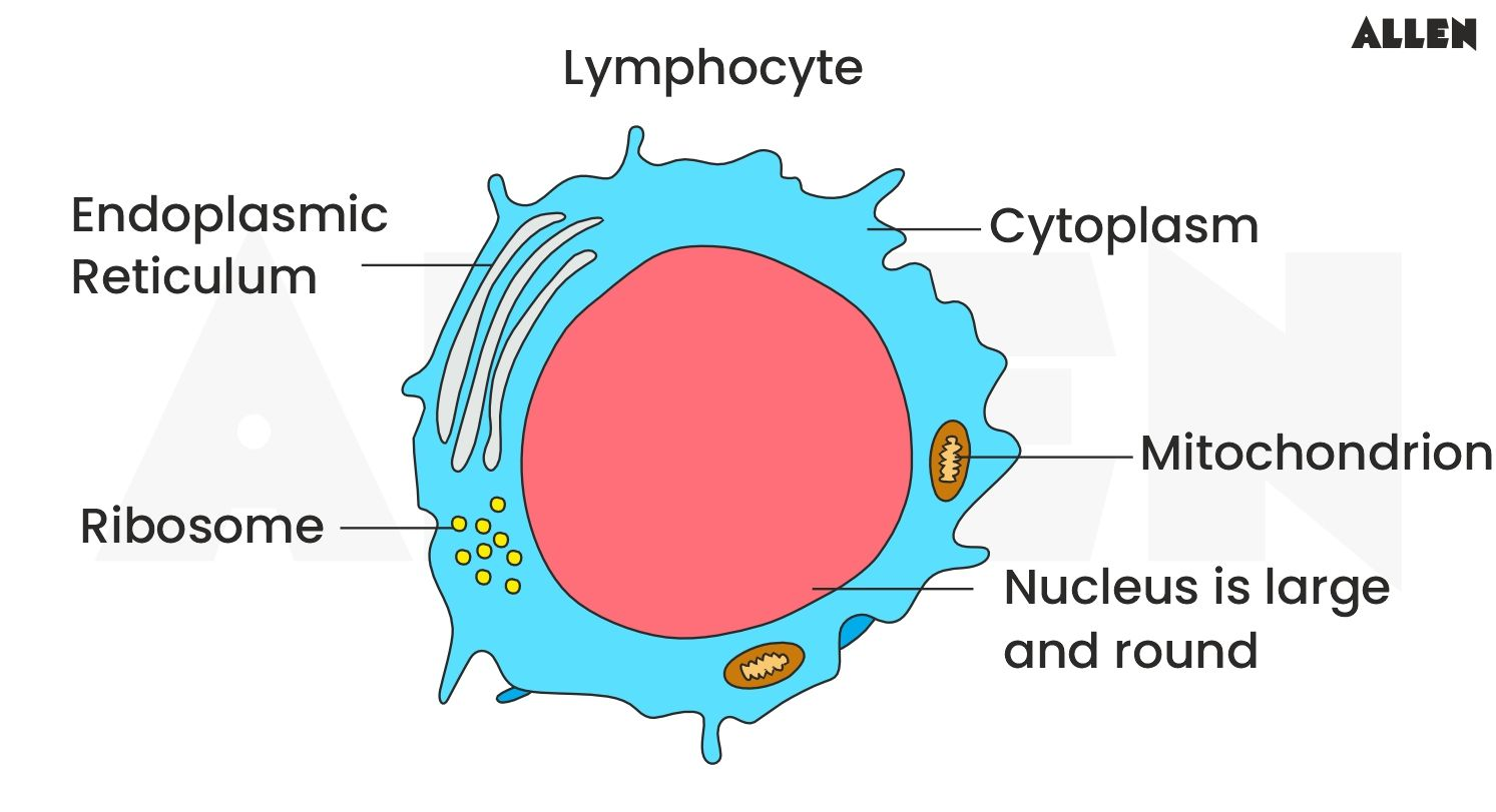
State the location of lymphocytes in the body.
C3.2.6: Lymphocytes as cells in the adaptive immune system that cooperate to produce antibodies.
Lymphocytes circulate in blood and are contained in lymph nodes.
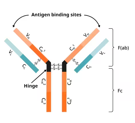
Define antibody.
C3.2.6: Lymphocytes as cells in the adaptive immune system that cooperate to produce antibodies.
Y-shaped proteins with binding sites at the end of branches which attach to antigens (on pathogens).
Outline the role of B-lymphocytes to produce antibodies.
C3.2.6: Lymphocytes as cells in the adaptive immune system that cooperate to produce antibodies.
B-lymphocytes synthesize one specific antibody when activated by a specific antigen. Each antibody recognizes and binds to a specific antigen.
Outline how antibodies respond to pathogens.
C3.2.6: Lymphocytes as cells in the adaptive immune system that cooperate to produce antibodies.
Antibodies have 2 binding sites. Thus, they can bind to 2 antigens (on 2 different pathogens) at the same time. As multiple antibodies bind, they form a cluster of pathogens. This cluster is easier for phagocytes to find and engulf.
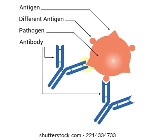
Define antigen.
C3.2.7: Antigens as recognition molecules that trigger antibody production.
A protein (typically on the surface of pathogens) that induces the immune system to produce antibodies.
Describe the structure of antigens.
C3.2.7: Antigens as recognition molecules that trigger antibody production.
Antigens are glycoproteins or other proteins. They are embedded in the outer membrane on pathogenic organisms (like the plasma membrane of bacteria, outer cells of protists or fungi, or viral capsids) or even in the body’s ow immune system.
Describe the cause and consequence of an antibody binding to an antigen.
C3.2.7: Antigens as recognition molecules that trigger antibody production.
It is caused when a B-cell makes a specific type of antibody in response to being activated by one specific antigen.
This specific antibody binds to that specific antigen. Each antibody has 2 binding sites, and they form a cluster of pathogenic molecules. This cluster makes it easier for phagocytes to engulf the pathogenic molecule.
Outline the difference between antigens on the surface of erythrocytes.
C3.2.7: Antigens as recognition molecules that trigger antibody production.
There are 3 types of antigens possible on the surface of erythrocytes:
A protein
B protein
Rh protein
An O blood type has neither A nor B proteins, and a (-) blood type is lacking the Rh protein.
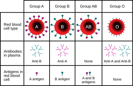
Describe the consequence of mismatched blood transfusions, including agglutination.
C3.2.7: Antigens as recognition molecules that trigger antibody production.
If a mismatched blood transfusion occurs, the body identifies the incompatible antigen as a threat. As a result, the production of antibodies is stimulated. Antibodies bind to these donated erythrocytes, resulting in agglutination (clumping).

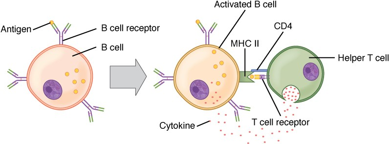
Outline the specific relationship between B-cells and T-cells.
C3.2.8: Activation of B-lymphocytes by helper T-lymphocytes.
There are many type of helper T-lymphocytes. A specific antigen activates a specific helper T-cell. This helper T-cell displays this antigen on their own plasma and activates a specific B-cell. This B-cell (activated by a specific antigen and T-cell) produces a specific antibody that targets that specific antigen.
Describe activation of helper T-lymphocytes.
C3.2.8: Activation of B-lymphocytes by helper T-lymphocytes.
T cells are activated when they interact directly with the specific antigen. As a result, they display that antigen on their own plasma membrane.
Outline the role of helper T-lymphocytes in the activation of B-lymphocytes.
C3.2.8: Activation of B-lymphocytes by helper T-lymphocytes.
Helper T-lymphocytes, once activated, display antigens on their own plasma membrane. They release cytokines once they find an antigen, which activates a specific B-cell.
B-cells must be exposed to an antigen of the pathogen and the activated T-cell that display the antigen and releases the cytokines.
Both T-cells and B-cells can become memory cells (long-lived).
Describe how B-lymphocytes are activated to form clones.
C3.2.9: Multiplication of activated B-lymphocytes to form clones of antibody-secreting plasma cells.
There are a wide variety of B-lymphocytes (because they are numerous types of antigens), so the immune system can only have a relatively low number of each type of cell.
As a result, when B-cells are activated, they undergo numerous mitotic cell divisions. The B-cells create clones of cells so that it can synthesize mass amounts of the antibody.
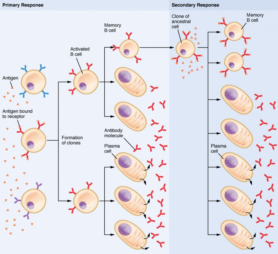
State when B-cells produce antibodies.
C3.2.9: Multiplication of activated B-lymphocytes to form clones of antibody-secreting plasma cells.
B-cells only produce antibodies after they have grown and are differentiated for protein synthesis.
Describe the development of B-lymphocytes.
C3.2.9: Multiplication of activated B-lymphocytes to form clones of antibody-secreting plasma cells.
B-cells undergo repeated mitosis to create numerous types of the same B-cell.
Activated B-cells grow larger, they develop many ribosomes, ER, and Golgi bodies to produce and secrete antibodies.
These B-cells produce antibodies in body tissues or circulate in the blood stream. Others become memory cells that won’t produce antibodies for the current infection.

Define immunity.
C3.2.10: Immunity as a consequence of retaining memory cells.
The body’s ability to eliminate an infectious disease from the body.
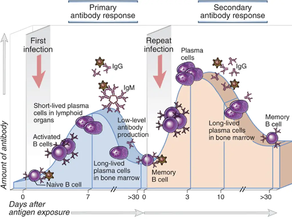
Outline the role of memory B cells in maintaining immunity.
C3.2.10: Immunity as a consequence of retaining memory cells.
Memory B cells are long-lived. They are produced during the primary infection and circulate in the bloodstream. When activated, they replicate to be in large numbers, so they respond to the same pathogen very quickly. This allows one to maintain immunity after a first infection.
Compare the primary and secondary immune responses to a specific pathogen in regards to speed and duration.
C3.2.10: Immunity as a consequence of retaining memory cells.
The primary immune response is the initial exposure to the antigen. The secondary immune response is the second/subsequent exposure to the same pathogenic antigen.
The primary immune response is relatively long (slow) because there are no memory cells yet, and they haven’t been activated or cloned. Symptoms show. But a secondary immune response is much faster because memory cells for that antigen already exist.
List mechanisms of HIV transmission.
C3.2.11: Transmission of HIV in body fluids.
Sexual intercourse
Blood transfusion (receiving infected blood)
Sharing hypodermic needles
From mother to baby through the placenta
During birth, through cuts or tears
Through breast milk
Describe the consequences of HIV on the immune system.
C3.2.12: Infection of lymphocytes by HIV with AIDS as a consequence.
HIV infects and destroys CD4 cells, a type of lymphocyte (T cells).
HIV viruses inject DNA into other cells to produce more HIV proteins. As a result, they destroy even more CD4 cells.
Losing these CD4 cells weakens the immune system, limiting the production of antibodies, and making it more difficult to fight infection.
Outline the relationship between HIV and AIDS.
C3.2.12: Infection of lymphocytes by HIV with AIDS as a consequence.
AIDS (Acquired Immunodeficiency Syndrome) is caused by HIV.
Outline the natural function of antibiotics against bacteria but not in eukaryotic cells.
C3.2.13: Antibiotics as chemicals that block processes occurring in bacteria but not in eukaryotic cells.
Antibiotics are chemicals that stop bacteria from growing or kill them. They block metabolic processes in bacteria (especially those affecting cell walls).
Antibiotics do not target eukaryotic cells because animal cells do not have cell walls.
Outline why antibiotics fail to control viral infections.
C3.2.13: Antibiotics as chemicals that block processes occurring in bacteria but not in eukaryotic cells.
Antibiotics don’t work on viruses because antibiotics affect the metabolism of living cells.
Viruses are not alive though, so they don’t have a metabolism. As a result, antibiotics don’t affect viruses.
Describe how natural selection leads to the development of antibiotic resistance in bacteria.
C3.2.14: Evolution of resistance to several antibiotics in strains of pathogenic bacteria.
Offspring of bacteria must compete for resources because bacteria produce more offspring than the environment can support.
Antibiotics are a limiting factor for bacteria.
An antibiotic will kill most of the bacterial population, but those that survive have a mutation in their DNA that allows them to be resistant to the antibiotic.
These surviving bacteria reproduce with each other, and they become more common. Eventually the proportion of resistant bacteria increases.
Discuss the medical cause and consequences of evolution of antibiotic resistance.
C3.2.14: Evolution of resistance to several antibiotics in strains of pathogenic bacteria.
The medical cause of antibiotic resistance is due to the over-prescription of antibiotics, incomplete courses of treatment, and the use of antibiotics in livestock.
Consequences of this include increased healthcare costs, prolonged hospital stays, and higher mortality rates.
This leads to current healthcare challenges to manage resistant infections, develop new antibiotics, and implement effective stewardship programs.
Define zoonosis.
C3.2.15: Zoonoses as infectious diseases that can transfer from other species to humans.
An infectious disease that can transfer from other species to humans.
List modes of infection/transmission of zoonoses.
C3.2.15: Zoonoses as infectious diseases that can transfer from other species to humans.
COVID-19: Through contact with animals
Tuberculosis: Through direct contact with animals
Rabies: Through bites from infected animals
Japanese Encephalitis: Through animals (like pigs) to humans through mosquito bites
Define immunization.
C3.2.16: Vaccines and immunization.
Making a person or animal resistant to an infectious disease using vaccines.
Outline the mechanism of vaccines.
C3.2.16: Vaccines and immunization.
Vaccines contain antigens or nucleic acids (DNA or RNA) with sequences that code for antigens.
Vaccines stimulate the development of immunity to a specific antigen without causing the disease.
Define herd immunity.
C3.2.17: Herd immunity and the prevention of epidemics.
When a disease has a hard time spreading in a population.
Explain how herd immunity limits the potential of disease transmission.
C3.2.17: Herd immunity and the prevention of epidemics.
Members of a population are interdependent in building immunity. If many people are immune (either from past infection or vaccination).
The more people who are immune, the less likely the disease can spread. If a sufficient percentage of a population is immune to a disease, transmission is impeded.
Distinguish between a pandemic and an epidemic.
C3.2.18: Evaluation of data related to the COVID-19 pandemic.
An epidemic is a disease that affects many people in one area.
A pandemic is a type of epidemic that spreads to multiple countries or continents.
State how to calculate percentage difference.
C3.2.18: Evaluation of data related to the COVID-19 pandemic.
Percentage Difference = (Final - Initial) / Average x 100
Compares 2 values
State how to calculate percentage change.
C3.2.18: Evaluation of data related to the COVID-19 pandemic.
Percentage Change = (Final - Initial) / Initial x 100
Measures change over time
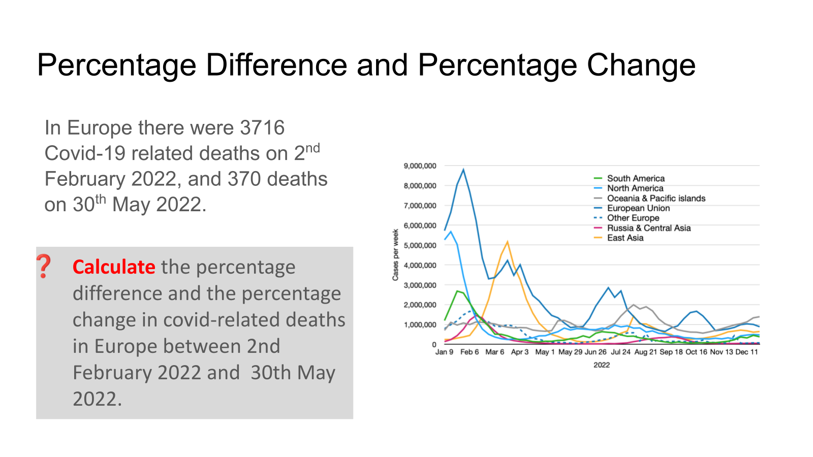
Calculate the percentage difference and the percentage change in COVID-related deaths in Europe between 2 February 2022 and 30 May 2022.
C3.2.18: Evaluation of data related to the COVID-19 pandemic.
Percentage Difference = (370-3716)/(2043) x 100 = 163.8%
Percentage Change = (370-3716)/3716 × 100 = 90% decrease