BMS1052 week 2
 Call Kai
 Learn
Learn Practice Test
 Spaced Repetition
 Match
Match Flashcards
 Knowt Play
1/72

There's no tags or description
Looks like no tags are added yet.
Name | Mastery | Learn | Test | Matching | Spaced | Call with Kai | Chat |
|---|
No analytics yet
Send a link to your students to track their progress
73 Terms
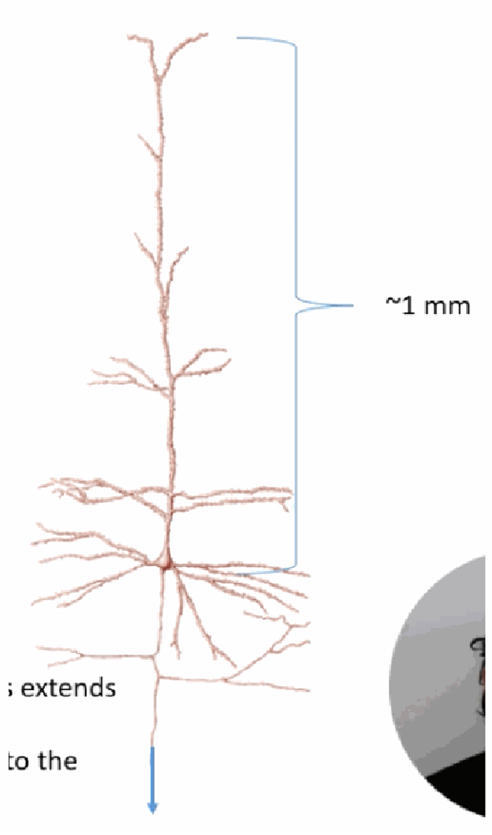
Why do neurons need to communicate both rapidly and reliably? Describe the example given.
A motor neuron's axon controlling the legs extends about 1 m from the brain down to the base of the spinal cord; a second neuron then carries the signal on to the leg muscles. The nervous system must perform many computations and still signal reliably over these long distances.
What four topics does the lecture video on membrane-potential calculation cover?
(1) Biological “wires” — the role of leaky membranes and ion flow. (2) How ionic concentration affects ion movement across a membrane. (3) The Nernst equation (the hypothetical equilibrium potential). (4) The Goldman equation (the measurable membrane potential).

Compare electrical wiring to a neuron as a “biological wire” — substrate, charge carrier, and insulation.
Wiring: metal substrate, charge carried by free electrons, usually very well insulated so only charge flow along the wire's length matters. Biological systems: intracellular fluid/cytosol substrate, charge carried by ions (Na+, K+, Ca2+, Cl−); poor insulation between intra- and extracellular space means charge flow both along the neurite AND across the membrane matters.
What three properties do electrical wires and axons have in common regarding conduction?
Insulation is important; thicker wires/axons conduct faster (lower resistance); and temperature affects conduction — warmer axons conduct faster.
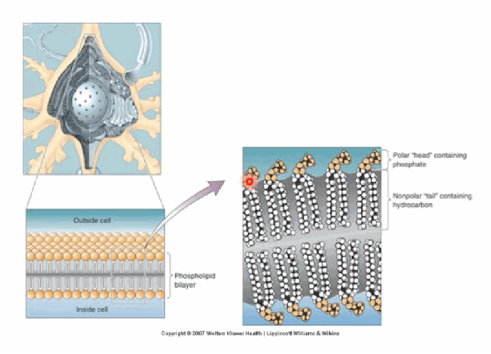
What structure separates intracellular and extracellular fluid, and what are its two key structural features?
The phospholipid bilayer membrane. It has hydrophilic “heads” (containing phosphate) facing outward on both sides, and hydrophobic “tails” (hydrocarbon chains) forming the interior. This dynamic (not static) bilayer makes it hard for both charged and uncharged particles, including water, to cross.
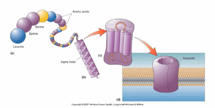
How do ion channels form, starting from individual amino acids?
Amino acids link via peptide bonds into polypeptide chains → multiple polypeptide chains associate into subunits (which have hydrophobic and hydrophilic regions letting them span the membrane) → multiple subunits coalesce together to form a membrane-spanning ion channel.
How does a sodium channel achieve selectivity for Na+ despite the entry to the pore being very narrow?
The channel has specific Na+ binding sites; sodium must shed its associated water molecules before it can fit through and bind. Selectivity arises from both the physical shape of the pore and the chemical properties (amino acid residues) lining it.
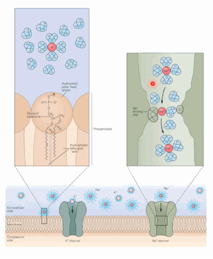
What does the ion-size comparison diagram illustrate?
It compares the size of the sodium channel's selectivity filter with the size of a partially hydrated Na+ ion and a partially hydrated K+ ion (all at roughly the 0.5 nm scale) — showing why the filter is precisely matched to sodium.
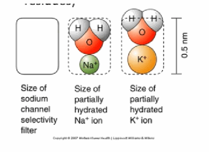
What is Aquaporin 1, and what can happen if these channels are blocked?
A specialised water-channel protein that allows relatively free flow of water across the membrane (plain diffusion of water through the lipid bilayer alone is very slow). If aquaporins are blocked, it can lead to disease such as brain oedema (swelling of cells).
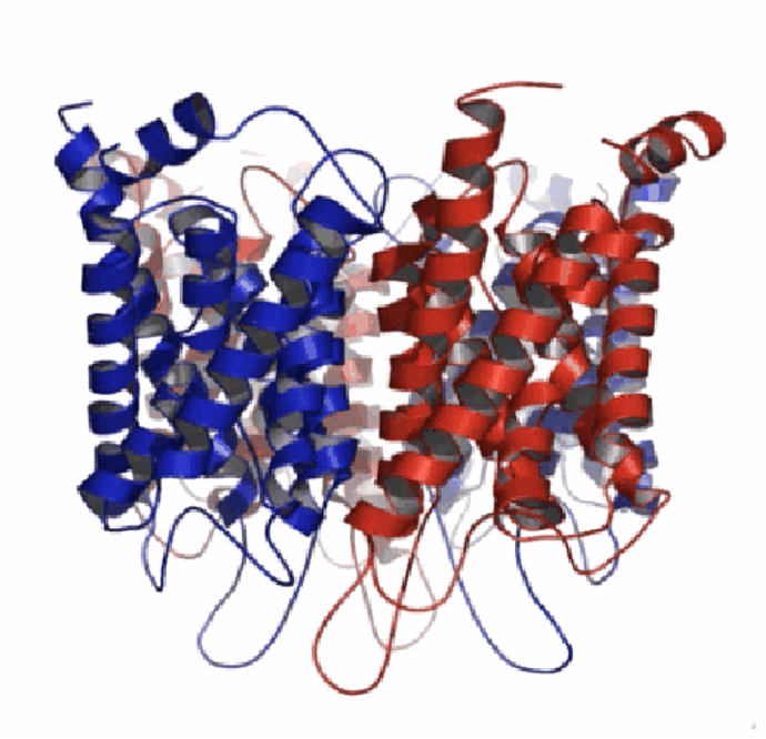
What two factors does ion movement across a membrane depend on?
The concentration gradient across the membrane, and the electrical field across the membrane (assuming, in these diagrams, that water itself moves freely).
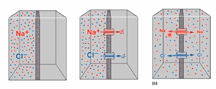
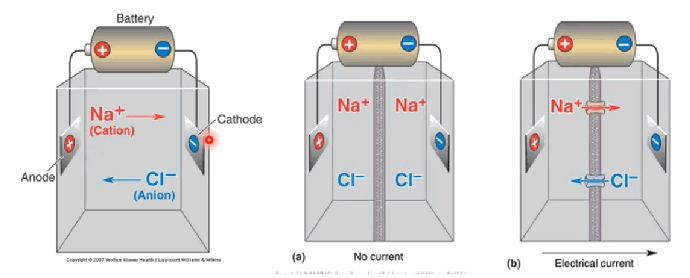
In the battery-driven ion-movement diagram, what happens with no current applied versus with an electrical current applied?
With no current, Na+ (cation) and Cl− (anion) simply distribute according to concentration. With current applied, Na+ moves toward the cathode and Cl− moves toward the anode. At equilibrium there is no net flow, although ion movement is still occurring.
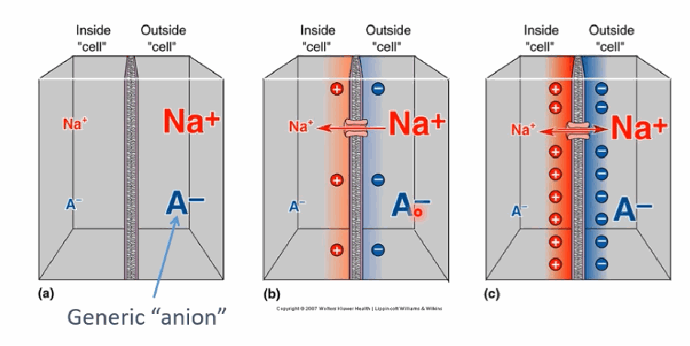
Using the “generic anion” cell diagram, explain how a charge imbalance develops when only Na+ (not the anion A−) can cross the membrane.
Na+ initially moves into the cell down its concentration gradient. Because only Na+ (not A−) can cross via the transporter, this creates a charge imbalance (slightly more positive charge on one side), which counteracts further net Na+ influx. At equilibrium, the concentration gradient balances the electrical field so there is no net movement of Na+.
What does the Nernst equation calculate, and what two pieces of information do you need to apply it?
It predicts the membrane potential at equilibrium if the membrane were permeable to a single ion (rarely the real case in a cell). You need: (1) the ionic concentrations inside and outside the cell, and (2) the ionic charge (z). Practical form: E_ion = (61.5/z) × log10([ion]out / [ion]in), in mV.
![<p>It predicts the membrane potential at equilibrium if the membrane were permeable to a single ion (rarely the real case in a cell). You need: (1) the ionic concentrations inside and outside the cell, and (2) the ionic charge (z). Practical form: E_ion = (61.5/z) × log10([ion]out / [ion]in), in mV.</p>](https://assets.knowt.com/user-attachments/f82c8ded-6459-471c-bb5a-72687d6cfbd3.png)
List the five different ways to think about an ion's equilibrium potential (E_ion).
(1) It's a “theoretical” potential — the observed Vm depends on multiple ions and their permeabilities. (2) E_ion is the voltage that counteracts an ion's movement due to its concentration gradient. (3) If a membrane is permeable to only one ion, Vm moves toward that ion's E_ion. (4) If Vm equals E_ion, there is no net movement of that ion. (5) It is independent of membrane permeability and ionic conductance.

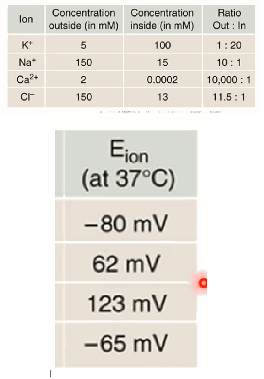
Using the Nernst equation and the standard neuronal ion concentrations, what are the approximate equilibrium potentials (at 37°C) for K+, Na+, Ca2+, and Cl−?
K+ ≈ −80 mV (out 5 mM : in 100 mM); Na+ ≈ +62 mV (out 150 : in 15); Ca2+ ≈ +123 mV (out 2 : in 0.0002); Cl− ≈ −65 mV (out 150 : in 13).
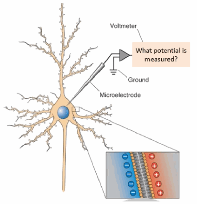
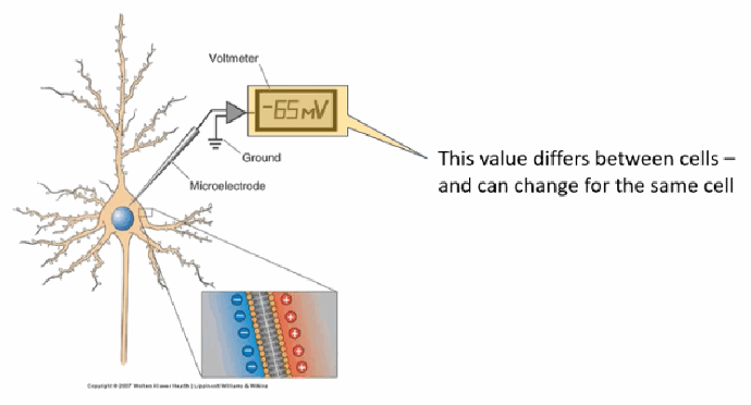
How is a neuron's membrane potential actually measured in the lab?
By inserting a fine microelectrode into the cell and connecting it to a voltmeter, referenced against a ground electrode placed outside the cell.
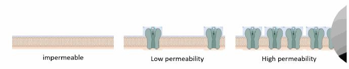
What does “membrane permeability” mean in terms of ion channels, from impermeable to highly permeable?
It reflects how many channels for that ion are present and open in the membrane: impermeable = no open channels; low permeability = few/rarely open channels; high permeability = many open channels.
Why is the Goldman (Goldman–Hodgkin–Katz, GHK) equation needed in addition to the Nernst equation, and what extra information does it require?
Real membranes are permeable to multiple ions at once, so the actual, measurable membrane potential depends on all of them together. The GHK equation needs the internal & external concentration of each ion AND the relative membrane permeability of each ion.

In the Goldman equation, why is Cl− treated differently from K+ and Na+?
Because Cl− is negatively charged, the position of its concentration terms is inverted — [Cl−]in appears in the numerator and [Cl−]out in the denominator, opposite to the cation terms.
If a membrane is only permeable to Na+, what do PK and PCl equal in the Goldman equation, and what does the equation simplify to?
PK = 0 and PCl = 0, so the Goldman equation collapses down to the Nernst equation for sodium alone: Vm = 61.5 log10([Na]out / [Na]in). This shows the Goldman equation reduces to the Nernst equation when only one ion is permeant.
![<p>PK = 0 and PCl = 0, so the Goldman equation collapses down to the Nernst equation for sodium alone: Vm = 61.5 log10([Na]out / [Na]in). This shows the Goldman equation reduces to the Nernst equation when only one ion is permeant.</p>](https://assets.knowt.com/user-attachments/4350df17-1017-4504-8bfd-924fa6de213d.png)
Calculate the membrane potential when the membrane is impermeable to Cl− and K+ permeability is 40× higher than Na+ permeability (standard K+/Na+ concentrations). Why is this a realistic scenario?
Vm = 61.5 log10[(40×5 + 1×150) / (40×100 + 1×15)] ≈ −65 mV. This is realistic because, at rest, real neuronal membranes are weakly permeable to Na+ and quite permeable to K+ — matching the typical −65 to −75 mV resting potential.
![<p>Vm = 61.5 log10[(40×5 + 1×150) / (40×100 + 1×15)] ≈ −65 mV. This is realistic because, at rest, real neuronal membranes are weakly permeable to Na+ and quite permeable to K+ — matching the typical −65 to −75 mV resting potential.</p>](https://assets.knowt.com/user-attachments/0aa739b3-e7cd-469b-942a-074610b1e26f.png)
What is the typical range for a neuron's resting membrane potential, and which side of the membrane is negative?
Typically between −65 and −75 mV; the inside of the cell is negative relative to the outside.
What two learning objectives does the video on specialised membrane proteins (pumps and channels) cover?
(1) Describe the function of the Na/K-ATPase (Na-K pump). (2) Describe how selectivity and gating occur in Na+ and K+ channels.

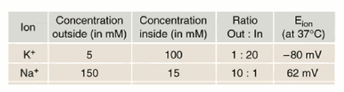
Recall the standard K+ / Na+ concentration and equilibrium-potential table used throughout these notes.
K+: out 5 mM, in 100 mM, ratio 1:20, EK ≈ −80 mV. Na+: out 150 mM, in 15 mM, ratio 10:1, ENa ≈ +62 mV.
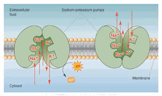
What does the Na/K-ATPase pump do, and what powers it?
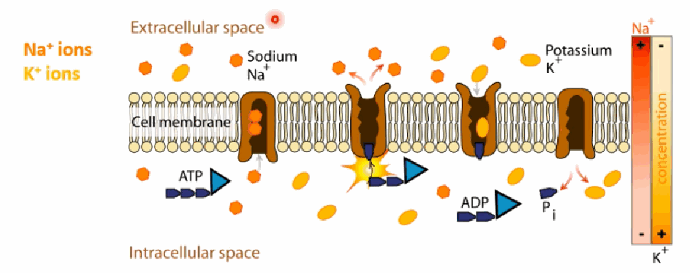
It actively transports Na+ out of the cell and K+ into the cell, both against their concentration gradients. It is powered by ATP hydrolysis.
Why is the Na/K pump important for the resting potential, and does it change Vm during an action potential?
The pump sets up and maintains the concentration gradients across the membrane that account for the resting membrane potential. However, the pump itself does NOT change the membrane potential during an action potential — that is driven by channels opening and closing.

Walk through the 5-step cycle of the Na/K-ATPase.
(1) While open intracellularly, the pump binds ATP and 3 intracellular Na+ ions, changing its conformation. (2) ATP is hydrolysed — the pump is phosphorylated and ADP is released. (3) The pump changes conformation again, releasing the 3 Na+ ions into the extracellular space. (4) The pump binds 2 extracellular K+ ions, triggering dephosphorylation and a second conformational change. (5) ATP binds and the 2 K+ ions are released intracellularly. This repeats for as long as ATP is available.
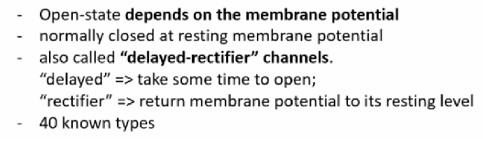
Describe the two-pore-domain (K2p) and voltage-gated (Kv) potassium channel families.
K2p channels: contain two pore-loop domains, are generally open, contribute to an ongoing K+ “leak” (high K+ permeability at rest), and help set the resting membrane potential (~15 known types). Kv channels: their open-state depends on membrane potential, they're normally closed at resting potential, and are also called “delayed rectifier” channels (“delayed” = take time to open; “rectifier” = return Vm to its resting level); ~40 known types.

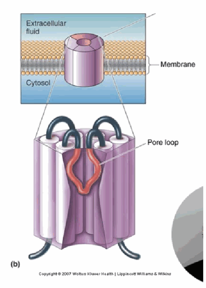
What structural feature do all potassium channels share, and what do calcium-activated and inward-rectifying K+ channels do?
All K+ channels have 4 protein subunits with pore loops that regulate permeability, and they don't require energy to open. Calcium-activated K+ channels are ligand-gated — the presence of Ca2+ triggers channel opening. Inward-rectifying K+ channels pass positive charge more easily into the cell than out of it.
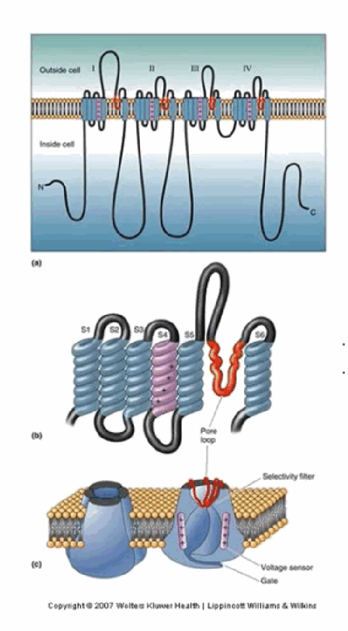
Describe the structure of a voltage-gated Na+ channel (domains, pore loop, and the intracellular blocking domain).
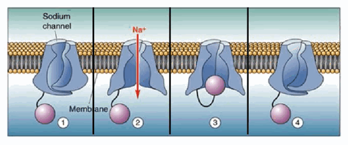
It's built from 4 homologous membrane-spanning domains (I–IV), each with 6 helices (S1–S6). The pore loop (shown in red) between S5/S6 acts as the selectivity filter, allowing only Na+ through. Extended intracellular domains can physically occlude the channel, giving a distinct inactivation mechanism separate from simply closing.
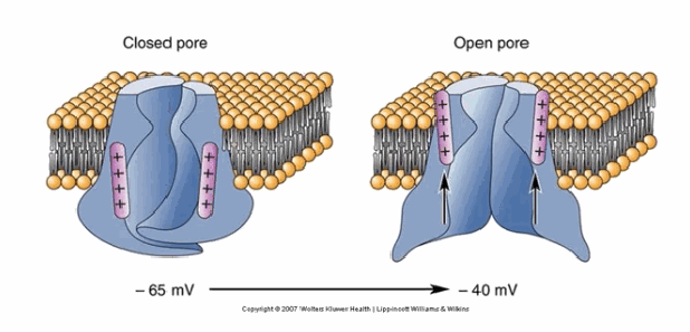
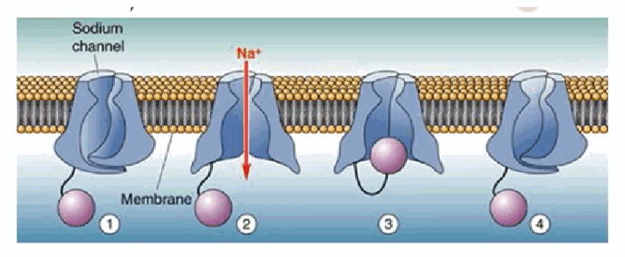
How does the sodium channel pore physically change between −65 mV (rest) and −40 mV?
At −65 mV the pore is closed. Depolarisation to about −40 mV causes a conformational change that opens the pore, allowing Na+ influx.
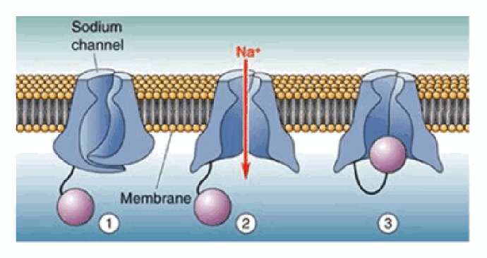
What are the three possible states of a voltage-gated Na+ channel, and how does the cycle proceed?
Closed (at rest, −65 mV) → open (~−40 mV, opens for ~1 ms allowing Na+ influx) → inactivated (after ~1 ms, the pore is occluded by a globular portion of the protein) → back to closed/reactivated once Vm returns to −65 mV. Channel opening is stochastic: higher voltages increase the probability of opening, but the exact timing can't be predicted.
What does tetrodotoxin (TTX) from the pufferfish (fugu) do, and why is it dangerous?
TTX blocks one class of voltage-gated sodium channel. It does not cross the blood–brain barrier, so a person remains conscious but loses the ability to generate action potentials (electrical communication) in their peripheral nerves.
What two learning objectives are covered in the “phases of an action potential” video?
(1) Describe the phases of an action potential, including which channels are involved and when. (2) Explain the action potential in terms of changes in membrane permeability and ionic concentrations.

What are the key general properties of action potentials?
They are “all-or-none” (they either happen fully or don't happen at all); they travel predominantly along the axon from the cell body to the axon terminals; they convey information via their timing and rate; and they can be explained completely by ionic concentrations and time-dependent changes in channel permeability.
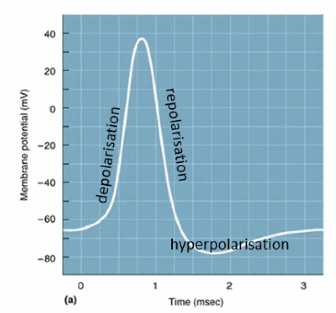
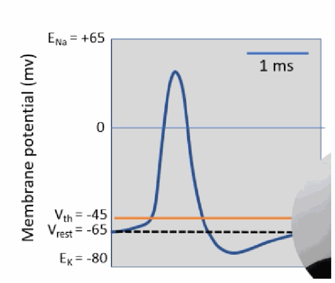
Label and describe the three main phases visible in the classic action-potential “spike” shape.
Depolarisation (the membrane becomes less polarised, rising toward or above 0 mV); repolarisation (the membrane potential becomes more negative again); and hyperpolarisation (it typically overshoots past the normal resting potential before returning to baseline).
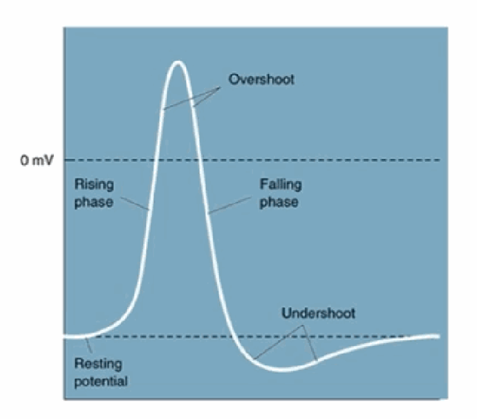
What alternative terminology is used for the same action-potential phases (rising/falling phase, overshoot/undershoot)?
Rising phase (≈ depolarisation) and falling phase (≈ repolarisation); overshoot = the peak that goes above 0 mV; undershoot = the dip below resting potential (≈ hyperpolarisation), before the trace returns to the resting potential.
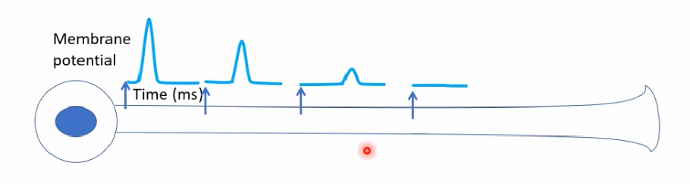
Why is “passive” transmission (simple diffusion of ions through the cytosol) inadequate for long-distance neuronal signalling?
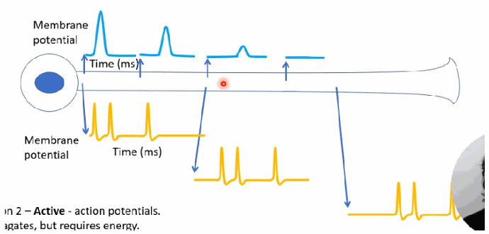
It requires no additional energy but is slow, and the signal amplitude breaks down (decays) over long distances, as shown by the diminishing spikes further along the axon.
How does “active” transmission via propagated action potentials overcome the limitations of passive diffusion?
It requires energy but faithfully recreates the full action-potential sequence at each point along the axon, so the signal doesn't decay — though there is a slight delay before the signal is seen further down the axon (e.g., near the axon terminal).
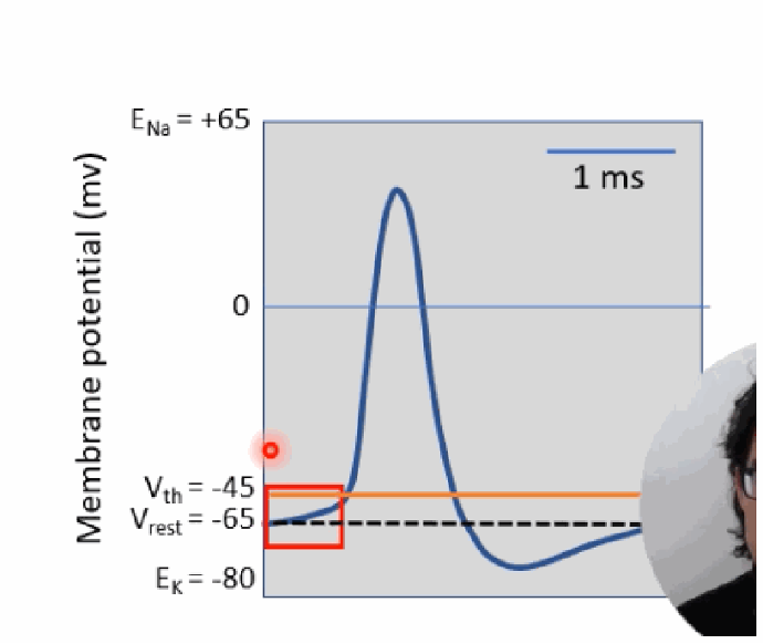
Phase 1 — What triggers an action potential, and roughly how long does the whole event take?
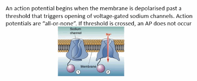
An action potential begins when the membrane is depolarised past a threshold (~−45 mV) that triggers voltage-gated Na+ channels to open. Action potentials are all-or-none — if threshold isn't crossed, no AP occurs. The entire event takes about 1–2 milliseconds.
What two general sources can cause the initial depolarisation that pushes the membrane toward threshold?
Physically-gated Na+ channels opening (e.g., stretch-sensitive Na+ channels in the skin), or depolarisation “inherited” (spread passively) from elsewhere in the neuron.
In the “reaching threshold” diagram, why do the first two small depolarisations fail to trigger an action potential while the third succeeds?
The first two depolarisations don't reach Vthresh, so no AP is generated. Only once a depolarisation crosses the threshold does it trigger the full, all-or-none action potential.
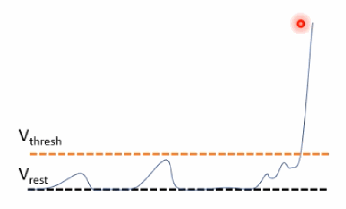
When Na+ channels open, why does the membrane potential rapidly approach ENa, and what assumption is usually made for these calculations?
Opening Na+ channels makes the membrane far more permeable to Na+ than K+, so by the Goldman equation Vm rapidly approaches ENa (+62 mV). For these calculations we usually assume the K+ and Na+ concentrations inside/outside the cell don't meaningfully change during a single action potential.
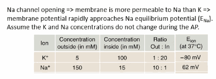
Write the Goldman-equation logic for why Vm → ENa during rapid depolarisation.
When voltage-gated Na+ channels open, PNa >> PK, so in the Goldman equation the Na+ terms dominate, and therefore Vm → ENa.
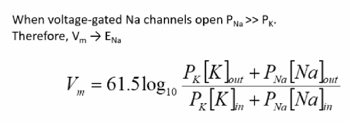
Recall the standard ion table used to compute ENa/EK during the depolarisation phase.
K+: out 5 mM, in 100 mM (EK ≈ −80 mV); Na+: out 150 mM, in 15 mM (ENa ≈ +62 mV).
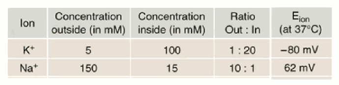
Phase 2 — What is happening during the rapid depolarisation phase, and why is it described as a positive feedback loop?
As the membrane depolarises, more and more voltage-gated Na+ channels open, letting in more Na+, which causes further depolarisation and opens still more channels — a positive feedback loop that continues until it drives Vm sharply upward toward ENa.
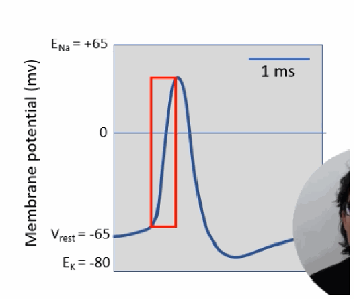
Phase 3 — What happens to voltage-gated Na+ channels after about 1 ms?
After being open for about 1 ms, the voltage-gated Na+ channels close and inactivate — the pore becomes occluded by a globular portion of the protein — which ends the Na+ influx.
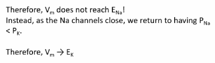
Once Na+ channels close/inactivate, why doesn't Vm reach ENa, and where does it head instead?
Because the Na+ channels close before full equilibrium is reached, Vm never actually reaches ENa. Instead, as PNa drops back below PK, Vm moves toward EK — this is repolarisation.
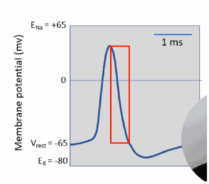
On the action-potential trace, which segment corresponds to repolarisation (Phase 3)?
The falling phase immediately after the peak, where the membrane potential drops rapidly from its peak (near ENa) back down toward and past the resting potential, as PNa falls and PK dominates again.
Phase 4 — Which second class of voltage-gated channels opens to cause hyperpolarisation, and what do K2p leak channels do throughout?
The “delayed rectifier” voltage-gated K+ (Kv) channels open. Meanwhile, the K2p leak channels remain open throughout the entire action potential, contributing background K+ permeability.
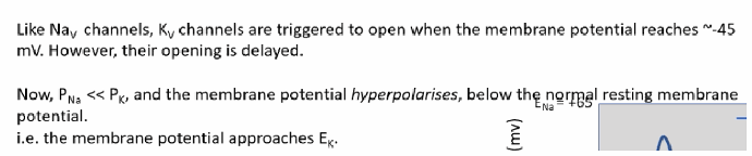
Using Goldman-equation logic, why does the membrane hyperpolarise below the normal resting potential once Kv channels open?
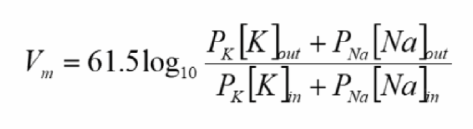
Like Nav channels, Kv channels are triggered to open at around −45 mV, but with a delay. Once open, PNa << PK, so Vm moves toward (and briefly overshoots below) the normal resting membrane potential, approaching EK.
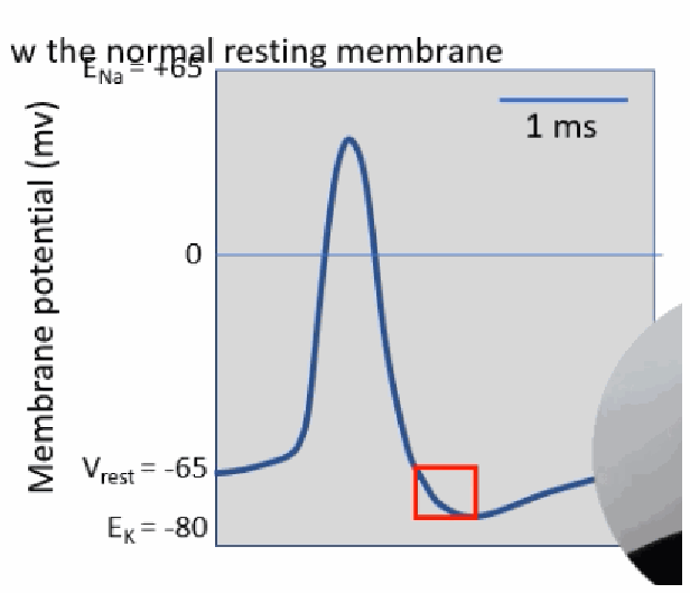
On the action-potential trace, which region corresponds to hyperpolarisation (Phase 4)?
The brief dip below the normal resting potential (below Vrest, approaching EK) that occurs just after repolarisation, before the trace returns to baseline.
Phase 5 — Why can't a second action potential occur for at least ~1 ms after the first (the absolute refractory period)?
Voltage-gated Na+ channels remain inactivated until the membrane potential is repolarised (de-inactivation requires Vm to drop below about −65 mV). This limits the theoretical maximum AP rate to <1000 spikes/s, though in practice AP rates rarely exceed ~100 spikes/s.
What needs to happen for a Na+ channel to reset from inactivated back to a closed/available state?
The membrane potential needs to drop below about −65 mV — producing the characteristic “undershoot” — resetting the sodium channel from an inactivated-and-closed state to an active, available closed state.
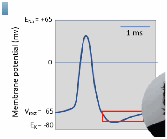
On the action-potential trace, what region is highlighted as the absolute refractory period?
The region spanning from the peak through the deepest point of the repolarisation/undershoot, during which Na+ channels are inactivated and no new action potential can be triggered, regardless of stimulus strength.
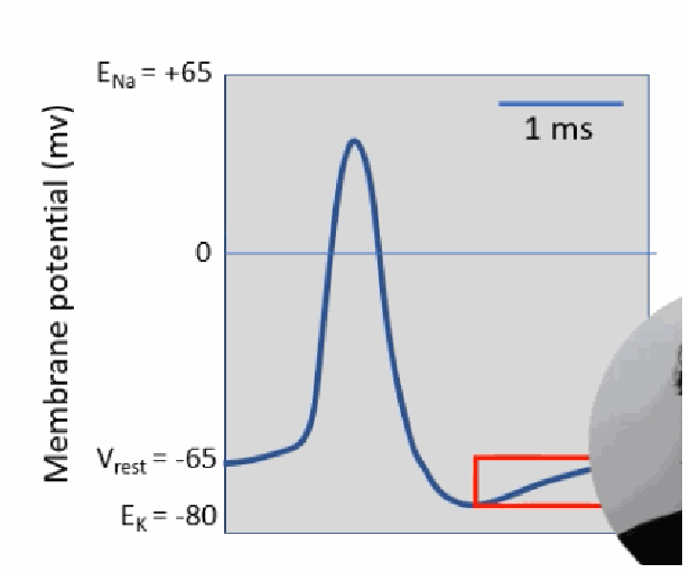
Phase 6 — Why is it difficult (though technically still possible) to trigger another AP during the relative refractory period?
The delayed-rectifier voltage-gated K+ channels also take time to close, so PNa remains << PK for a while; the membrane stays slightly hyperpolarised and some Na+ channels may still be recovering from inactivation — both make it harder, but not impossible, to reach threshold again.

On the action-potential trace, what region corresponds to the relative refractory period?
The tail-end portion where the membrane potential is recovering back toward resting level but is still slightly hyperpolarised, as the Kv channels are still finishing closing.
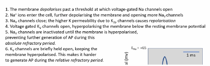
As the slow K+ channels finish closing during the relative refractory period, what happens to the membrane potential, and why does this make firing harder?
The membrane potential can briefly drop below its normal resting level, moving the voltage further away from the threshold needed to fire another action potential — on top of the sodium channels still recovering from inactivation.
Summarise all six phases of the action potential in order, with the channels responsible for each.
(1) The membrane depolarises past threshold, opening voltage-gated Na+ channels. (2) Na+ enters, further depolarising the membrane and opening more Nav channels (rapid depolarisation). (3) Nav channels close; the higher K+ permeability from K2p channels causes repolarisation. (4) Voltage-gated Kv channels open, hyperpolarising the membrane below resting potential. (5) Nav channels stay inactivated until the membrane hyperpolarises — the absolute refractory period. (6) Kv channels are briefly held open, keeping the membrane hyperpolarised and making it harder to fire again — the relative refractory period.
What two learning objectives are covered in the “action potential propagation” video?
(1) Account for the stochastic nature of action potentials. (2) Describe factors affecting the speed and nature of action potential conduction.

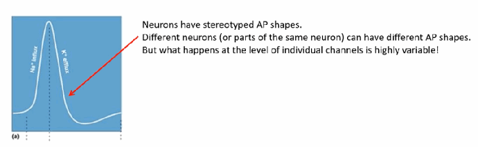
What does it mean that “channel openings are stochastic,” and why do whole-neuron action potentials still look like a clean, stereotyped spike?
Individual channels open at unpredictable times and durations (stochastic), and different neurons (or different regions of the same neuron) can even have different overall AP shapes. But because there are typically very many (up to several thousand per µm² at the nodes of Ranvier) channels opening together, these individually random events sum together and smooth out into the clean, reproducible AP trace we observe.
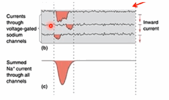
How does the summed sodium current relate to the individual, stochastic currents through single Na+ channels?
Each individual voltage-gated Na+ channel opens at a slightly different time, producing a variable inward current (different Na+ flux). Summing across all the open channels (potentially thousands) produces a smooth, clean summed Na+ current trace.
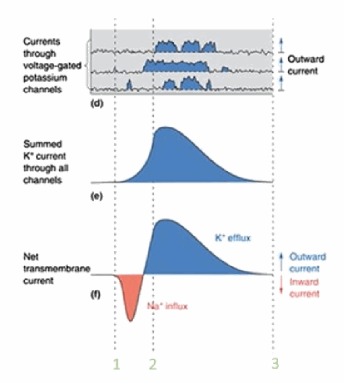
How does the summed potassium current combine with the sodium current to give the overall transmembrane current?
Individual Kv channel openings are also stochastic, each producing a variable outward current; summing across channels gives a smooth summed K+ current trace. The net transmembrane current is simply the sum of the (inward) Na+ current and the (outward) K+ current at each moment.
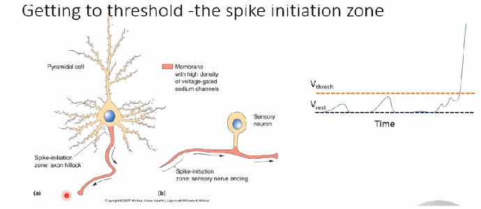
What is the “spike initiation zone,” and what three sources can bring the membrane there to threshold?
It's the location where the action potential is actually first triggered — usually the axon hillock for a pyramidal cell, or the sensory nerve ending for a sensory neuron. Depolarisation reaching this zone can be “inherited” from an adjacent axonal region, “inherited” from dendritic depolarisations caused by other neurons' inputs, or caused directly by physically-gated Na+ channels opening (e.g., stretch-sensitive Na+ channels in sensory nerve endings).
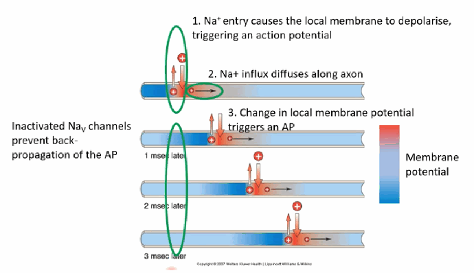
How does an action potential propagate along the axon once triggered, and what prevents it from propagating backward?
Na+ entry depolarises the local membrane, triggering an AP; the resulting Na+ influx diffuses a short distance along the axon, changing the local membrane potential enough to trigger an AP in the adjacent region, and so on toward the axon terminal. Inactivated Nav channels in the region just “behind” the moving spike prevent back-propagation, and also cap the maximum firing rate of a single neuron.
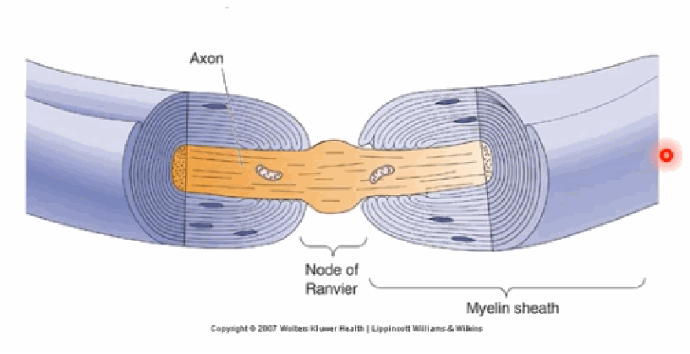
What are myelin sheaths and nodes of Ranvier, and which cells produce myelin?
Myelinating glia — oligodendrocytes in the CNS and Schwann cells in the PNS — wrap axons in fatty, insulating myelin sheaths. The small unmyelinated gaps between sheaths are the nodes of Ranvier, where ion channels are densely concentrated.
What is saltatory conduction, and why does it occur only at the nodes of Ranvier and not the myelinated regions?
It's the “leaping” of action potentials from one node of Ranvier to the next. Nodes have very high densities of Na+/K+ channels, giving high permeability when open, while the myelinated internodal regions are insulated, making it very difficult for ions to cross between the intracellular and extracellular space there.
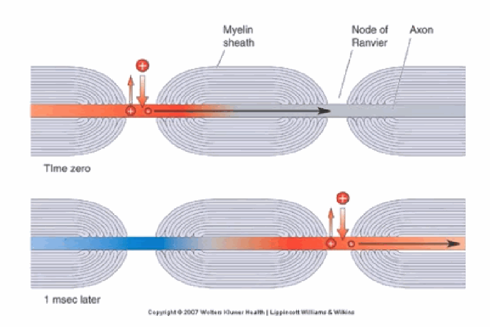
Why must the gaps between nodes of Ranvier be appropriately spaced (roughly 0.2–2 mm), and what happens if the myelin is too thick or nodes too far apart?
An unmyelinated axon gets reliable but slow AP propagation, because every patch of membrane must depolarise in turn. Correctly-spaced nodes allow fast “leaping” (saltatory) conduction. If myelin is too thick or nodes are spaced too far apart, passive diffusion between nodes may be insufficient to depolarise the next node past threshold, risking failure to propagate.
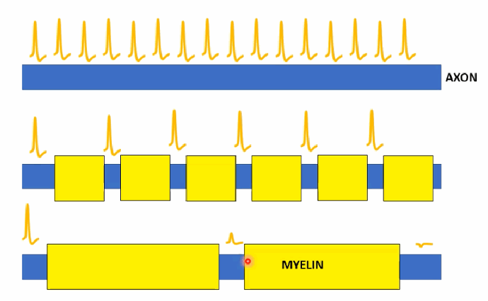
What three factors affect axonal conduction velocity, and how does each affect speed?
(1) Myelination — more myelin increases speed by providing insulation, lowering membrane capacitance, and enabling saltatory conduction. (2) Axon/myelin thickness — thicker myelin further improves insulation and speeds signal jumps between nodes. (3) Temperature — colder temperatures generally slow conduction, but this matters little for most human nerves since blood supply keeps nerve temperature fairly constant.
Why does an intravenous injection of KCl affect the heart quickly, but not the brain?
IV KCl causes a large rise in extracellular K+ in the blood, depolarising the resting membrane potential of excitable cells (neurons, cardiac cells), making them more likely to fire — this quickly stops the heart from beating. The brain is protected by (1) the blood–brain barrier, which prevents free movement of blood K+ into the brain's extracellular space, and (2) astrocytic buffering, which spatially redistributes potassium to prevent excessive extracellular build-up.

Describe the relationship between extracellular [K+] and membrane potential shown in the KCl graph, and what tool predicts this curve?
As extracellular [K+] rises, the membrane potential becomes progressively more depolarised (less negative). This dose–response curve can be calculated using the Goldman equation.
![<p>As extracellular [K+] rises, the membrane potential becomes progressively more depolarised (less negative). This dose–response curve can be calculated using the Goldman equation.</p>](https://assets.knowt.com/user-attachments/274e3344-4af9-4a1f-8786-1b059878ad60.png)
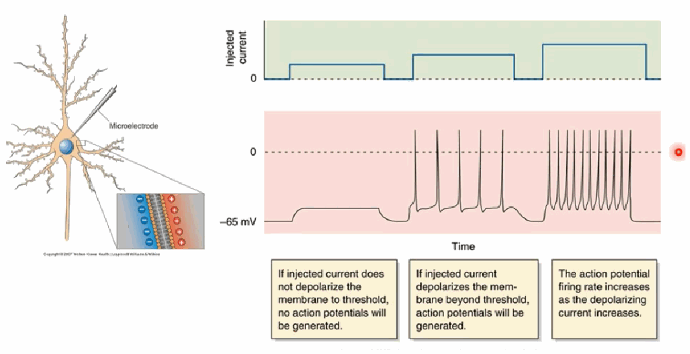
Describe “Method 1” for experimentally manipulating neurons (single-electrode current injection), and what determines whether an action potential is generated.
A single electrode is placed in or on a neuron to inject current (charge), depolarising the membrane — mimicking the effect of Na+ channels opening. If the injected current isn't large enough to depolarise the membrane past threshold, no AP is generated; if it does cross threshold, APs are generated, and the firing rate increases as the depolarising current increases.
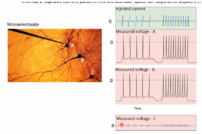
Describe “Method 2” for experimentally manipulating neurons (extracellular biphasic stimulation), and give a real-world application.
A larger microelectrode is placed in the extracellular space and delivers biphasic (alternating positive/negative) current pulses, which cause local ionic movements that can depolarise nearby neurons enough to trigger action potentials — the closer a neuron is to the electrode, the more strongly it is affected. This principle underlies many neuroprostheses, such as devices that electrically stimulate the retina to help restore sight in blind patients.