Chapter 6 Anatomy
 Call Kai
 Learn
Learn Practice Test
 Spaced Repetition
 Match
Match Flashcards
 Knowt Play
1/230

There's no tags or description
Looks like no tags are added yet.
Name | Mastery | Learn | Test | Matching | Spaced | Call with Kai | Chat |
|---|
No analytics yet
Send a link to your students to track their progress
231 Terms
What is the axial skeleton and its jobs?
Forms the long axis of the body
supports the head, neck and trunk
and protects the brain, spinal cord and thoracic organs

Three major regions of the axial skeleton
The skull
The vertebral column
The bony thorax
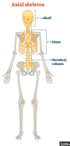
Most complex bony structure of the skeleton made of cranial and facial bones
Skull

Immovable joints connecting most bones of the skull, made of mostly FLAT bones
Sutures

The braincase portion of the skull.
Cranium

Skull cap or roof of the skull
Cranial vault (calvarium)
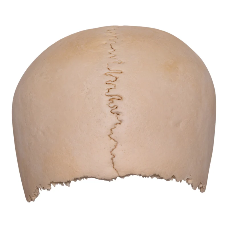
Inferior portion of the cranium divided into anterior, middle and posterior cranial fossae
Cranial base

Depression in a bone
Fossa

Opening through a bone that allows nerves, blood vessels, and other vital structures to travel from one area to another
Foramen

Number of bones that comprise the skull
22 skull bones
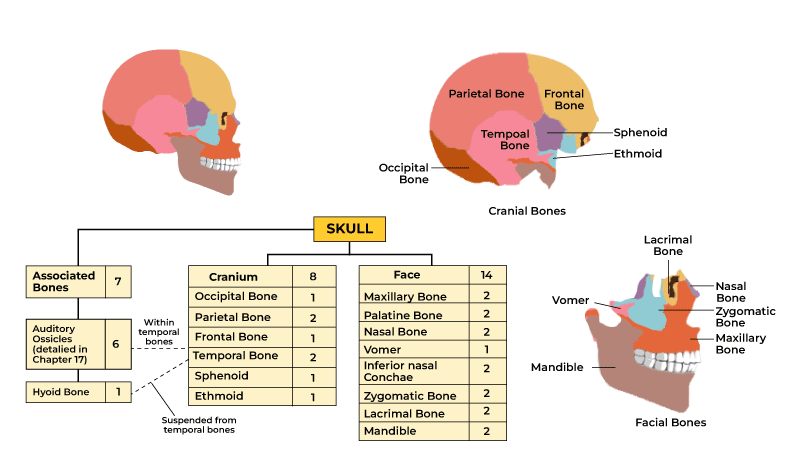
Number of cranial bones in the skull
8 cranial bones

Number of facial bones in the skull
14 facial bones

What CRANIAL bone forms the forehead and roofs of the orbits
Frontal bone (1)
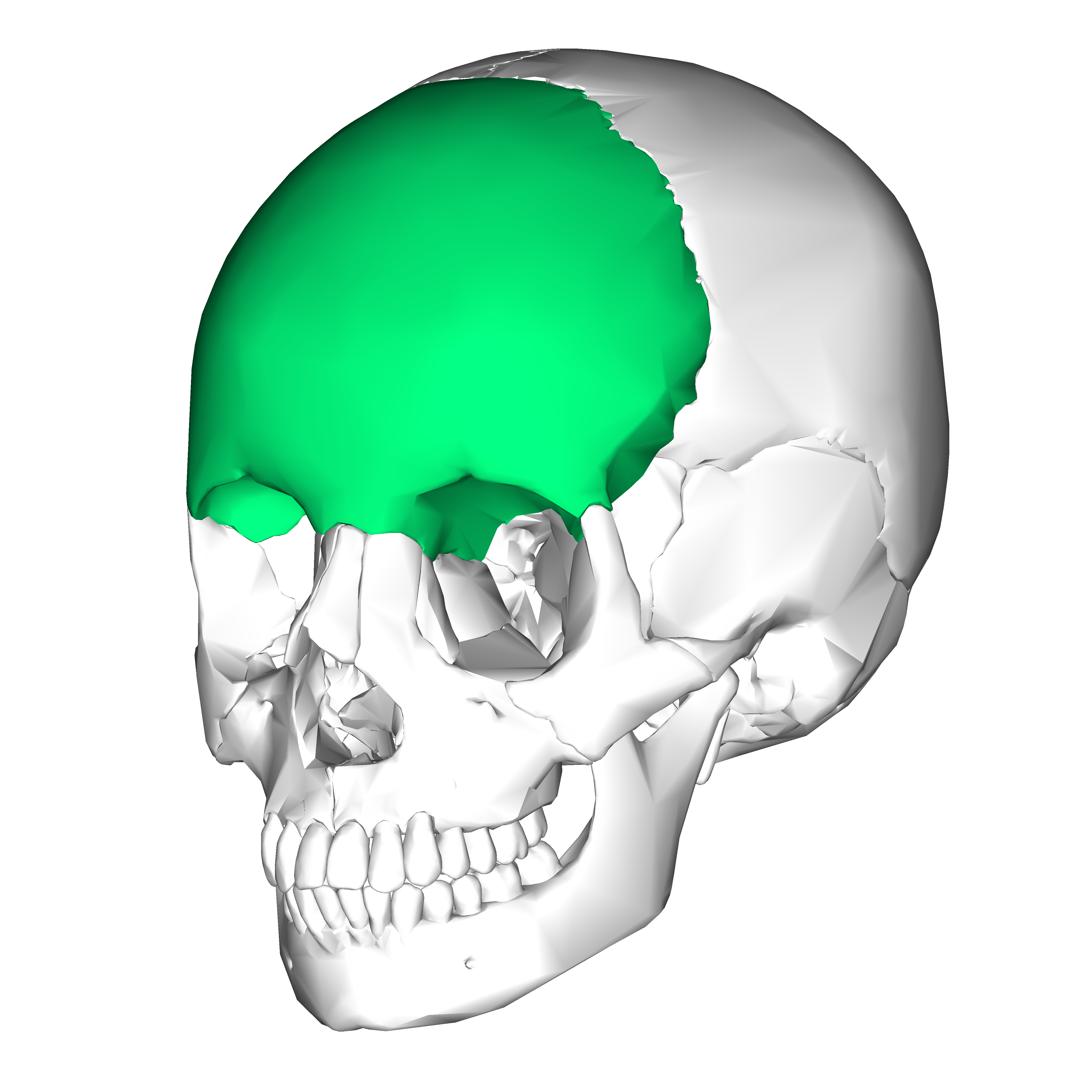
Smooth midline area of the frontal bone between the eyebrow ridges
Glabella

Vertical anterior portion of the frontal bone forming the forehead
Frontal squama

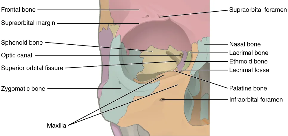
Superior limits of the eye orbits. Supports and protects the eyeballs
Supraorbital margins

Opening above each orbit transmitting the supraorbital artery and nerve
Supraorbital foramen


Depression containing the tear gland (lacrimal gland) that lubricates the eye
Lacrimal fossa

Paired bones forming much of the sides and roof of the cranium
Parietal bones (2)

Ridges on the parietal bones marking attachment of the temporalis muscle (closes the mouth)
Temporal lines
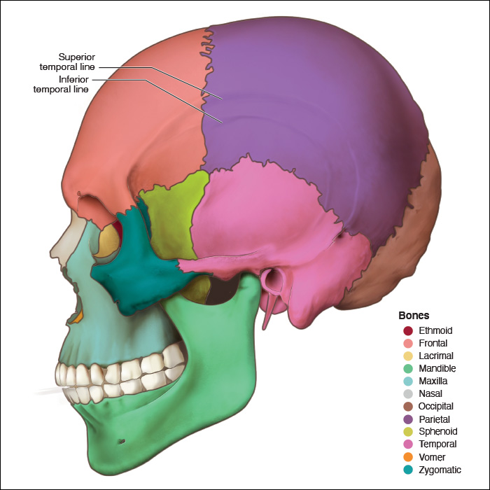

Smooth parietal surface superior to the temporal lines
Parietal eminence

Most posterior cranial bone forming the floor and back wall of the skull
Occipital bone (1)

Large opening in the occipital bone through which the spinal cord connects with the brain
Foramen magnum

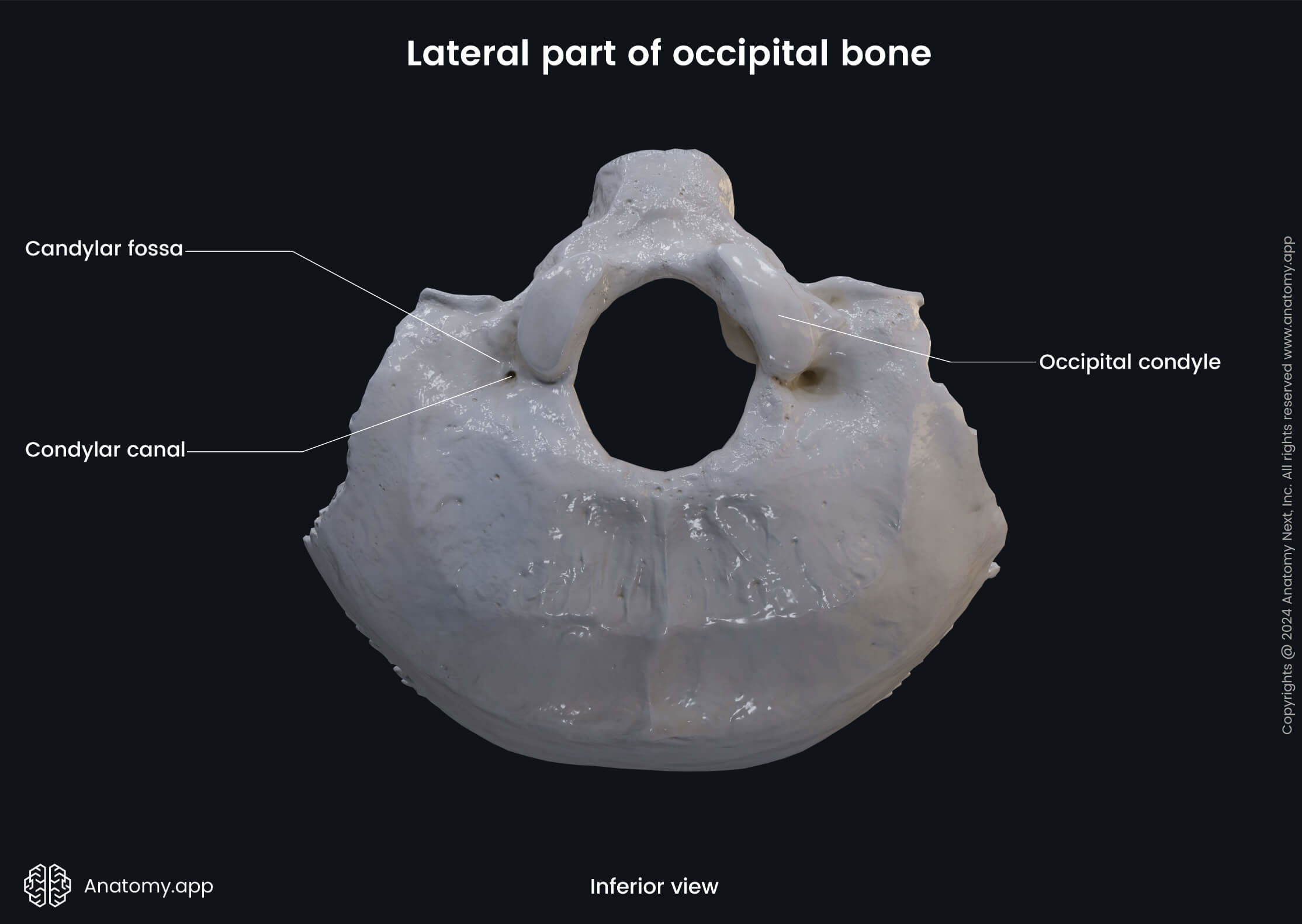
Structures of the occipital bone that articulate with the atlas
Occipital condyles


Passageways for cranial nerve twelve. begins at the lateral base of each occipital condyle
Hypoglossal canals


Band of bone anterior to the foramen magnum. the point of articulation between occipital bone and sphenoid
Basioccipital (basilar part)

Midline ridge on the posterior occipital bone.
AND
Small midline bump on the posterior occipital bone
The crest extends posteriorly from foramen magnum, ending in the protuberance. (list 2)
External occipital crest
AND
External occipital protuberance

Horizontal ridges marking muscle and ligament attachment on the occipital bone (list 2)
These stabilize the articulation between first vertebra C1 and occipital condyles
Superior AND inferior Nuchal lines

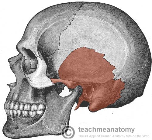
Paired cranial bones located inferior to the parietal bones on the lateral skull
Temporal bones (2)
Four major regions of the temporal bone
Squamous part: A large, flat, and translucent plate of bone that forms the anterior and superior part of the temporal bone. It assists in forming the lateral wall of the cranium and features the zygomatic process, which helps form the cheekbone.
Tympanic part: A small, curved plate of bone located below the squamous part and anterior to the mastoid process. It forms the bony walls of the external acoustic meatus (ear canal).
Mastoid part: The posterior portion of the temporal bone that sits behind the ear. It features the prominent, conical mastoid process (an attachment point for neck muscles) and contains hollow mastoid air cells.
Petrous part: A dense, wedge-shaped, pyramidal mass located at the base of the skull between the sphenoid and occipital bones. It houses the internal acoustic meatus, as well as the delicate structures of the middle and inner ear (cochlea and vestibular system).

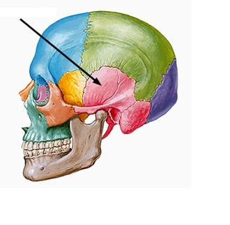
Lateral region of the temporal bone bordering the squamous suture
Squamous region
A squama in a squamous region refers to a thin, flat, scale-like plate of bone that forms part of the skull.
contains: cerebral surface, zygomatic process.


Projection of the temporal bone that joins the temporal process of the zygomatic bone
Zygomatic process

Arch formed by the temporal and zygomatic bones which forms cheekbone. also apart of zygomatic process
Zygomatic arch


Region surrounding the external acoustic meatus
Tympanic region
Contains: styloid process

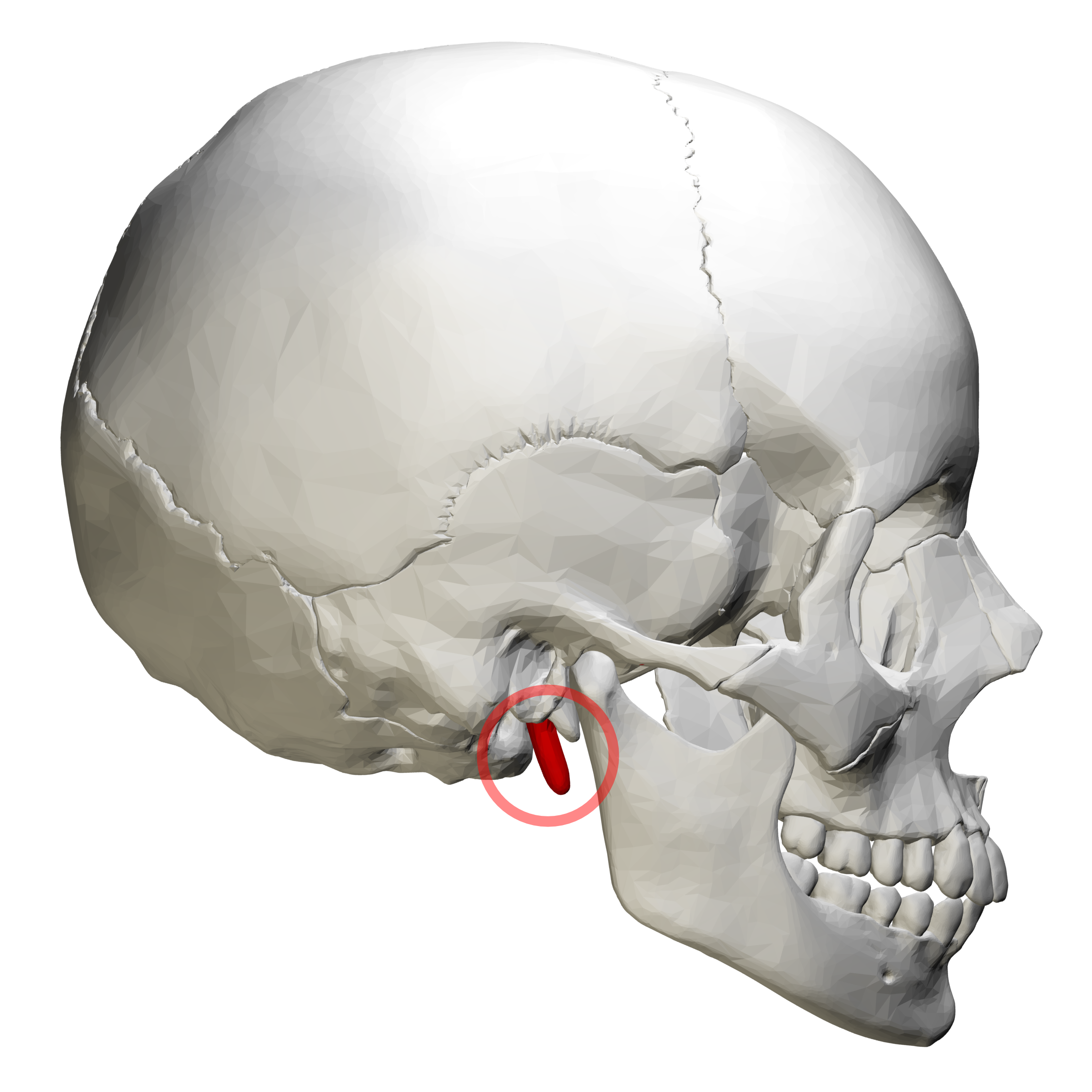
Needlelike projection inferior to the external acoustic meatus
Styloid process


Rough projection behind the ear that anchors neck muscles.
Mastoid process

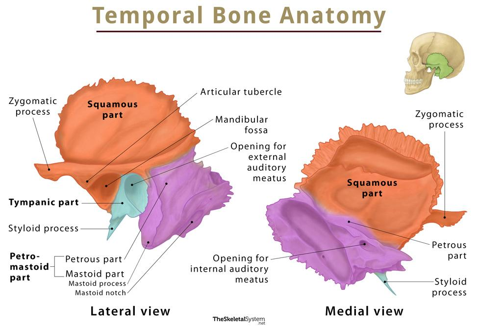
Most massive region of the temporal bone contributing to the cranial base
Petrous region
Contains: jugular foramen, carotid canal, foramen lacerum, internal acoustic meatus


Opening transmitting the internal jugular vein and cranial nerves nine ten and eleven
Jugular foramen

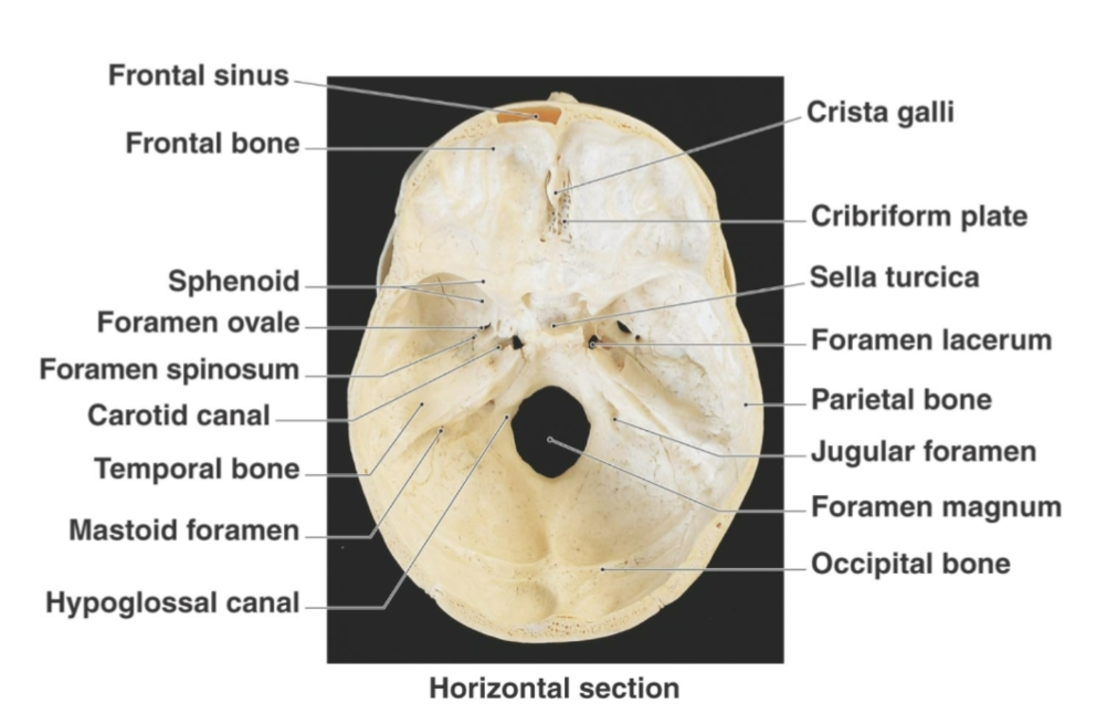
Passageway for the internal carotid artery within temporal bone
Carotid canal

Opening between the temporal and sphenoid bones that is mostly closed by cartilage in life
Foramen lacerum

Passageway transmitting cranial nerves seven and eight
Internal acoustic meatus
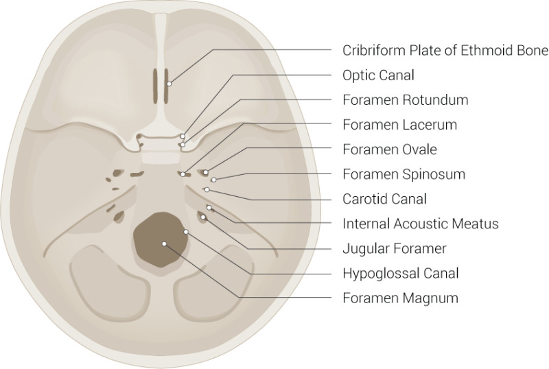

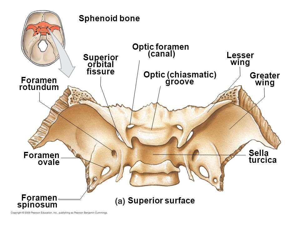
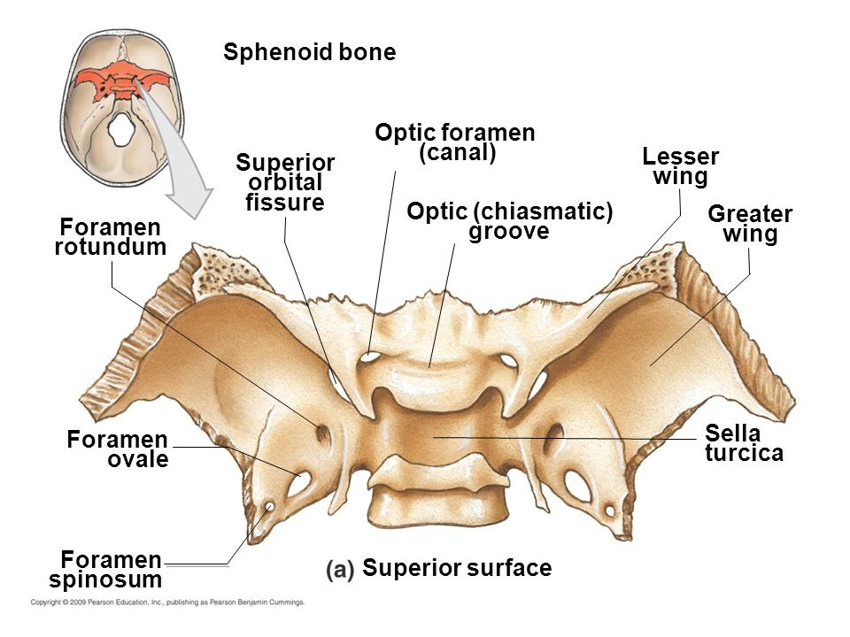
Bat shaped cranial bone spanning the width of the cranial floor and forming anterior plateau of middle cranial fossa
Sphenoid bone (1)
Has 3 PAIRS of PROCESSES (projections)
(greater wings, lesser wings, pterygoid processes)

Large processes of the sphenoid that contribute to the eye orbits
Greater wings

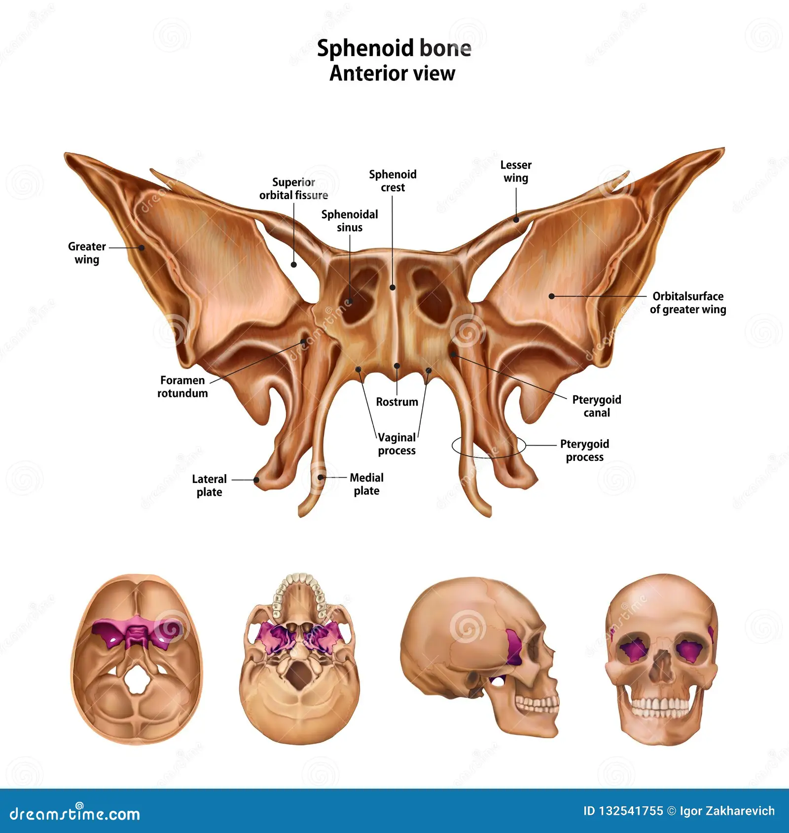
Smaller bat shaped portion located anterior to the sella turcica of the sphenoid bone
Lesser wings

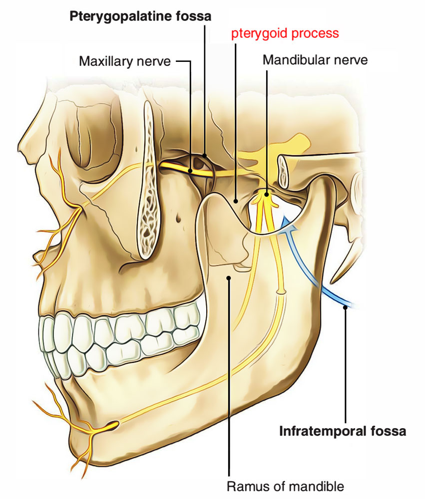
Vertical sphenoid projections providing attachment for muscles that move the lower jaw and soft palate
Pterygoid processes

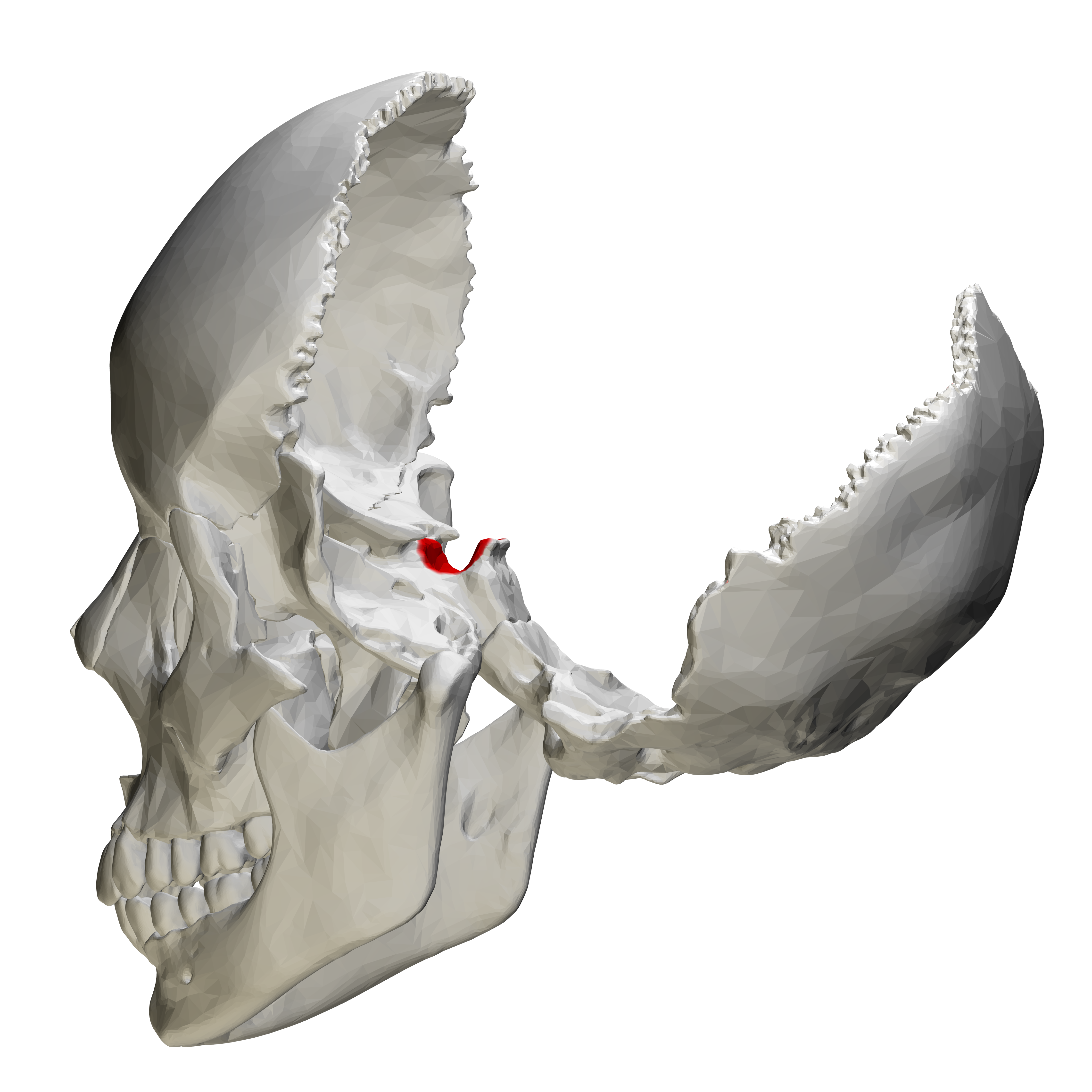
Turkish saddle shaped structure on the superior sphenoid
Sella turcica
Contains: anterior clinoid processes, tuberculum sellae, posterior clinoid processes, optic groove (foramen), optic canal
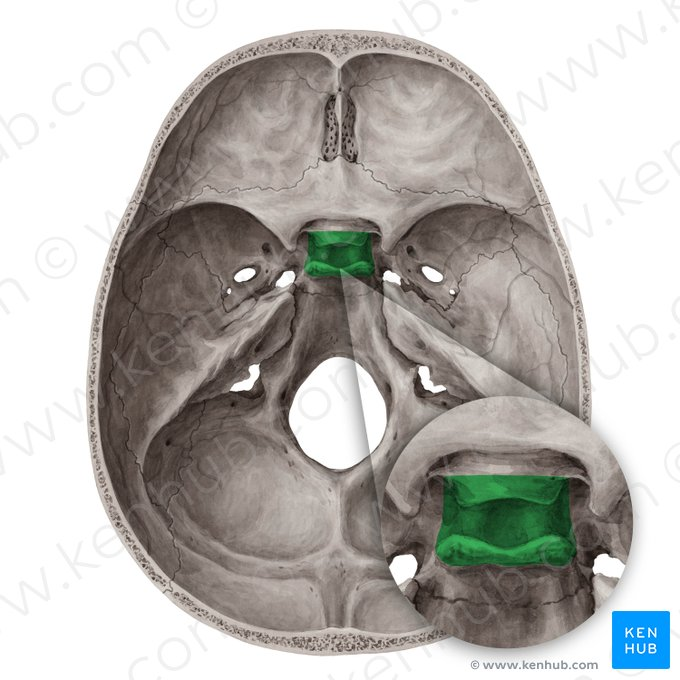
Depression within the sella turcica holding the pituitary gland
Hypophyseal fossa

Anterior border of the sella turcica
Tuberculum sellae


Posterior border of the sella turcica
Dorsum sellae


Groove located anterior to the sella turcica in the sphenoid bone
Optic groove

Openings at either end of the optic groove
Optic canals

Opening transmitting cranial nerve two in the sphenoid bone
Optic foramen
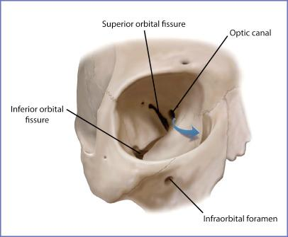
Long slit between the greater and lesser sphenoid wings transmitting cranial nerves three four and six
Superior orbital fissure (cleft in bone)

Opening transmitting a branch of cranial nerve five
Foramen rotundum
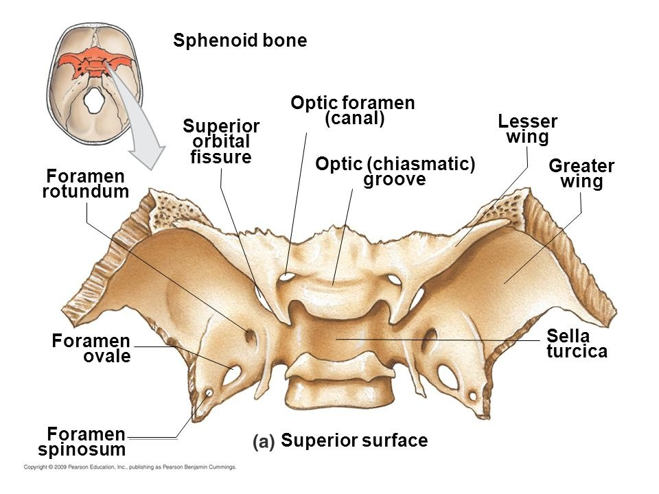

Opening posterior to the sella turcica transmitting a branch of cranial nerve five
Foramen ovale
Opening transmitting the middle meningeal artery within the sphenoid bone
Foramen spinosum

Deeply situated cranial bone anterior to the sphenoid
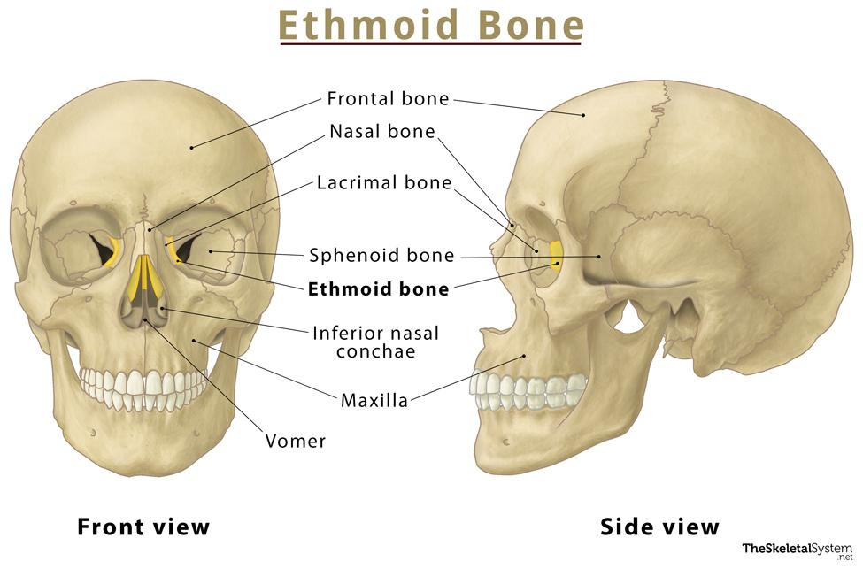
Forms part of orbital wall, anteromedial floor of the cranium, roof of nasal cavity, and part of nasal septum
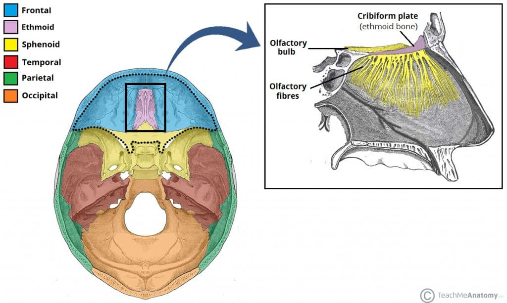
Ethmoid bone (1)

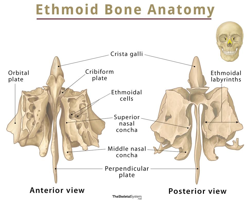
Three major parts of the ethmoid bone
Cribriform plates
ethmoidal labyrinth
perpendicular plate

Within ethmoid bone. Structures forming part of the roof of the nasal cavities and floor of the anterior cranial fossa
Cribriform plates
Contain: Olfactory Foramina, Crista Galli
Apart of Cribriform plates, Small openings transmitting olfactory fibers of cranial nerve one
Transmits sense of smell
Olfactory foramina

Apart of Cribriform plates. Superior projection between the cribriform plates in the ethmoid bone that helps secure the brain
Crista galli

Within ethmoid bone. Network of interconnected ethmoidal air cells. Dominated by superior and middle nasal conchae
Ethmoidal labyrinth

Scroll shaped structures of the ethmoid (labyrinth) extending into the nasal cavity
Superior and middle nasal conchae
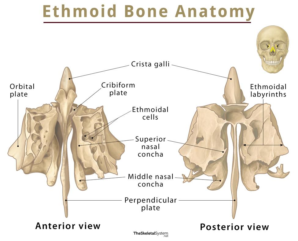
Within ethmoid bone. Structure forming the superior part of the nasal septum
projects inferiorly in the medial plane and superior portion is covered by olfactory epithelium
Perpendicular plate

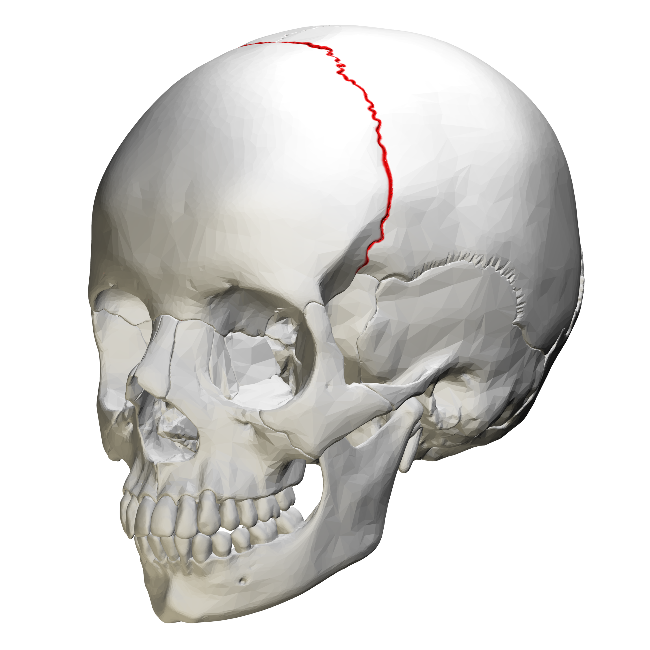
Suture between the frontal and parietal bones
Coronal suture
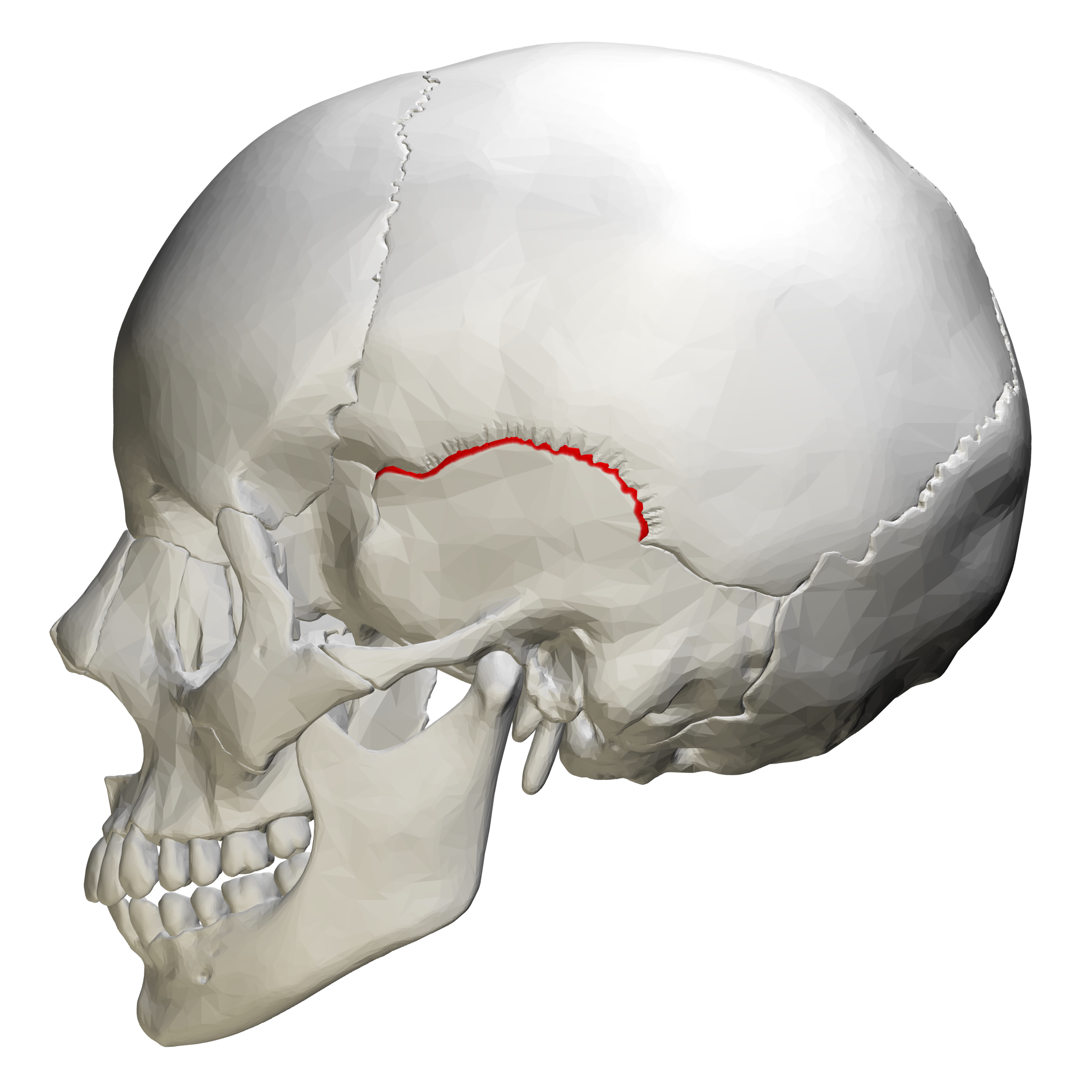
Suture between the parietal and temporal bones
Squamous suture
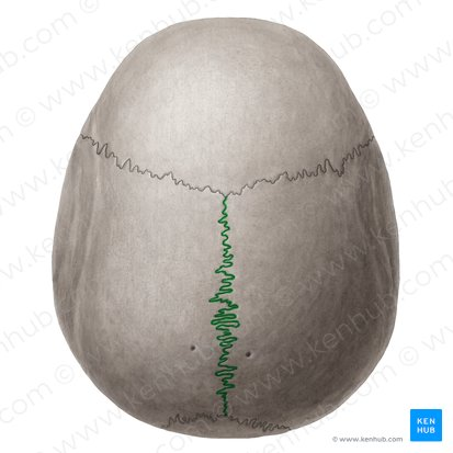
Suture where the right and left parietal bones meet
Sagittal suture
Suture between the parietal and occipital bones
Lambdoid suture

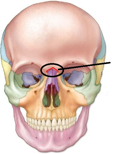
Suture between the frontal bone and nasal bones
Frontonasal suture
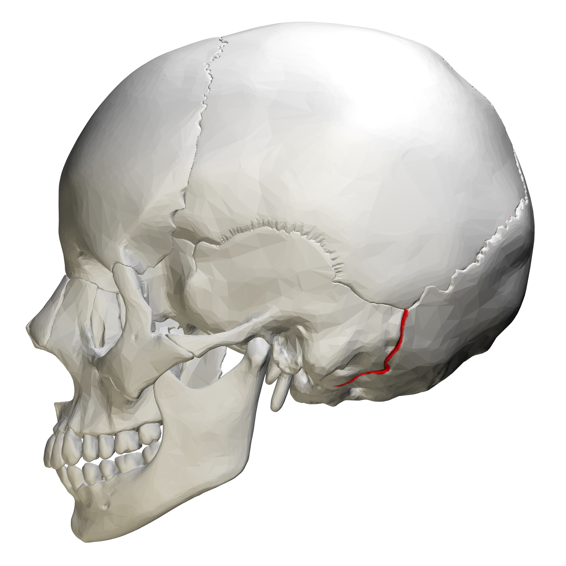
Suture between the occipital and temporal bones
Occipitomastoid suture
Lower jawbone and only movable skull bone
Mandible

Horizontal portion of the mandible supporting the teeth
Mandibular body
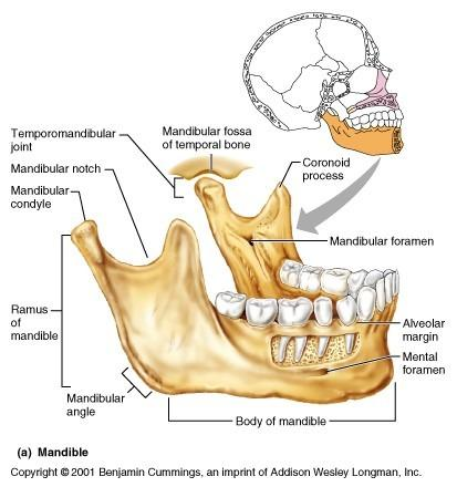
Ascending portions on each side of the mandibular body
Rami

Areas where the mandibular rami meet the body
Mandibular angles

Thickened mandibular area containing the sockets and roots of the teeth
Alveolar margin

Posterior processes forming the heads of the mandible
Condylar processes

Heads of the mandible articulating with the temporal bones
Mandibular condyles
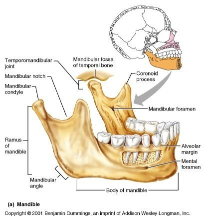
Anterior projections of the mandibular rami serving as attachment sites for the temporalis muscle
Coronoid processes
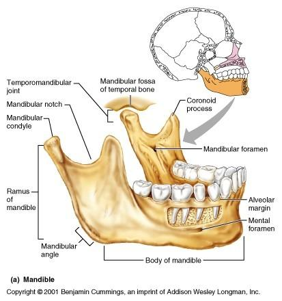
Depression between the condylar and coronoid processes
Mandibular notch
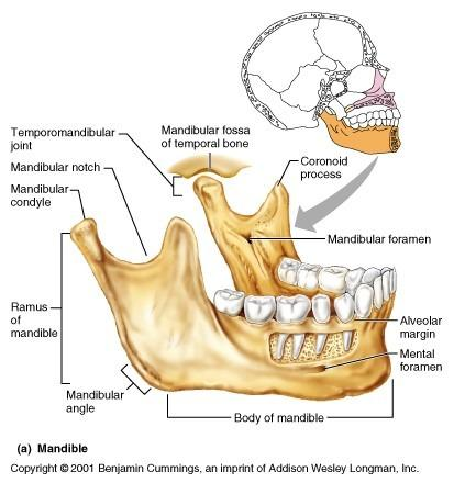
Openings on the anterior mandible transmitting mental nerves and blood vessels
Mental foramina
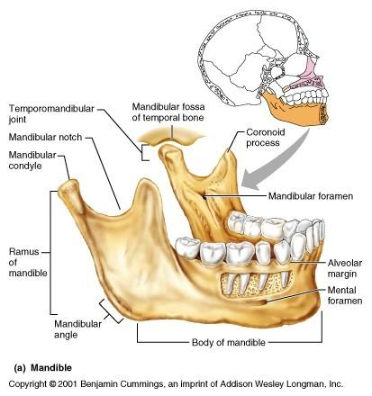

Line on the medial mandible marking the origin of the mylohyoid muscle
Mylohyoid line

Depression containing the submandibular salivary gland
Submandibular fossa
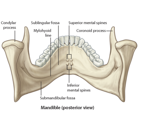
Openings on the medial mandibular rami transmitting branches of cranial nerve five
Mandibular foramina

Paired bones forming the upper jaw and central facial skeleton
Maxillae (2)

Processes of the maxillae forming the anterior hard palate
Palatine processes

Processes of the maxillae forming part of the bridge of the nose
Frontal processes
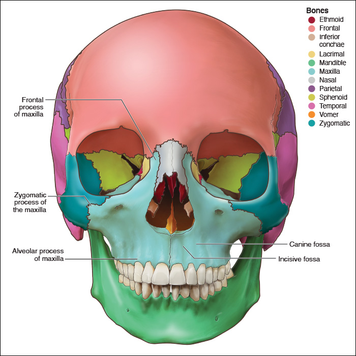
Processes of the maxillae articulating with the zygomatic bones
Zygomatic processes

Opening beneath each orbit transmitting infraorbital nerves and vessels
Infraorbital foramen

Margins of the maxillae containing the upper teeth
Alveolar margins
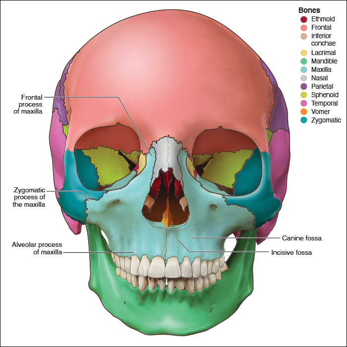
Bones forming the cheekbones and part of the lateral orbital rims
lateral to maxillae, contributes to orbital wall
Zygomatic bones (2)

Process of the zygomatic bone that joins the temporal bone
Temporal process

Opening transmitting a sensory nerve supplying the cheek
Zygomaticofacial foramen

Small rectangular bones forming the bridge of the nose
Articulates with the frontal bone at the frontonasal suture
Nasal bones (2)

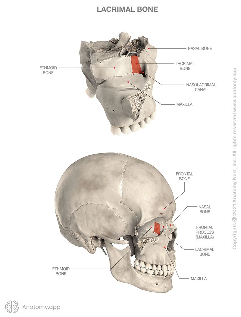
Delicate fingernail shaped bones in the medial portions of the orbits
Articulates with the frontal bone at the frontonasal suture
Lacrimal bones (2)


Groove in the lacrimal bone leading toward the nasolacrimal canal
Lacrimal groove

Canal enclosing the tear duct as it passes toward the nasal cavity
Nasolacrimal canal

Small L shaped bones forming the posterior portion of the hard palate
Palatine bones (2)


Horizontal portions of the palatine bones forming the posterior hard palate
Horizontal plates

Slender plow shaped bone forming the posterior and inferior nasal septum
Articulates with both the maxillae and palatine bones along the midline
Vomer (1)


Thin curved scroll shaped bones projecting into the nasal cavity
Perform same functions as the conchae of the ethmoid
Inferior nasal conchae (2)
