Integumentary anatomy bold terms
 Call Kai
 Learn
Learn Practice Test
 Spaced Repetition
 Match
Match Flashcards
 Knowt Play
1/67

There's no tags or description
Looks like no tags are added yet.
Name | Mastery | Learn | Test | Matching | Spaced | Call with Kai | Chat |
|---|
No analytics yet
Send a link to your students to track their progress
68 Terms
Integument (skin)
Functions include protection, prevention of water loss, temperature regulation, metabolic regulation, immune defense, sensory reception, excretion by secretion
How many layers does the epidermis have?
4-5 specific layers (thick skin - 5; thin skin - 4)
What is the epidermis?
A keratinized stratified squamous epithelium (also composed of dead cells). Thicker in the areas of the body like palms and soles of feet.
What is the Stratum Corneum?
Most superficial layer of the epidermis, composed of dead cells (keratinocytes) on the surface of the epithelium
What does the Stratum Corneum look like?

What is the Straum Lucidum?
Seen only in thick skin (palms, soles, digits), appears clear or white and is superficial to the stratum granulosum
What does the Stratum Lucidum look like?

What is the Stratum Granulosum
three to five layers of thin flattened cells which contain numerous dark granules. Acts as a transition zone where living skin cells turn into tough waterproof barrier by secreting lameller bodies filled with lipids to create spaces between cells hence creating a hydrophobic seal.
What does the Stratum Granulosum look like?

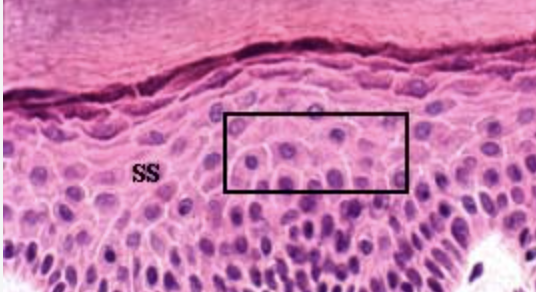
What is the Stratum Spinosum
several layers of cells which at high magnification look like spines on their surface connecting to cells. Provides strength and flexibility (to prevent tearing and blistering under physical stress) to the skin while supporting immune defense.
What does the Stratum Spinosum look like?
What is the Stratum Basale
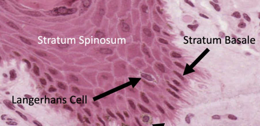
deepest stratum, consists of layer of dark stained cells. continuously produce new skin cells and regenerate the epidermis. These cells constantly divide through mitosis to create new keratinocytes that push towards surface.
What does the Stratum Basale look like
What is the dermis?
Mostly Made up of dense irregular connective tissue; some areolar connective tissue, split up into 2 layers, vascular
Two layers of dermis
Papillary layer, Reticular layer
What is the papillary layer?
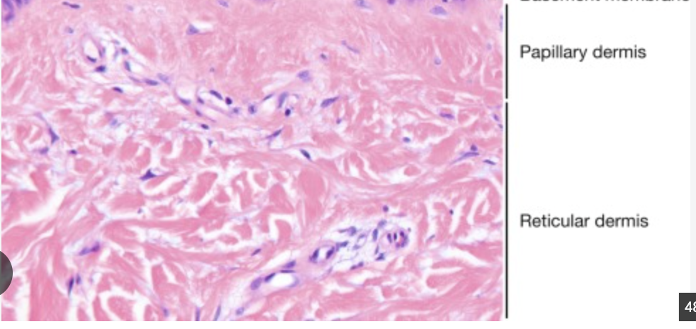
Deep to the stratum basale of the epidermis; most superficial layer of dermis. Named after the structures that protude from this layer.
What tissue type is the papillary layer?
areolar connective tissue
What is the dermal papillae and what layer is it in?
Finger like projections that lock epidermis to strengthen the skin. Contain nerve endings that allow detection of light touch, texture, and pressure. Also creates our fingerprints. In the papillary layer of dermis.
What is the Reticular layer
Deep in the papillary layer and thicker, contains large coarse bundles of collagen fibers, usually dark staining
What is in the Reticular layer
This layer contains secretory portion of sweat glands, oil glands, and hair follicles
What is the subcutaneous layer?
The layer that stores fat and anchors integument to underlying tissue.
What cells does areolar connective tissues have?
fibroblasts, with lots of ground stubstance
is areolar connective tissue vascular?
true
what cells do dense irregular connective tissue have?
fibroblasts, collage protein fibers, little ground substance
functions of areolar connective tissue
This tissue acts as the body's packaging material, connecting skin to underlying muscles and holding organs in place. Its fluid-filled, gel-like matrix stores water and salts, allowing nearby cells to absorb nutrients and release wastes. Loosely arranged collagen and elastic fibers let tissues stretch and return to their original shape while cushioning organs against impact and it has white blood cells (like macrophages and mast cells) to fight off bacteria, and fibroblasts to repair damaged
functions of dense irregular connective tissue
this tissue allows strength because it is interwoven, random collagen fibers let the tissue withstand tension from any angle. It stabilizes organs, anchors skin layers, and resists tearing. Tough capsules shield delicate internal structures from excessive stretching or distension
What cells are adipose tissue made out from?
adipocytes
is dense irregular tissue vascular?
poorly vascularized
is adipose tissue vascular
true
which layer is the dense irregular tissue found in?
dermis
which layer is adipose found in?
subcutaneous layer
Where is the nuclei pushed to in adipose connective tissue?
the periphery of the cell by fat droplet
functions of adipose tissue
energy storage, temperature regulation, cushion, hormone regulation by releasing adipokines to signal hunger/appetite
what tissues does the subcutaneous layer consists of?
areolar connective tissue and adipose connective tissue.
hair follicle
a tunnel-shaped skin organ that anchors and grows an individual strand of hair, found deep in epidermis, dermis, and subcutaneous layer
arrector pili muscle
pulls on hair follicle to shift how hair stands “arrector" = to straighten, aka goosebumps

Where are nails formed?
Stratum corneum
Nail functions
protects tips of fingers and toes, to help grasp objects
epidermal accessory organs are formed by what?
epidermis and invaginate deeper (dermis, sub)
sebaceous gland function
produce and secrete sebum
where are sebaceous glands located
located in the dermis (the second layer of the skin) and are found all over the body except on the palms of the hands and soles of the feet
hair function
protection against UV rays, sensory receptors, heat retention.
exocrine gland location
located in secretory portion in reticular layer of dermis
where do exocrine glands release their secretions
through ducts that open onto the surface of the epidermis
sebaceous oil gland’s two types
holocrine glands and sweat glands
holocrine gland
assoicated with hair follicles, secrete sebum, lubrication/waterproofing of epidermis, prevents hair from becomin brittle
sweat glands
excrete water products (sodium/urea), release perspiration to cool body
what is the immune defense of the integument?
epidermal dendritic cells
epidermal dendritic cell functions
also known as Langerhans cells, detects foreign invaders, capture antigens, and trigger body’s immune response. they phagocytize bacteria/debris
The integument system consists of
skin, exocrine glands in the skin, hair, nails
Functions of the integument
Protection to keep pathogens out and act as barrier to UV rays
to lose/dissipate heat
blood vessels dilate and sweat glands secrete
to conserve heat
blood vessels in dermis constrict and blood is shunted deeper
What tissues are found in the epidermis
keratinized stratified squamous epithelium, thicker in parts like soles of feet/palms, superficial
what tissues make up the dermis?
mostly dense irregular CT and some areolar connective tissue, deep
which layer has nerve endings, hair follicles, and glands?
dermis
what tissues make up the subcutaneous layer?
adiposes CT and areolar connective tissue
Which layer of epidermis is found in thick skin only?
Stratum Lucidum
What is keratinization?
The process by which cells form fibrils of keratin and harden, begins in deepest layer (basal), then as new keratinocytes form they move upward then as these cells rise, they fill with a tough protein called keratin, flatten out, and lose their internal cell parts, The cells die and form a hard, protective outer layer known as the stratum corneum in completion
epidendritic cells location
stratum spinosum
what kind of cells reside in the epidermis
keratinocytes, melanocytes, Langerhans cells, and Merkel cells
keratinocyte function
produce keratin, form a protective barrier against environmental damage, germs, and water loss, found throughout all layers, most abundant
melanocytes function
produce melanin, pigmentation is result of melanocyte activity as pigment is transferred to keratinocytes
melanocytes is found in what layer
stratum basal only
langerhan’s cells
epidermal dendritic cells
Tactile cells AKA Merkel cells function
connect to nerve endings, tactile (touch) sensation, located in stratum basal only
Reticular layer function
provides structural strength , elasticity, and support to skin while housing important glands, hair follicles, and nerve receptors while housing important glands, hair follicles, and nerve receptors
papillary layer function
It features finger-like projections (dermal papillae) that interlock with the epidermis to anchor the skin layers, house capillaries to feed the avascular epidermis, and contain Meissner's corpuscles for light touch sensation. supplies the outer layer of skin (epidermis) with vital nutrients and oxygen while firmly connecting the two layers together
