Immune Regulation & Tolerance, Autoimmunity
 Call Kai
 Learn
Learn Practice Test
 Spaced Repetition
 Match
Match Flashcards
 Knowt Play
1/110

There's no tags or description
Looks like no tags are added yet.
Name | Mastery | Learn | Test | Matching | Spaced | Call with Kai | Chat |
|---|
No analytics yet
Send a link to your students to track their progress
111 Terms
Immune regulation
the check and balance to our normal immune response; prevents overuse of the immune response which would damage an animal's own cells and tissues
The normal immune response must be __________ and be able to ______ ________ following clearance of a pathogen
regulated; calm down
Adaptive immune system
Responds to specific antigens; Responds by remembering signature molecules, called antigens, from pathogens to which the body has previously been exposed
Following clearance of a pathogen, animals have tens of millions of antigen-specific ___ and ___ cells circulating
B & T cells (plasma cells and CD8+ T cells, respectively)
B cells
create antibodies for isolating and destroying invading bacteria and viruses.
T cells
adaptive immune cells responsible for coordinating immune responses, killing infected cells, and providing immunologic memory
CD4⁺ helper T cells
release cytokines that activate macrophages, B cells and T cells; the generals
CD8⁺ cytotoxic T cells
directly kill virus-infected, damaged, or abnormal cells; the foot soldiers of the battlefield, initiating direct cell killing
Once an infection is cleared, there is no need for T cells or B cells any longer. Therefore, following infection, T cells and B cells undergo...
contraction
contraction of T cells and B cells
massive reduction in their numbers by way of apoptosis, killing 98% of T cell and B cell populations
Once contraction occurs, remaining B and T cells are..
memory cells, which will stick around and wait for future infection
Three players of immune regulation:
1.) immunoregulatory cytokines
2.) immunoregulatory cells
3.) programmed cell death 1
immunoregulatory cytokines
anti- inflammatory and immunosuppressive cytokines that function to suppress or shut-down immune responses
There are two immunoregulatory cytokines:
1.) IL-10
2.) TGF-β
There are two immunoregulatory cells:
1.) regulatory T cells (Tregs)
2.) M2 macrophages
regulatory T cells (Tregs)
unique subset of CD4+ T cells that actively suppress the immune response via physical contact with other immune/inflammatory cells
How do regulatory T cells (Tregs) suppress the immune response?
they engage in various surface ligand/receptor interactions as well as secrete IL-10 and TGF-β
regulatory T cells (Tregs) are produced in the same manner as all other T cells in the ________, having a TCR that is _______-specific
thymus; antigen
What role do regulatory T cells (Tregs) have in preventing autoimmune disease?
by suppressing self-reactive immune cells.
M2 macrophages
suppress inflammation via the production of the cytokines IL-10 and TGFβ
programmed cell death 1
aka PD-1; an inhibitory receptor located on many immune cells that results in suppression of those cells
Six cells with the PD-1 receptor:
1.) CD4+ T cells
2.) CD8+ T cells
3.) B cells
4.) macrophages
5.) NK cells
6.) dendritic cells
What cells are especially repressed with the PD-1 receptor?
T cells
What ligands bind to PD-1 receptors (2)?
1.) PD-L1
2.) PD-L2
PD-1 in cancer
In many cancers, neoplastic cells may upregulate their expression of the ligands PD-L1 and PD-L2; when tumor-specific T cells come to kill the cancer cells, the PD-L1/PD-L2 expressed on the cancer cells engage the PD-1 on the T cell surface and essentially shut down the T cell, preventing them from killing the cancer cells!
Immunological tolerance
the immune system's ability to recognize specific antigens without mounting an immune response against them, especially the body's own self-antigens
Failure of immunological tolerance leads to...
auto-immune disease
The body ensures tolerance via two broad mechanisms:
1.) central tolerance
2.) peripheral tolerance
central tolerance
self-reactive T and B cells that recognize self-antigen are eliminated during development in the thymus and bone marrow
The process of self-reactive T and B cells being eliminated during development is called...
negative selection
The process of negative selection is not perfect and some self-reactive T and B cells enter _______
circulation;
Three reasons why negative selection may fail to get rid of self-reactive T and B cells:
1.) not all self-antigens are presented in the thymus. Some are presented outside
the thymus.
2.) some proteins (antigens) are not expressed until later on in development/life,
long after thymic selection has occurred.
3.) some self-antigens are revealed secondary to inflammation
cryptic antigens
self-antigens that are revealed secondary to inflammation
However, even though some self-reactive T and B cells slip through the cracks, there are additional means to preventing autoimmunity such as...
peripheral tolerance
peripheral tolerance
second line of defense in tolerance; self-reactive T and B cells that escape central tolerance are inactivated, suppressed, or eliminated in peripheral tissues
There are three mechanisms of peripheral tolerance:
1.) peripheral deletion
2.) anergy
3.) regulatory T cells
peripheral deletion
T or B cells that recognize self antigen in lymph nodes are triggered to undergo apoptosis
anergy
self-reactive T cells and B remain alive but unresponsive; process by which self-reactive T cells and B cells are "shut down"
Normally what is required for T cell activation?
three signals in addition to antigens
1.) binding of the T cell TCR to the antigen/MHC complex
2.) co stimulation via molecular interactions (CD4 or CD8 w/ MHC)
3.) cytokine signaling
Normally what is required for B cell antibody production?
Helper T cells
If T cells or B cells don't receive the signals they need to activate, they undergo ________
anergy
Therefore, if a self-reactive T or B cell encounter a self-antigen under normal, non-inflammatory conditions...
there is often little or no costimulation, and the cells will not be activated (anergy); the body realizes it's a false alarm, and shuts down the T cell
Regulatory T cells and peripheral tolerance
Regulatory T cells (Tregs) help maintain peripheral tolerance by suppressing self-reactive immune cells that escaped central tolerance, preventing them from attacking the body's tissues
How do Regulatory T cells (Tregs) suppress self-reactive immune cells?
-release anti-inflammatory cytokines, especially IL-10 and TGF-β
-inhibit activation and proliferation of other T cells
-reduce the activity of antigen-presenting cells
Fetal tolerance
process by which a developing fetus accidentally learns that a pathogen is part of its own body; the fetus will see an exogenous antigen (pathogen) as endogenous antigen (self!)
How does fetal tolerance occur?
Normally, developing T cells that strongly recognize self-antigens are deleted through negative selection. This prevents autoimmune disease later in life.
A problem occurs if a fetus is infected with a pathogen during this critical period of immune development. The pathogen's antigens may be present while the immune system is learning what is "self."
The process then looks like this:
Fetus infected early in development
→ pathogen antigens are present during immune development
→ developing lymphocytes that recognize those pathogen antigens are deleted/inactivated
→ fetus becomes immunologically tolerant to the pathogen
→ after birth, the immune system doesn't recognize the pathogen as something it should attack
→ pathogen can persist in the animal
If fetal tolerance occurs in utero...
the fetus's immune system will no longer develop an immune response to an infectious agent, resulting in catastrophic consequence
Fetomaternal and fetal tolerance
both maternal and fetal mechanisms in place to prevent the foreign DNA of a developing fetus from being perceived as an antigen
How does fetomaternal tolerance work?
during pregnancy, the maternal immune response shifts towards a Treg and Th2 immune skewing; this reduces the Th1 immune response that may otherwise attack the fetus
How does fetomaternal tolerance work on the placenta?
there are Tregs present within the placenta itself, and the placental tissue has upregulation of immunosuppressive cytokines such as IL-10 and TGF-β
Dizygotic twins
aka fraternal twins; develop from two separate ova fertilized at the same time
Why don't Dizygotic twins attack each other in utero despite being genetically different?
since the placental circulation is shared, self- antigens from both fetuses are "shared" between each other; therefore, for ex, calf A sees the
"self" antigens of calf B, and vice versa. The result is that each calf sees the others
calf's antigens as "self", and each calf is tolerant of the other calf's antigen
Oral tolerance
there is an immense amount of "foreign" antigens that your body is exposed to in the food that you eat; these antigens induce Tregs and anergy and deletion of reactive T cells, thereby preventing an immune response to these molecules
The two fundamental goals of the immune response are:
1.) discriminate self from non-self
2.) remove an infection (non self) with as little damage to the host as possible
Autoimmunity
an immune response directed at "self" antigens and often a failure of tolerance
How is autoimmunity multifactorial?
involving factors such as genetics, other internal factors and external factors
There are two types of autoimmune diseases:
1.) primary autoimmune disease
2.) secondary autoimmune disease
primary autoimmune disease
aka true autoimmune disease; genetic susceptibility with no obvious "trigger"
secondary autoimmune disease
aka immune mediated disease; develops secondary to some specific and known trigger
Examples of triggers for secondary autoimmune diseases
drug administration, vaccination, viral infection, and cancer
For a secondary autoimmune disease, once the trigger/inciting cause is removed...
the autoimmune response ends (though it is very difficult to identify the exact trigger)
Six factors that influence autoimmune disease:
1.) genetics
2.) age
3.) hormones
4.) cancer
5.) drugs
6.) pathogen infection
How do genetics influence autoimmune disease?
autoimmunity occurs at higher rates in genetically similar animals, such as littermates, or a breed associated autoimmune disease
ex: cocker spaniels and IMHA
How does age influence autoimmune disease?
autoimmune diseases tend to have a higher incidence in middle age to older individuals
Why are older animals more likely to have autoimmune disease?
As animals age, there is a general reduction in the function of cell-mediated immunity (CD8+ T cells) and CD4+ T cells; among these reduced CD4+ T cells are Tregs, and their reduced numbers likely lead to reduced suppression of autoimmune diseases
How do hormones influence autoimmune disease?
In humans, women are more predisposed to autoimmune diseases. This seems to be less clearly known in domestic species, in part due to the ubiquity of neutering.
How does cancer influence autoimmune disease?
Benign or malignant neoplasms may rarely be associated with immune-mediated diseases, though the mechanisms are not well known
*removal of cancer = regression of autoimmune disease
How do drugs influence autoimmune disease?
drugs that normally do not elicit an immune response may act as a hapten and bind to carrier proteins to form a new molecule that is then targeted by the immune response
How does pathogen infection influence autoimmune disease?
Pathogen infections can trigger or worsen autoimmune disease by causing the immune system to mistakenly begin attacking the body's own tissues.
Four mechanisms for how pathogens influence autoimmune disease:
1.) superantigens
2.) innocent bystander
3.) cryptic epitopes
4.) molecular mimicry
superantigens
bacteria may produce specific toxins, called superantigens, that activate massive numbers of T cells in an antigen non specific manner
How do superantigens activate T cells in an antigen non specific manner?
The structure of the superantigen allows for it to bridge the MHC II molecule on an antigen-presenting cell and the TCR of the T cell, even though the TCR cannot directly bind the molecule

What bacteria particularly acts as a superantigen?
Staphylococcus sp.
innocent bystander
infection causes inflammation and cytokine release, which can accidentally activate nearby self-reactive lymphocytes that normally would remain inactive

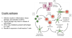
cryptic epitopes
self antigen that may otherwise be hidden, may be presented accidentally by antigen-presenting cells, resulting in T cell activation and an immune response to those epitopes
Four steps of cryptic epitopes
1.) infection results in inflammation and activation of antigen-presenting cells
2.) tissue damage results in release of self antigen
3.) antigen-presenting cells may mistakenly present self antigen to T cells
4.) results in expansion of self reactive T cells
molecular mimicry
Microbial antigens may rarely share peptide similarity to self
antigens, so when the immune response to the pathogen develops, the self is also targeted

Example of molecular mimicry
Equine recurrent uveitis (ERU); example of molecular mimicry between Leptospira interrogans serovar Pomona and intraocular proteins
When we consider autoimmune disease, one of the most important clinical considerations is what? Why?
identifying what is triggering the autoimmune disease; while not all triggers can be removed (like genetic diseases), if the trigger can be removed, this leads to the autoimmune disease being cured
Idiopathic autoimmune disease
when the cause of trigger of an autoimmune disease is unknown
How are idiopathic autoimmune diseases often treated?
immunosuppression
Two common autoimmune diseases in animals:
1.) immune mediated hemolytic anemia
2.) myasthenia gravis
immune mediated hemolytic anemia
disease in which the immune system mistakenly targets and destroys the body's own red blood cells, causing anemia
What species commonly have immune mediated hemolytic anemia?
most common in dogs, but can be found in cats, horses, and ruminants
Three canine breeds with higher incidences of immune mediated hemolytic anemia
1.) cocker spaniels
2.) english springer spaniels
3.) old english sheepdogs
Most cases of immune mediated hemolytic anemia have an __________ cause
idiopathic
What causes the immune system to attack the body's own RBCs during IMHA?
the body will produce antibodies against the antigens located on erythrocytes, making them a target of the immune system
Structure of antibodies
Y shaped proteins with two arms that bind antigens (making them capable of binding two antigens at once)
Five clinical/diagnostic findings of IMHA:
1.) regenerative anemia
2.) hyperbilirubinemia
3.) spherocytes
4.) autoagglutination
5.) Coomb's test positive
Why does hyperbilirubinemia occur with IMHA?
excessive destruction of red blood cells (hemolysis) within the vessels releases large amounts of hemoglobin, which is broken down into bilirubin
Why do spherocytes occur with IMHA?
macrophages partially remove the membranes of antibody-coated red blood cells without destroying the entire cell
Why does autoagglutination occur with IMHA?
in IMHA, antibodies bind two RBCs at once (one on each arm), allowing them to cross-link multiple RBCs together to form visible clumps

What test is used to diagnose IMHA?
Coombs test
Coombs test
diagnostic test that detects the presence of erythrocyte-directed auto-antibodies
Coombs reagent
Contains Anti-IgG and Anti IgM antibodies
Anti-IgG and Anti IgM antibodies
antibodies that specifically recognize and bind to other antibodies (IgG and IgM); in the Coombs test, an anti-IgG antibody binds to IgG antibodies that are already attached to the patient's red blood cells, while an anti-IgM antibody binds to IgM antibodies on the RBCs
How does the Coombs test work?
-Coombs reagent, which contains antibodies against the patient's immunoglobulins and/or complement, is added to the RBCs
-This reagent binds to the antibodies coating different RBCs and cross-links the cells together, causing visible agglutination
-Therefore, a positive Coombs test indicates that the patient's RBCs are coated with immune components
myasthenia gravis
autoimmune disease caused by auto-antibody targeting of the acetylcholine (Ach) receptors located on skeletal muscles at the neuromuscular junctions
What happens when antibodies target acetylcholine (Ach) receptors in myasthenia gravis?
results in both destruction of the receptors as well as blocking of the Ach binding sites
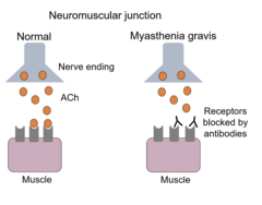
What happens as a result of Ach receptors being destroyed/blocked?
This prevents the normal Ach-triggered signaling, and the result is reduced and/or inconsistent firing of the post-synaptic muscle fibers