A&P II ~ Exam 1
 Call Kai
 Learn
Learn Practice Test
 Spaced Repetition
 Match
Match Flashcards
 Knowt Play
1/52

There's no tags or description
Looks like no tags are added yet.
Name | Mastery | Learn | Test | Matching | Spaced | Call with Kai | Chat |
|---|
No analytics yet
Send a link to your students to track their progress
53 Terms
anterior mediastinum
small space in front of the heart
composed mainly of CT, fat, lymph nodes, & thymus (in children)
middle mediastinum
heart, pericardium, roots of the great vessels
posterior mediastinum
esophagus, descending aorta, thoracic duct, azygos & hemiazygos veins (drain the wall of the chest & abdomen), and the sympathetic trunk
dextrocardia
a congenital defect of the heart where the apex sits on the right side instead of the left
also called situs inversus
epicardium (outer layer)
a thin, smooth membrane that reduces friction between the beating heart and the surrounding structures
contains CT, fat, blood vessels, lymphatics, and nerves
also called the visceral layer of the serous pericardium
myocardium (middle layer)
a thick, muscular layer made of specialized cardiac muscle cells (cardiomyocytes)
responsible for pumping action of the heart
endocardium (inner layer)
a thin, smooth lining of the heart chambers and valves
made up of endothelium & connective tissue
wall of the heart
pericardium
double-walled sac that surrounds & protects the heart
serous fluid
thin film of fluid that enables the heart to move and beat in a frictionless environment
fibrous pericardium (outermost layer)
connective tissue that keeps the heart from expanding too much
desmosomes
anchor cardiac cells together
intercalated discs
part of sarcolemma
gap junction
allow quick propagation of APs
endocarditis
inflammation of one of the heart valves
auricles
wrinkled, flap-like extensions of the atria
act as reservoirs which increase volume capacity of the atrium
crista terminalis
separates the ridged muscular part from the smooth part of the atrium
moderator band
a prominent trabecula in the right ventricle
helps obstetrician identify the right vs. left ventricle in the developing fetus
hypoxia
insufficient oxygen reaching the tissue
hypoxemia
abnormally low level of oxygen in the blood and cause hypoxia as the blood is not supplying enough oxygen to tissues in the body
due to respiratory disorders such as pneumonia or blood disorders such as iron deficiency anemia
ischemia
insufficient blood flow to the tissue
such as coronary artery disease where there is blockage of a coronary artery preventing blood flow to heart muscle
necrosis
tissue death
infarction
when necrosis is due to hypoxia
ventricular systole
when ventricles are contracting and pumping blood
ventricular diastole
when ventricles are relaxed and filling with blood
stenosis
thickening/narrowing of a heart valve that restricts blood flow
insufficiency
failure of a valve to close completely
prolapse
valve leaflets balloon upward as the ventricle contracts
regurgitation
valve leaflets do not properly close, forcing blood back into the atrium
cardiac cycle
from one heartbeat to the next heartbeat
automaticity
can generate own action potential
cardiac pacemaker cells
found in SA node, AV node, and purkinje fibers
cardiac conducting fibers
1%, intrinsic conduction system of the heart
initiate and spread APs through the heart, triggering each contraction
demonstrate automaticity
cardiac contractile fibers
99%, make up most of the myocardium
responsible for heart contractions that pump blood through the heart into the rest of the body
do not demonstrate automaticity
sinus rhythm
electrical impulses initiated in the SA node coordinate your heart beats at regular intervals
arryhthmia/disrhythmia
irregular heart rhythm
EDV
volume of blood in the ventricle at the end of ventricular diastole
SV
volume of blood pumped out of each ventricle after each contraction
ejection fraction
% of EDV that was ejected with each contraction
p wave
atrial depolarization
qrs complex
ventricular depolarization
t wave
ventricular repolarization
pq interval
delay of AV node to allow filling of ventricles
ST elevation
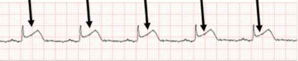
acute myocardial infarction

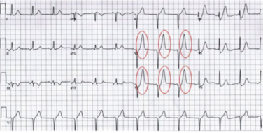
peaked t waves
hyperkalemia
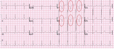
inverted (flipped) t waves
myocardial ischemia/onset of myocardial infarction
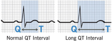
long qt syndrome
certain medications can cause
phase 4
potassium leakage channels maintain the inside of the membrane at a relative negative charge
phase 0
rapid sodium influx due to opening of voltage-gated channels
phase 1
sodium channels begin to close & potassium voltage-gated channels open, allowing potassium to efflux out of the cell
phase 2
calcium channels open
calcium influx into cell
potassium channels still open
potassium effluxing out of the cell balances calcium influxing into the cell
plateau phase
phase 3
calcium channels are closed
potassium channels remain open with continued efflux of potassium out of the cell
phase 4 (after phase 3)
once excitation reaches the ventricles through the cardiac conduction system, the electrical signal passes from one contractile cell to another contractile cell via gap junction