Portage Learning Anatomy and Physiology Module 2
1/234
There's no tags or description
Looks like no tags are added yet.
Name | Mastery | Learn | Test | Matching | Spaced | Call with Kai |
|---|
No analytics yet
Send a link to your students to track their progress
235 Terms
Tissue
group of cells that work together to perform one or more specific functions
Intracellular
internal environment inside the cells
Extracellular
environment outside the cells
Extracellular matrix
substance to help regulate their extracellular material and fluids
Histology
the microscopic study of the structure of tissues and their surrounding extracellular matrix
Epithelial
covers exposed surfaces, lines, pathways/cavities and can produce glandular secretions
Connective tissue
supports and underlines other structures and fills the internal space of the body
Muscle tissue
contract to enable movement
Nervous Tissue
carries electrical impulses and translates information to control activities of the body
Polarity
unique surfaces with specialized features
Apical (free)
facing external environment or inside of a lumen (opening). Allows cells to secrete or absorb materials from lumen.
Microvilli
finger-like extensions on apical that add extra surface area to cells, allowing for extra secretion and absorption
Cilia
longer cytoplasmic extensions that have an internal structure that allows for their movement (found in trachea and bronchi)
Avascular
do not have a direct blood supply
Basal surface
connects the basement membrane (noncellular - protein rich, allowing for cells to anchor to it for support)
Regenerative
the ability of epithelial tissue cells to regenerate through constant division of stem cells.
Cell Junction
connected between adjoining cells with specialized intercellular connections with unique proteins
Hyaluronan
polysaccharide derivative that helps lock epithelial cells together called intercellular cement
Gap junctions
permit the free diffusion of ions and small molecules between cells.
Tight junctions (occluding junctions)
serve as an impermeable barrier that do not allow solutes and fluids to diffuse between the cells in these locations.
Adhesion belt
forms deep to the tight junction, forming a handlike structure between cells.
Spot desmoses
help tie adjacent cells together and provide strong attachment between cells using CAM's.
Hemidesmos
connect half of the cell to the basal membrane
Stem cells
constantly dividing to replace lost or damaged cells.
High regeneration
Because of constantly damaged due to location in high-traffic areas.
Simple Epithelial Tissue
Cells that are only one layer thick.
Stratified Epithelial Tissue
Cells that have two or more layers.
Squamous Cells
Cells are wider than the height; thin and flat.
Cuboidal Cells
Cells have the same height, width and depth; square and cube.
Columnar Cells
Height of the cell is greater than width; tall, thin, rectangular.
Simple Squamous Epithelium
One layer thick, allows for materials to be exchanged through diffusion through cells.
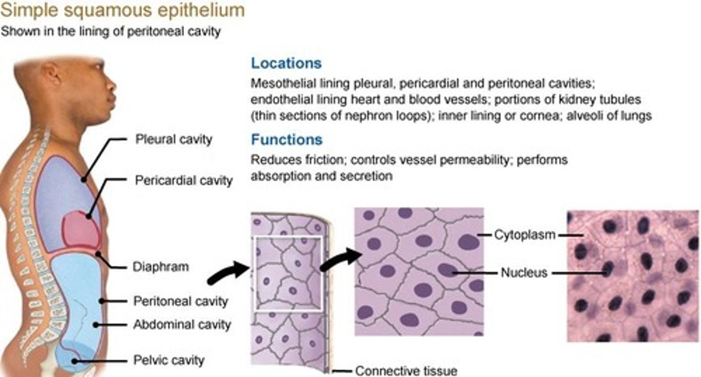
Endothelium
Lining of the blood and lymphatic vessels.
Endocardium
Lining of the ventricles and atria of the heart.
Mesothelium
Lining the walls and closed cavities of the body (abdominal, pericardial and pleural cavities).
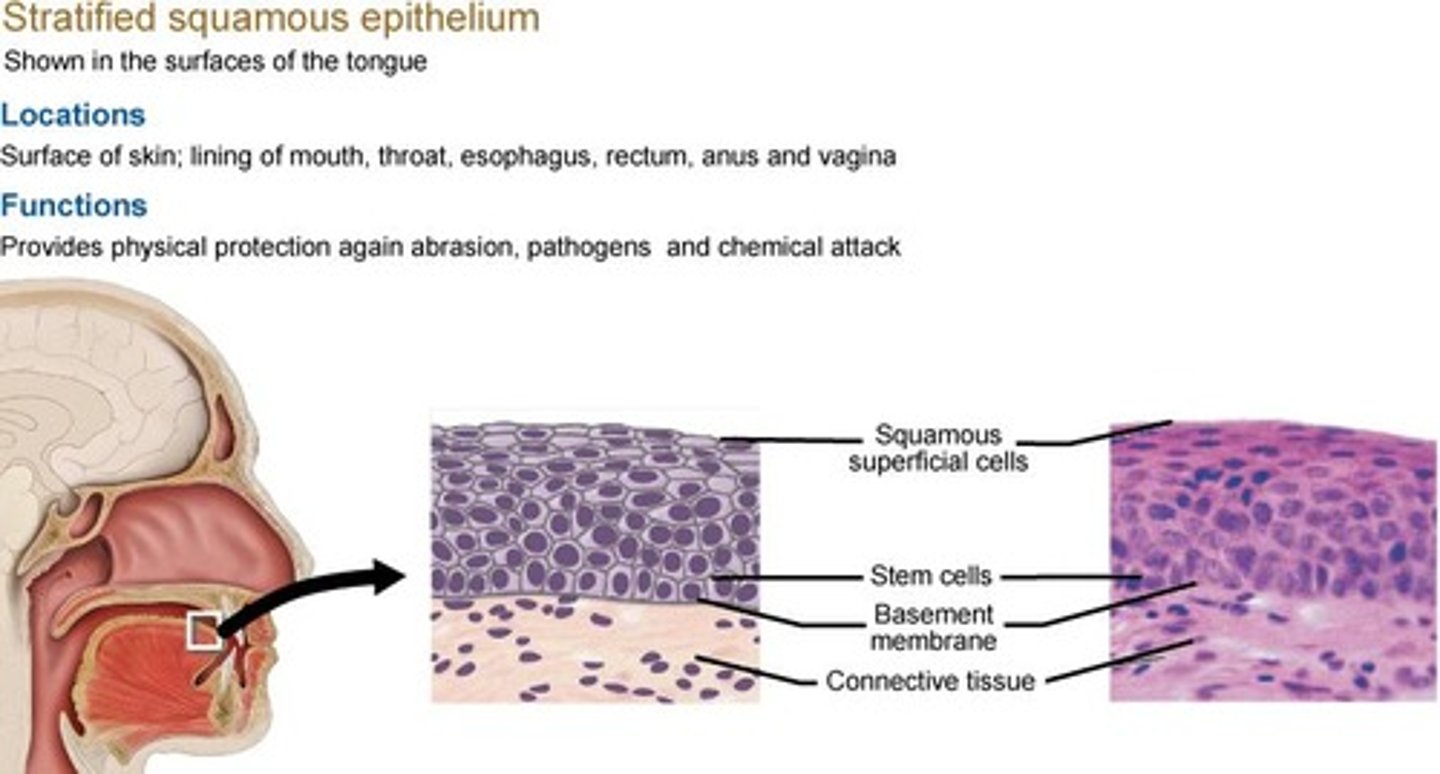
Stratified Squamous Epithelium
Forms the epidermis; found in oral cavity, esophagus and vagina.
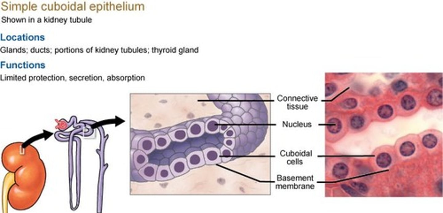
Simple Cuboidal Epithelium
Absorption and secretion of substances; found in small ducts of exocrine glands, kidney tubules and thyroid gland.
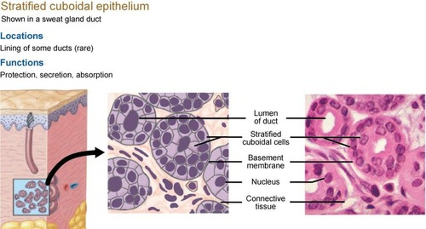
Stratified Cuboidal Epithelium
Acts as a physical barrier for protection and specializes in secretion and absorption; found in lining of ducts, sweat glands and larger ducts of exocrine glands.
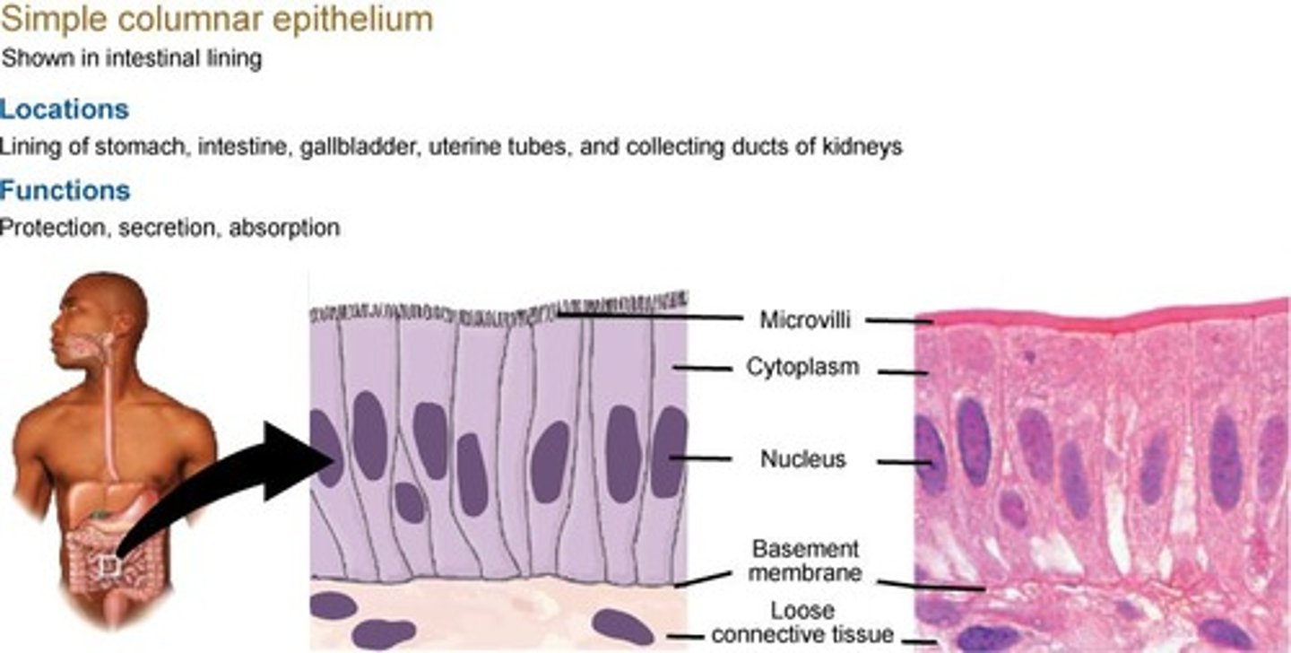
Simple Columnar Epithelium
Absorption and secretion; found in the small intestine and colon.
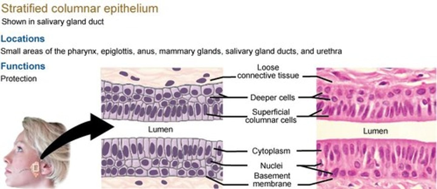
Stratified Columnar Epithelium
Serves mostly for protection; found in largest ducts of exocrine glands, pharynx, and epiglottis.
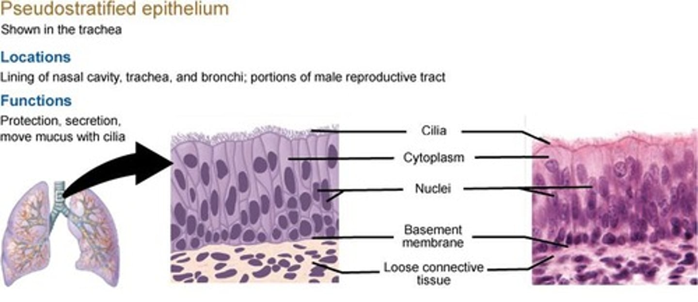
Pseudostratified Epithelium
Cells appear to be stratified but are actually one layer of cells made up of different sizes; functions for protection and secretion.
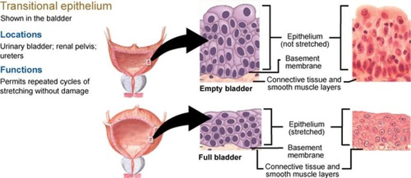
Transitional Epithelium
Allows cells to expand and stretch repeatedly to absorb fluid without causing cellular damage; found in the urinary tract (bladder and ureters).
Exocrine Glands
Release their products directly onto epithelial surface through duct or tube.
Endocrine Glands
Secrete their products directly into connective tissue; ductless.
Hormones
Products of an endocrine gland that enter bloodstream to reach target cell.
Paracrine Glands
Secrete substances that do not reach bloodstream but only affect other cells nearby in the same epithelium.
Merocrine Secretion
Product is delivered via membrane-bound vesicles to apical surface; vesicle fuse with plasma membrane and release via exocytosis.
Apocrine Secretion
Product is released from the apical side of the cell.
Holocrine secretion
Product accumulates within the maturing cell and at the appropriate time undergoes apoptosis (programmed cell death).
Unicellular glands
Simplest structure because it is one single secretory cell among other non-secretory cells.
Goblet cells
Found in ciliated columnar epithelium in the lining of intestines and trachea; function to secrete mucus to provide a protective layer.
Multicellular glands
Structure of more than one cell, classified by the arrangement of the secretory cells and type of branching the gland exhibits.
Simple exocrine glands
Duct is unbranched.
Simple tubular gland
Shaped like a tube.
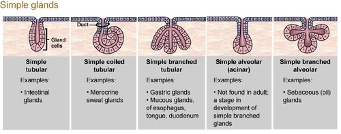
Simple coiled tubular gland
Shaped like a coil.
Simple branched tubular gland
Multiple secretory branches.
Simple alveolar (acinar) gland
Outpouching of secretory portion.
Simple branched alveolar gland
Branches of multiple acinar-shaped secretary portions.
Compound glands
Duct is branched.
Compound tubular gland
More than one branch with tube-like secretary portion.
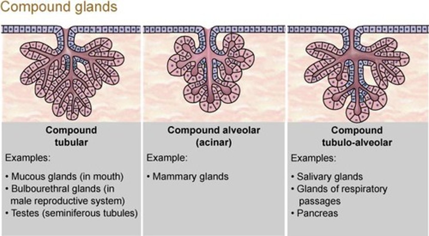
Compound alveolar gland
More than one branch with outpouching of secretary portions.
Compound tubulo-alveolar gland
More than one branch with a combination of tubelike and alveolar-type secretory portions.
Basal lamina
Created by connective tissue to connect with all other structures of the body.
Connective tissue fibers
Include collagen fibers, reticular fibers, and elastic fibers.
Collagen fibers
Long and straight with no branches, high tensile strength, flexible and strong when pulled lengthwise.
Reticular fibers
Thinner and have branching to form a mesh-like network, important for holding organs in place.
Elastic fibers
Branched and wavy, composed of elastin, allows return to original shape after stretching.
Ground substance
Clear, thick syrup substance with a high amount of water content, fills spaces between cells and fibers.
Fibroblast
Secretes hyaluronan and proteins, helps lock epithelial cells together.
Macrophages
Phagocytize (ingest) pathogens, damaged cells, or debris to rid them of the body.
Adipocytes
Fat cells that push the nucleus and other organelles off to the side.
Mesenchymal cells
First connective tissue to develop in an embryo, can divide and turn into other types of connective tissue cells.
Mast cells
Move around and tend to reside near blood vessels, release chemicals histamine and heparin after an injury.
Neutrophils and eosinophils
Phagocytic cells that move through connective tissue, attracted to the area to help fight infection.
Lymphocytes
Travel through connective tissues and throughout the entire body, can develop plasma cells to produce antibodies.
Areolar tissue
High number of elastic fibers in a loose, open network, acts as a shock absorber.
Adipose tissue
Provides padding, absorbs shocks, helps insulate the body from heat loss, and stores energy.
Reticular tissue
Contains reticular fibers that form a complex network of tissues to support functional cells of organs.
Dense regular connective tissue
Arranged as tightly packaged, parallel collagen fibers to withstand forces.
Tendons
Connect muscle tissue to bone.
Ligaments
Connect bone tissue to other bones or stabilize internal organs.
Aponeuroses
Flat sheets of fibrous connective material like a tendon.
Dense irregular connective tissues
Support stress from many different angles, interwoven network of fibers.
Elastic connective tissue
Primarily composed of elastic fibers, supports vertebral column.
Blood
Contains plasma and formed elements, including erythrocytes and leukocytes.
Erythrocytes
Transport oxygen and carbon dioxide in the blood.
Leukocytes
White blood cells, including neutrophils, eosinophils, basophils, lymphocytes, and monocytes.
Platelets
Tiny membrane-bound cytoplasm with special proteins and enzymes that help in blood clotting.
Lymph
Formed from interstitial fluid entering lymphatic vessels.
Cartilage
Made up of chondrocytes, avascular, surrounded by perichondrium.
Hyaline cartilage
Most common type of cartilage, provides stiff but somewhat flexible support.
Elastic cartilage
contains a high number of elastic fibers
Fibrocartilage
Prevents bone-to-bone contact and limits movement.
Neurons
Conduct electrical impulses, allowing for propagation of information.
Skeletal muscle tissue
Contains large cells, moves or stabilizes the position of the skeleton.
Cardiac muscle tissue
Only found in the heart, produces movement to pump blood.
Pacemaker cells
Regulate the contraction of cardiac tissue at regular intervals.
Common characteristics of Connective tissue
Provide support, structure, protection and insulation; basal lamina created by connective tissue to connect with all other structures of the body; transport materials and provide storage and energy reserves.
Antibodies
Specialized proteins that help defend the body against pathogens.
Smooth muscle tissue
involuntary. Small and spindle shaped with ends that become gradually smaller at each end; moves food, urine and reproductive tract secretion, controls diameter of respiratory passageways; regulates diameter of blood vessels.
Smooth muscle tissue characteristics
Found throughout the body; nonstriated involuntary muscle - nervous system does not provide control; contract on their own with gap junctions.