intro to mycology + lab procedures for fungal cultures
 Call Kai
 Learn
Learn Practice Test
 Spaced Repetition
 Match
Match Flashcards
 Knowt Play
1/48

There's no tags or description
Looks like no tags are added yet.
Name | Mastery | Learn | Test | Matching | Spaced | Call with Kai | Chat |
|---|
No analytics yet
Send a link to your students to track their progress
49 Terms
favic chandeliers
resemble deer antlers

nodular organs
knots of twisted hyphae

pectinate
comb-shaped
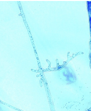
racquet hyphae
resembles tennis racquets placed end to end

spiral hyphae
may be flat or corkscrew shaped

sexual reproduction
produce fruiting bodies (ascomata/ascocarp). ascomata → asci → ascospores. ex: chaetomium spp, pseudallescheria boydii
perithecium
type of ascomata (fruiting body). disperses spores through an opening called an ostiole.

cleistothecium
type of ascomata (fruiting body). lacks specialized opening. splits under pressure, releasing spores

asexual spores
produced from cytoplasmic cleavage. ex: sporangiospores
conidiophore
specialized hypha upon which the conidia develop. can refer to a conidiogenous cell or specialized structure supporting a conidiogenous cell or cells
conidiogenous cells
specialized cells that produce conidia. ex: annellides & phialides
conidium (conidia)
unicellular or multicellular fungal element specialized to detach from the mycelium & disseminate
blastoconidia
seen in molds or yeasts. molds: bud at apex or side of conidiophore or from another conidium. yeasts (candida): elongate to form pseudohyphae

phialoconidia
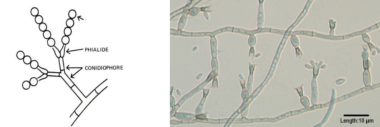
bottle shaped conidiogenous cells. apices produce conidia in masses or in chains. some apices have collarettes
anelloconidia
produce conidia in masses or in chains. deep staining truncated bases. series of annular rings at apex (not easily seen with light microscopy.

arthroconidia
arise from transformation of a series of cells long length of hypha. contiguous is simple. intercalated have separating cells (alternate)

chlamydoconidia
environmentally resistant resting cells that are formed from pre-existing cells within a hypha. inflated round cells with thick walls. seen in older cultures. ID marker for T. verrucosum

aleuroconidia
terminally or laterally located. truncated bases w/ small annular frills. macroconidia: larger, produced by same fungus, often multicellular. microconidia: smaller, produced by same fungus, unicellular

glabrous
smooth surface because they do not produce aerial mycelium. ex: yeasts

rugose
deep furrows irregularly radiating from center of culture

umbronate
button-like central elevation. may also have rugose furrows around elevation

verrucose
wrinkled & convoluted

dermatophytosis
diseases caused by microsporum, epidermophyton, trichophyton
common organisms on skin
dermatophytes, candida spp, malassezia spp, sporothrix schenkii
common organisms on hair
dermatophytes, candida spp, trichosporon spp, piedraia hortae
common organisms on nails
dermatophytes
common pathogens in respiratory
aerobic actinomycetes, aspergillus spp. dimorphic fungi: histoplasma capsulatum, blastomyces dermatitidis, coccidioides immitis
common organisms in blood
candida spp, cryptococcus neoformans, malassezia spp, histoplasma capsulatum
common organisms in sterile body fluids
histoplasma capsulatum, cryptococcus spp
common organisms in tissue specimens
yeast & filamentous fungi. aerobic actinomycetes
KOH prep
dissolves keratin & cellular debris while fungal elements remain visible
calcofluor white stain
binds fungal cell wall components (chitin). highly sensitive. fungi fluoresce bright white or blue-white

india ink stain
detection of encapsulated yeasts. classically used for cryptococcus neoformans from CSF. clear halo surrounding yeast cell

gram stain
yeasts stain gram-positive. demonstrates budding yeast & pseudohyphae
giemsa stain
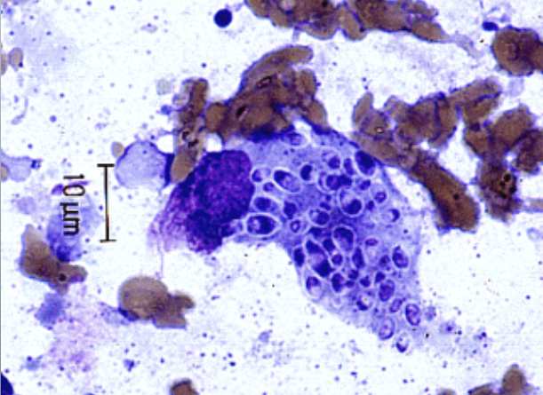
blood & bone marrow specimens. used to look for histoplasma capsulatum, an intracellular organism that appears as small oval to pear-shaped yeast-like cells
modified acid-fast stain
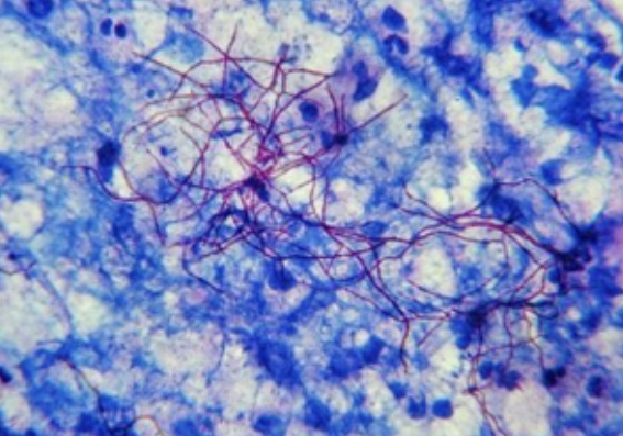
differentiate acid-fast nocardia (bacteria) from other true fungal elements
mucicarmine stain
highlights cryptococcal capsule. capsule stains red

sabouraud’s dextrose agar (SAB or SDA)
primary isolation medium. grows non-patho & patho fungi. acidic pH inhibits bacteria. H. capsulatum & nocardia will not grow.
sabouraud’s dextrose with brain heart infusion agar (SABHI)
primary isolation medium superior to SAB. enriched for recovery of yeasts. chloramphenicol & gentamicin can be added to inhibit bacteria. cycloheximide can be added to inhibit rapidly growing fungi
mycosel agar
isolation of dermatophytes. chloramphenicol to inhibit bacteria. cycloheximide to inhibit saprophytic fungi & some yeasts
brain heart infusion slant (BHI)
more enriched than SAB. used in recovery of H. capsulatum
BHI with chloramphenicol & gentamicin
used for selective isolation of fungi from specimens heavily contaminated with bacteria
potato-dextrose agar & corn-meal agar
used in slide cultures. induces spore formation, aids in ID
caffeic acid agar (bird seed agar aka niger seed agar)
selective isolation & differentiation of cryptococcus neoformans & cryptococcus gatti. C. neoformans will produce melanin → black colonies. protect media from light

temperature fungal growth requirements
25-30C for most molds, some dimorphics prefer 37C.
35-37C for yeast
teased preparation
advantage: performed imediately after maturation of fungal colony
disadvantage: teasing disturbs hyphae & conidia, can make discerning morph difficult
slide culture techniques
advantage: undisturbed microscopic morphology
disadvantage: technical expertise, must wait for fungus to grow before examining
transpararent tape preparation
performed immediately after maturation of fungal colony
lactophenol cotton blue (LPCB)
lactic acid: clearing agent, helps preserve fungal structures
phenol: kills fungus, making it safe to remove slide from BSC
glycerol: slightly viscous, prevents drying of prepared slide
cotton blue: aniline dye, adds color to fungal prep, enhances & contrasts structures