(A&P 1) Lab 4: Bone Tissue
 Call Kai
 Learn
Learn Practice Test
 Spaced Repetition
 Match
Match Flashcards
 Knowt Play
1/36

There's no tags or description
Looks like no tags are added yet.
Name | Mastery | Learn | Test | Matching | Spaced | Call with Kai | Chat |
|---|
No analytics yet
Send a link to your students to track their progress
37 Terms
General Function of Bones
1. Support
2. Protection
3. Anchorage
4. Mineral and growth factor storage
5. Blood cell formation
6. Fat storage
Osteology
Study of bone and the skeletal system
Skeletal System
Composed of bones cartilages and ligaments.
forms the flexible framework
Cartilage
Forerunner of most bones, covers many joint surfaces of mature bones
Ligaments
Holds bones together at the joints (bone to bone whereas tendons are muscle to bone)
Tendons
Connective tissue that attach muscle to bone
Hyaline
Provide support with flexibility and resilience. With two locations:
Articular: cover the ends of most bones at movable joints.
Costal: connect the ribs to the sternum
Elastic
Better for repeating bending
Location: external ear and epiglottis
Fibrocartilage
Compressive with tensile strength made of bundles of collagen.
Location: Menisci, Pubic Symphysis and Intervertebral discs
Long Bone Structure
(Practice Exercise on iPad)
Diaphysis: Known as the shaft and provides leverage
Epiphysis: Expanded head at each distal end which provides strength to the joint and attachment area
Articular Cartilage: Hyaline cartilage located at the joint surface and acts as a bone-to-bone cushion

Long Bone Structure Continued
(Practice Exercise on iPad)
Bone is covered with a sheath called the periosteum (collagen)
A thin layer of reticular connective tissue called the endosteum lines the spongy bone

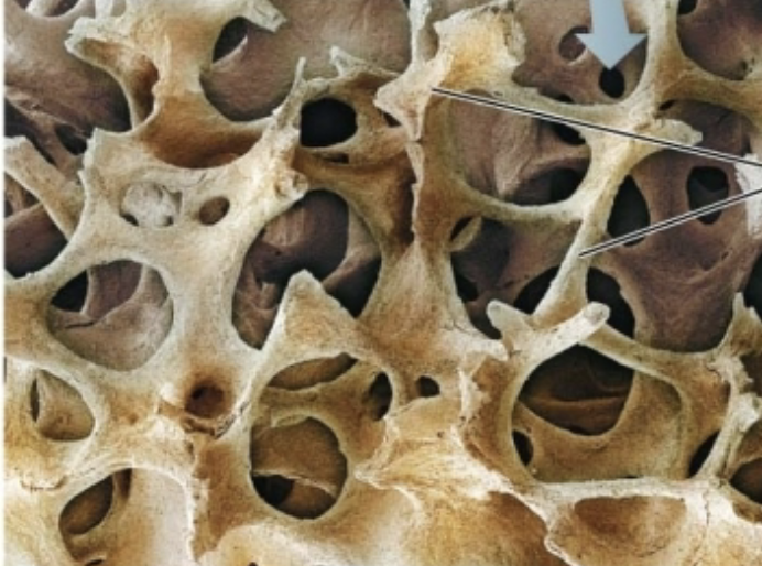
Spongy Bone
Contains a mesh of bondy spines called trabeculae.
It is occupied by the central space.
in flat bones the spongy bone is called diploe

Compact Bone
This will look smooth and solid.
Dense outer shell which encloses the medullary cavity
contains bone marrow.

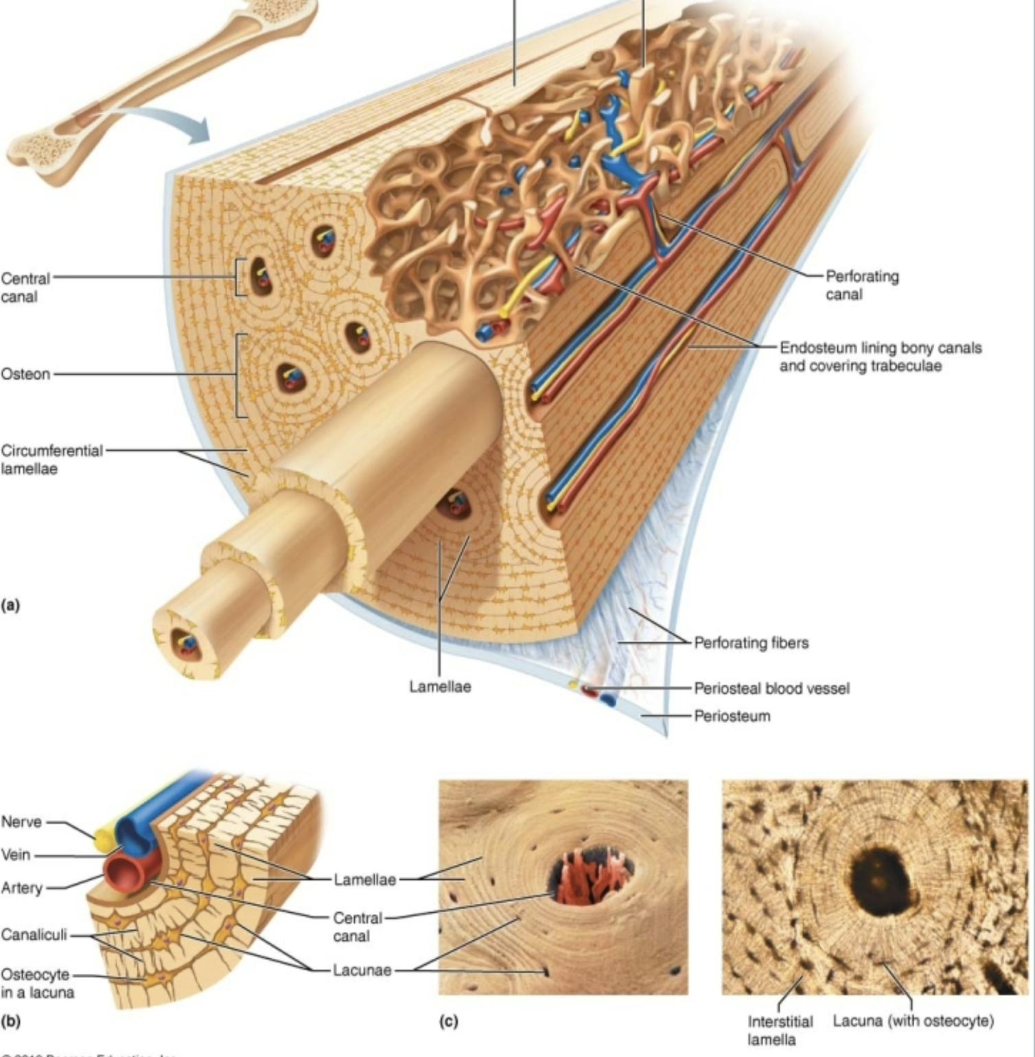
Bone Components (Labeling Exercise on iPad OSTEON MODEL)
• Central canal
• Osteon
• Circumferential lamellae
• Lamellae
• Lacunae
• Osteocyte
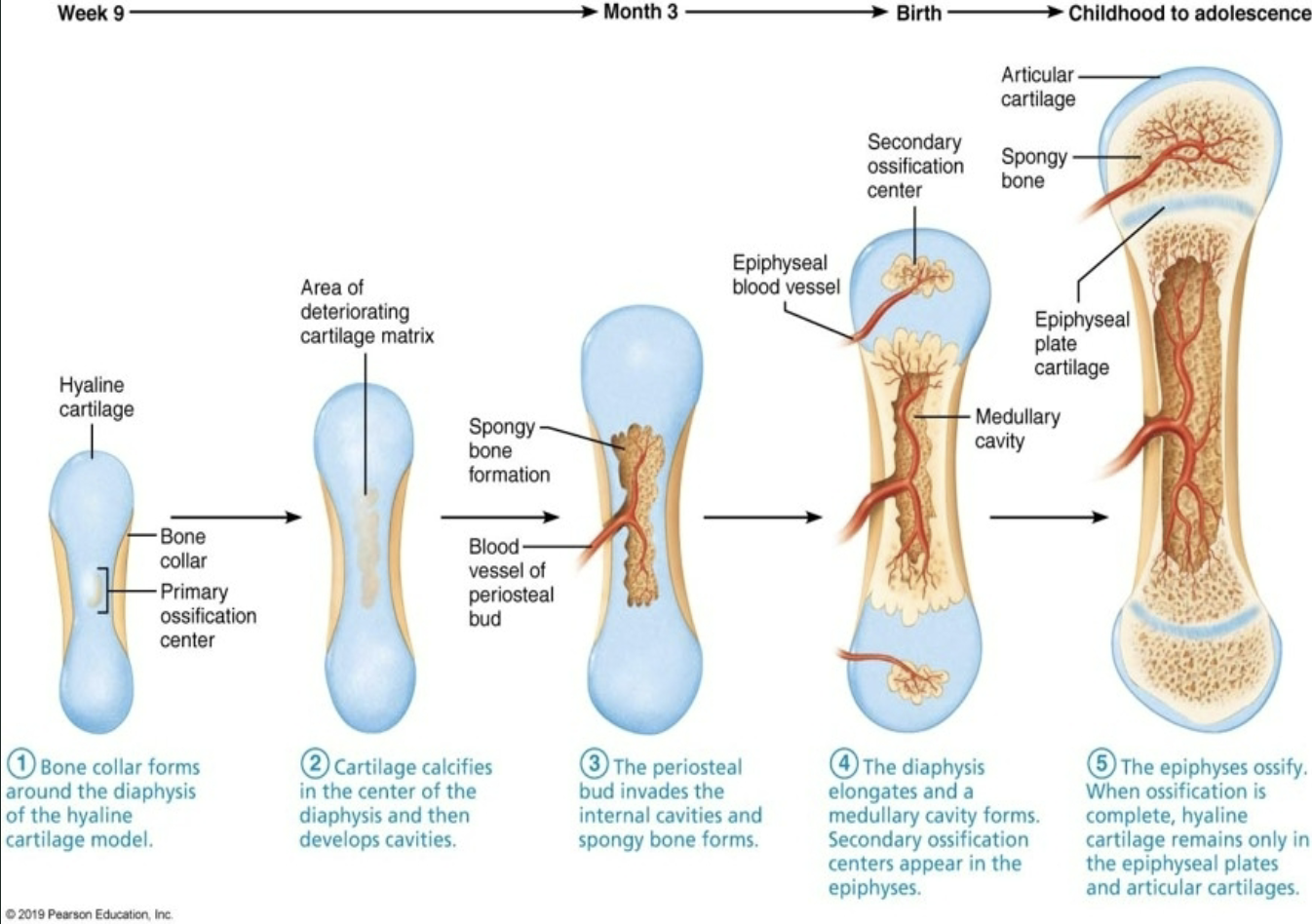
Epiphyseal Plate (Growth Plate)
Hyaline cartilage separates the marrow spaces of epiphysis and diaphysis: where the bones grow in length
Adults have an epiphyseal line which marks where the plate used to be
Epiphyseal Plates (X-ray)

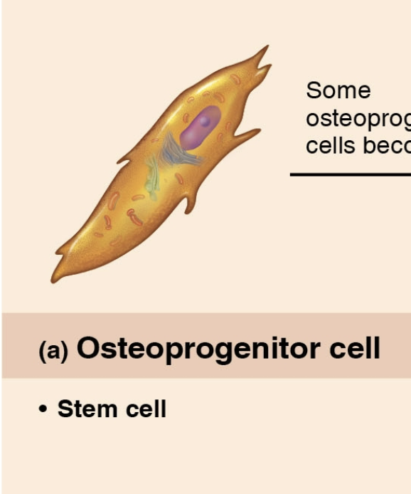
Osteoprogenitor Cell (Osteogenic)
A stem cell derived from mesenchyme that specializes to form bone tissue
Osteoblast
A specialized, non-mitotic bone cell responsible for bone formation (osteogenesis).

Osteocyte
A a mature, star-shaped (stellate) bone cell trapped within the mineralized bone matrix.
Regulation of bone remodeling
Bone maintenance

Osteoclast
A giant, multinucleated bone cell responsible for bone resorption—the process of breaking down and absorbing bone tissue. (multinucleated)

Bone Remodeling
A lifelong process where mature bone tissue is continuously resorbed by osteoclasts and replaced with new bone matrix by osteoblasts to maintain structural integrity and calcium homeostasis.

Osteoporosis
A condition in which overall tissue loss within bones results in bone pain, tenderness, and weakened and brittle bones

Bursae
A small, fluid-filled sac lined with a synovial membrane that reduces friction and cushions points of pressure between bones and surrounding soft tissues (tendons, muscles, or skin).
fibrous sac filled with synovial fluid

Regulation of Blood Calcium
Systemic blood calcium concentration is tightly maintained within a narrow physiological range of 8.5 to 10.5 mg/dL.
Negative feedback system relies on bones, kidneys, and small intestine
Hormones: Parathyroid Hormone (PTH), Calcitriol (active Vitamin D), and Calcitonin

Calcitonin
Stores Ca++ when we have too much.
Hormone released by the thyroid.
Lowers blood calcium levels only when present at high (therapeutic) levels
stimulates osteoclasts for bone border breakdown
Parathyroid Hormone
Gets Ca++ from bones when we need it.
Hormone released by the parathyroid glands that regulates blood calcium level.
Stimulates osteoblasts for bone formation
Flat Bones
Thin, flattened, and often slightly curved bones designed to provide protection for internal organs and broad surface areas for muscle attachment.
(e.g.) cranial bones (frontal, parietal, occipital), sternum, ribs, and scapulae.
Long Bones
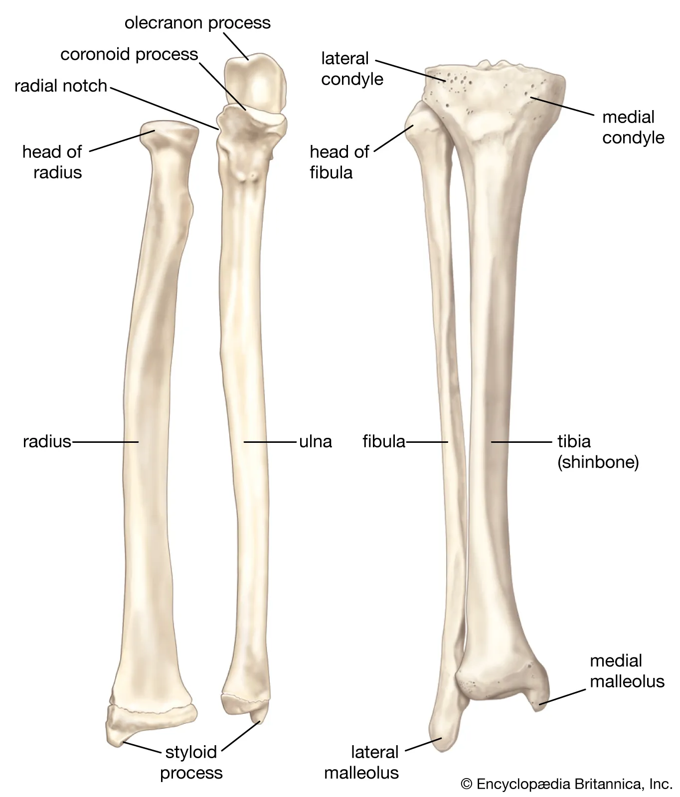
Longer than they are wide; consist of a central shaft (diaphysis) and two expanded ends (epiphyses).
(e.g.) Humerus, Radius and Ulna
Irregular Bones
Complex, elaborate shapes that do not fit into any other category. Vary in structure and composition.
(e.g.) Vertebrae, sacrum, coccyx, and facial bones (e.g., sphenoid, ethmoid, zygomatic).

Sesamoid Bones
Small, round/sesame-seed-shaped bones embedded directly within tendons where pressure and friction occur.
(e.g.) Patella (kneecap) and small sesamoid bones in the tendons of the hands and feet.

Short Bones
Roughly cube-shaped (equal length, width, and thickness). Consist of spongy bone surrounded by a thin layer of compact bone.
(e.g.) Carpals (wrist bones) and tarsals (ankle bones)

Synovial Joints
Synovial joints are characterized by a fluid-filled joint cavity that separates the articulating bones, allowing for free movement.
Enclosed by an articular synovial cavity

Fibroblast-like cells
Secrete fluid Macrophages that remove debris fom the joint cavity.
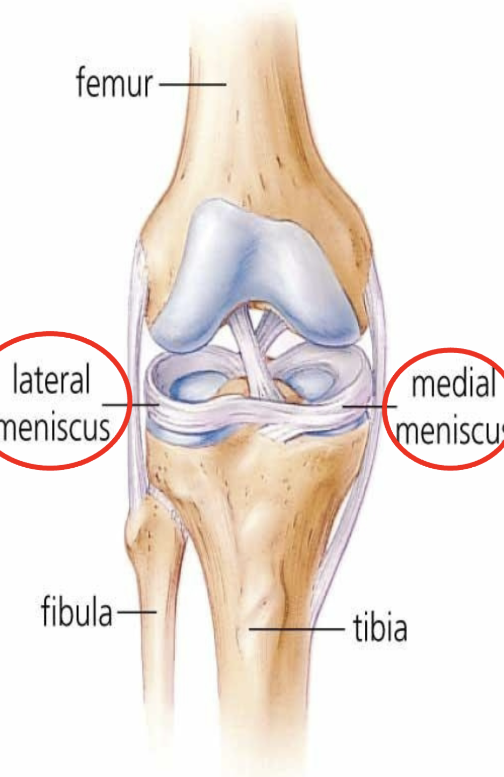
The Knee Joint (Labeling Exercise on iPad)
anterior aspect is covered by patellar ligament and the lateral and medial patellar retinacula (extensions tendon of the quadriceps femoris muscle)
knee stabilized quadriceps tendon and the tendon of the semimembranosus muscle

Knee Joint Continued (Labeling Exercise on iPad)
The joint cavity contains two C-shaped cartilages called the lateral and medial menisci joined by a transverse ligament
• The menisci absorb the
shock of the body weight
Knee Joint Continued (Labeling Exercise on iPad)
fibular (lateral) collateral ligament
tibial (medial) collateral ligaments
anterior cruciate ligament (ACL)
posterior cruciate ligament (PCL)
