NMS Lecture 6 - NMJ + Muscle Anatomy
 Call Kai
 Learn
Learn Practice Test
 Spaced Repetition
 Match
Match Flashcards
 Knowt Play
1/27

There's no tags or description
Looks like no tags are added yet.
Name | Mastery | Learn | Test | Matching | Spaced | Call with Kai | Chat |
|---|
No analytics yet
Send a link to your students to track their progress
28 Terms
3 types of muscle
Smooth muscle, Cardiac muscle, Skeletal muscle
Smooth muscle
Involuntary movement, seen in GI tract
Cardiac Muscle
Striated muscle found in walls of heart to propel blood. Involuntary control
Skeletal Muscle
Muscle attached to the skeleton (striated), voluntary contractions
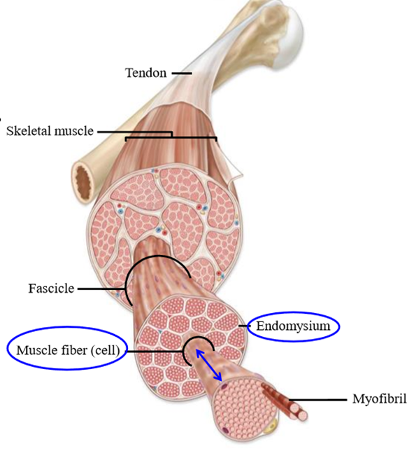
What is a skeletal muscle made out of
Number of muscle cells and fibers that run lengthwise down the muscle
Endomysium
Membrane that sits over each muscle cell and ioslates each muscle cell from another
Role of motor neuron
Activate the muscle it is attached to
Motor unit
The motor neuron, its axon, and all the muscle fibers it activates
How many synapses does a muscle fiber have
One - the neuromuscular junction
How many synapses does a post-synaptic cell have
10-40 000 inputs
Neuromuscular junction
Synapse between the efferent (motor) neuron and the muscle fiber
What type of synaptic transmission does a neuromuscular juction have
Direct-gated chemical synaptic tranmission
Motor end plate
Region of muscle fiber plasma membrane underneath the terminal portion of the axon
Steps of synaptic transmission to a muscle
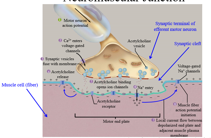
Action potential travels down efferent neuron to pre-synaptic terminal
Depolarized terminal causes opening of voltage-gated calcium channels
Calcium enters the terminal, causing synaptic vesicles to fuse to the membrane andn release neurotransmitter
Acetylcholine (ACH) released by the neuron
ACH binds to the post-synaptic nicotinic receptors on the muscle
Nicotinic receptors open and cause sodium to enter the muscle cells
Local depolarization occurs, the muscle is brought to threshold and opens voltage gated sodium channels.
Action potential produced in the muscle
Differences between synaptic transmission at the NMJ and a central synapse
One action potential in motor neuron generates one action potential in a muscle (no summation like in CNS)
Each muscle cell is innervated by 1 pre-synaptic axon (not 40 000)
No inhibitory NT, only ACH
Transverse tubule (T-tubule)
Muscle extracellular space - positively charged compared to intracellular space
Events at the neuromuscular junction (steps)
ACH from efferent neuron released into synaptic cleft and binds to muscle receptors
Muscle cell is depolarized at the motor end plate due to sodium entering the cell through chemically gated ion channels
Current flows between the end plate and the adjacent membrane at rest, which depolarizes the membrane to threshold and opens voltage gated Na channels
Action potential generated
Action potential travels along the surface of the muscle fiber, outwards in both directions towards the ends of the fiber, and down the T-tubules
Function of the T-tubule
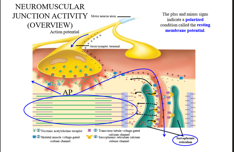
Site of excitation-contraction coupling
Excitation-contraction coupling
The process in which an action potential in the T-tubule initiates calcium release for muscle contraction
Dihydropyridine (DHP) receptor
Voltage gated calcium channel in the T-tubule that is physically bound to a ryanodine receptor
Ryanodine receptor
Physically connected to the DHP receptor and acts as a calcium channel
Steps of Excitation-Contraction coupling
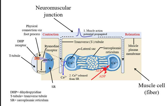
Action potential travels down the T tubule
Action potential activates DHP receptor
DHP receptor changes conformation, and opens the ryanodine receptor connected via the foot process
Calcium rushes out of the sarcoplasmic reticulum through the ryanodine receptor and enters the cytosol
Calcium interacts with the contractile elements of the muscle cell underneath the sarcoplasmic reticulum
Comparison of excitation-contraction coupling between skeletal and heart muscle
No physical coupling of receptors in heart muscle. The heart uses calcium-induced calcium release
Calcium induced calcium release (steps)
When an AP occurs, L-type calcium receptors open and release calcium
Calcium binds to ryanodine receptors, releasing sarcoplasmic reticulum calcium into the cytosol
Structure of a muscle cell/fiber
Composed of myofibrils, bundles of myofilaments. Myofilaments are made out of actin and myosin
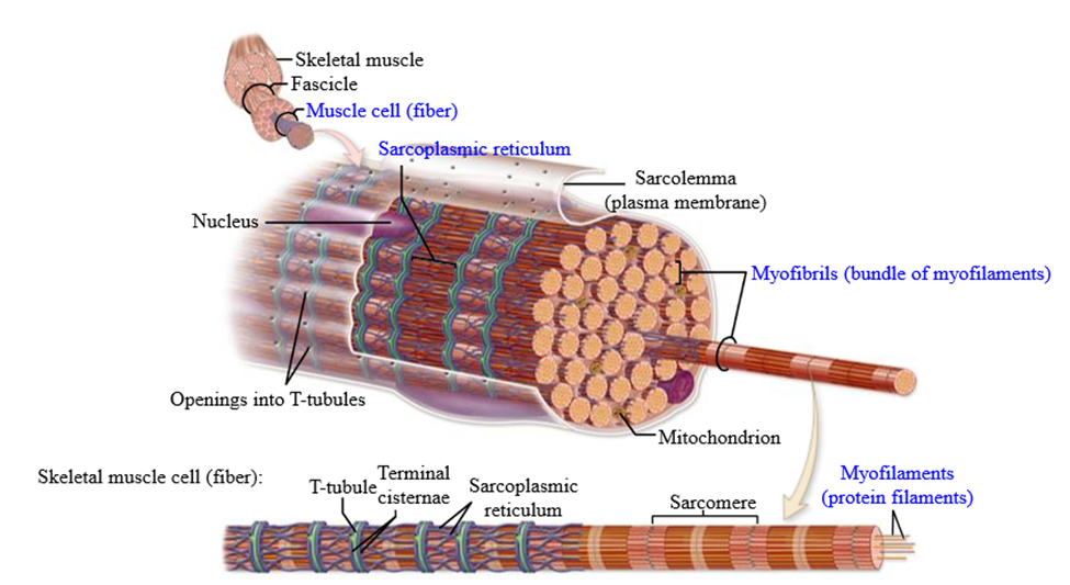
Structure of a myofilament
T-tubules and sarcoplasmic reticulum are wrapped around the sarcomere

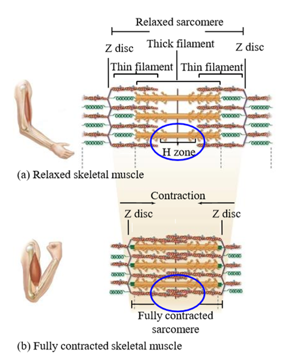
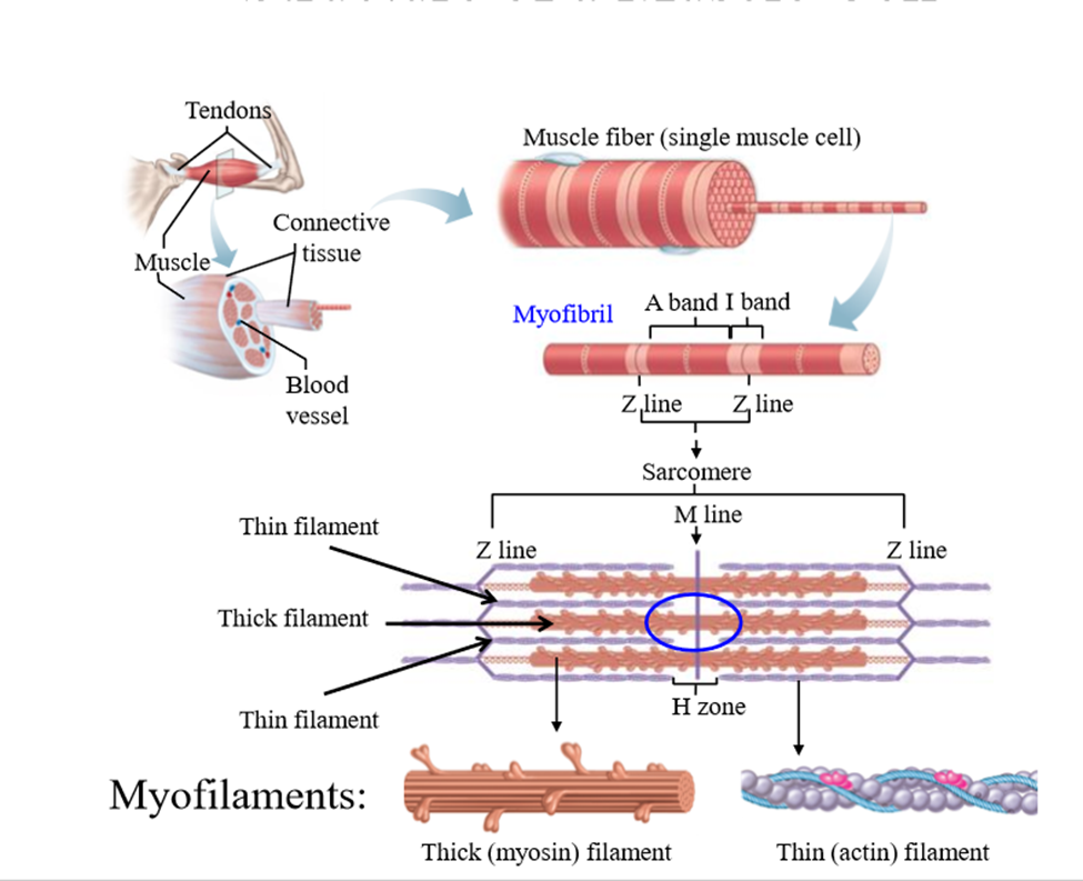
Structure of a sarcomere
Structural unit of a myofilament
Bound by Z lines (interconnecting protein networks)
Made up of actin (thick filament) and myosin (thin filament)
Actin and myosin are what allow the muscle to contract
H zone is the area in the middle of the sarcomere, only containing actin
What contracts in a muscle
The size of the actin/myosin filaments does not change. The H zone shrinks during contraction