Apical Windows
 Call Kai
 Learn
Learn Practice Test
 Spaced Repetition
 Match
Match Flashcards
 Knowt Play
1/45

There's no tags or description
Looks like no tags are added yet.
Name | Mastery | Learn | Test | Matching | Spaced | Call with Kai | Chat |
|---|
No analytics yet
Send a link to your students to track their progress
46 Terms
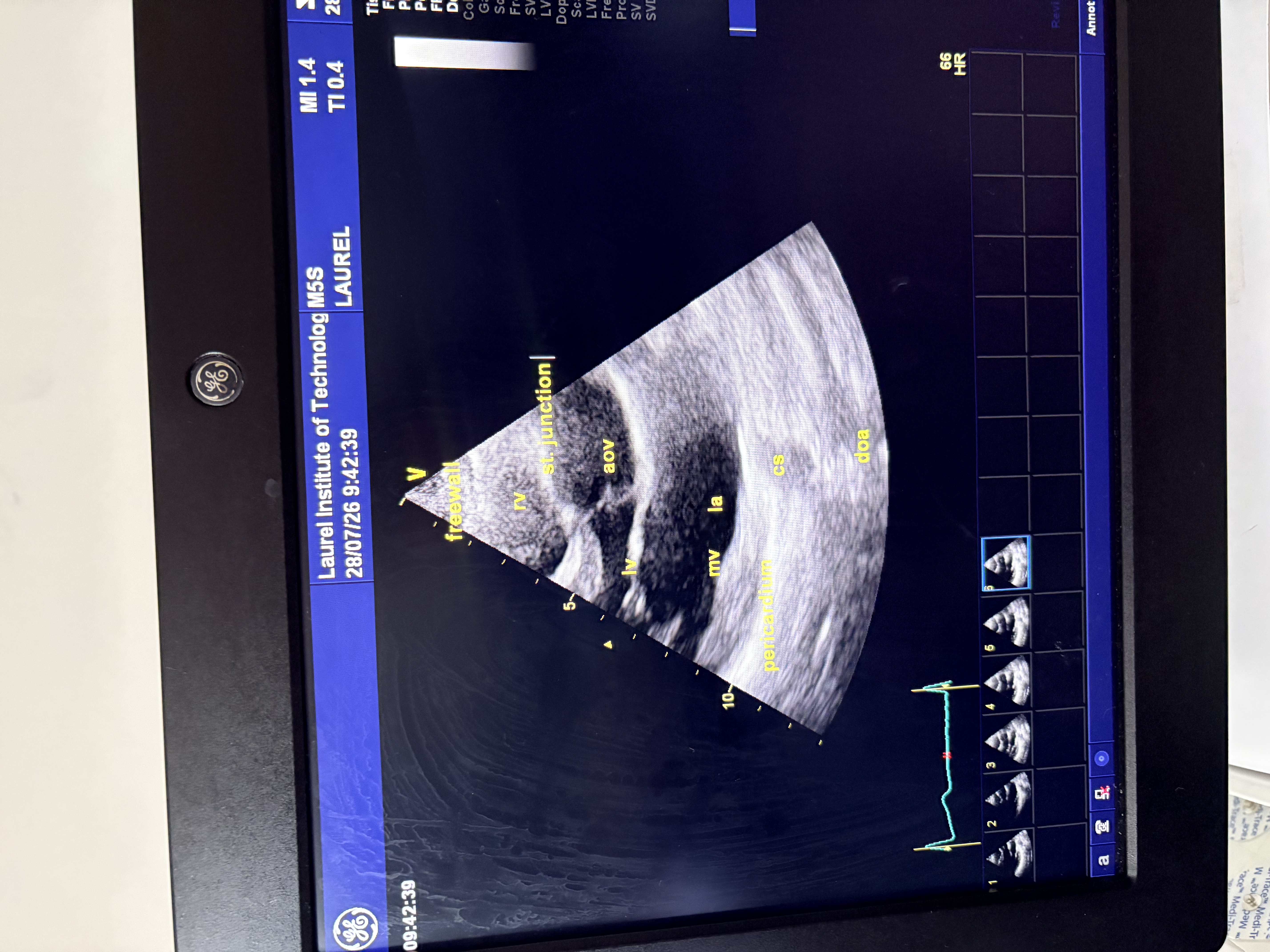
What is the cardiac antomy visible in PLAX?
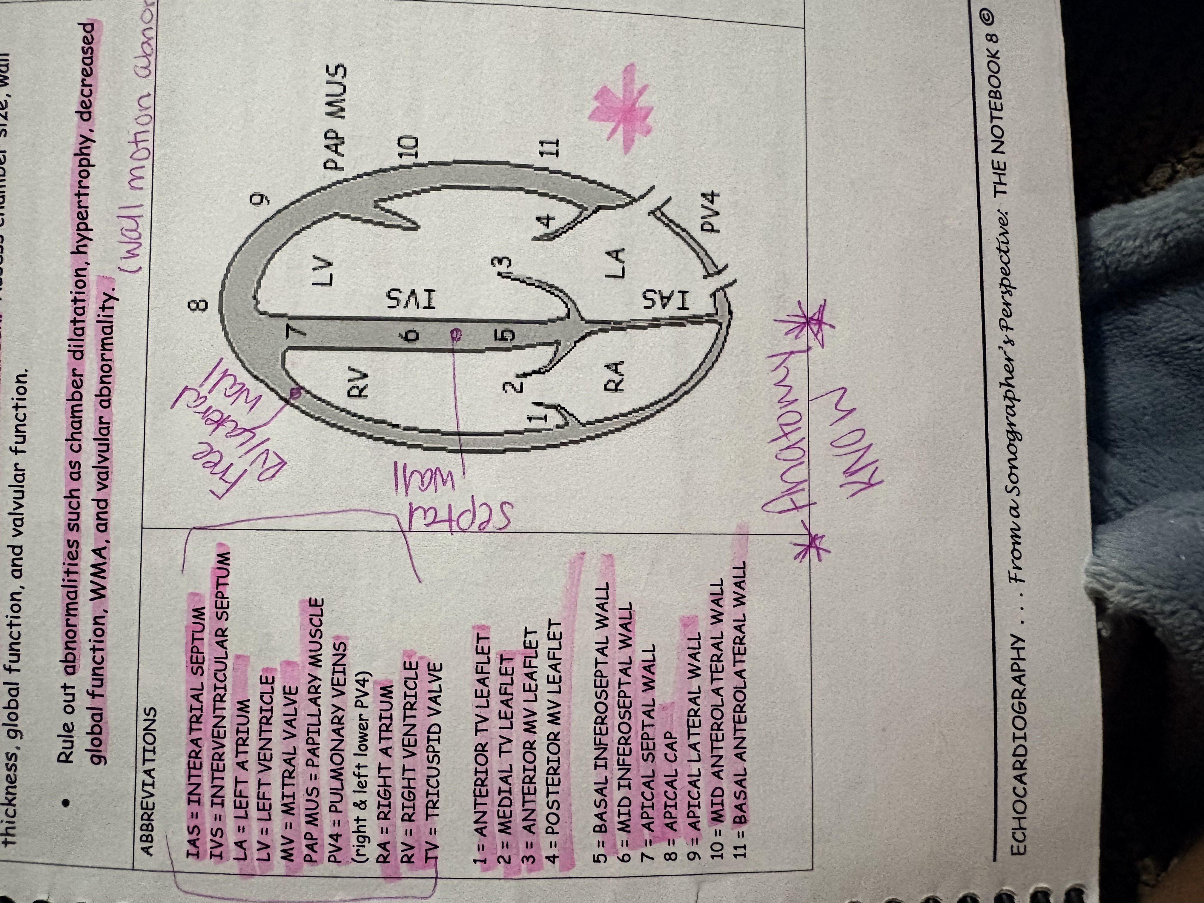
What is the cardiac anatomy visble in a 4C?
How should the transducer be placed for the Apical Four Chamber (4C)
The transducer should be placed at the apex of the heart, with the indicator directed towards the patient's right shoulder.
What is the proper protocol for assesing 4C?
Initially, increase the depth to acquire the entire cardiac structure and surroundings. This is to rule out extracardiac abnormalities such as pericardial effusion, plueral effusion and mass. Then decrease the depth, so the heart fills the screen.
From here, we rule out abnormalities such as chamber dilation, hypertrophy, decreased global function, WMA (wall motion abnormality) and valvular abnormality.
In 4C the lateral and septal RV walls are thin and squeeze concentrically during systole.
What is TAPSE? What is it used for?
Tricuspid Annular Plane Systolic Excursion. Used to provide a simple but good estimate of the RV systolic function.
Note, anything less than 1.7 cm or 17 mm indicates a decreased RVFX (right ventricular systolic function)
(meausre from the bottom of the hill to the top)
What is a moderator band? Where is it found?
A fibrous band that is a normal embryonic remnant only visible in the RV.
RV (Right Ventricle) vs LV (Left Ventricle)
The RV is smaller. Has the moderator band and more trabeculations. Size between the two should always be compared and if the RV is showing larger, it should be reported. Report any ICD (implantable cardiovertor defibrillator/pacemaker wires
When is the RV & LV measured and at its fullest?
End diastole
How many TV (tricuspid valve) leaflets can you see in 4C? What are they? What is ruled out from this view?
2 leaflets shown are the anterior and medial. They are thin and pliable with unrestricted opening, Posterior is not visible.
Rule out TVP (tricuspid valve prolapse), dilated TV annulus etc
RA (Right Atrium)
The smallest of the 4 chambers and acts as a reservoir for systemic venous return and assists in RV filling.
We view the RA to rule out RA enlargement, clot/mass and dilated CS (coronory sinus)
When is the RA and LA measured and at its fullest?
End systole
The LV in 4C View
Lateral and septal LV walls are thicker than RV walls and squeeze concentrically during systole. Here we can rule out LVH (left ventricular hypertyrophy)
LV is the largest of the 4 chambers and is longer and less trabeculated than the RV. We can also rule out LV dilation, decreased LVFX
Biplane Method of Disk Summation
Also known as Simpsons Biplane.
Associated with volume of blood. The endocardium is traced in 4C and 2C to correctly calculate and measure LVEF (left ventricular ejection fraction)
What is the equation to calculate LVEF?
(Left Ventricular Ejection Fraction)
EDV-ESV
————— x100
EDV
What is considered a normal LVEF% (Left Ventricular Ejection Fraction)
53-73% (20+ years)
AMVL (anterior) and PMVL (posterior) in 4C view
Thin and pliable with unrestricted opening. The AMVL is the more mobile of the two.
Viewed to rule out abnormalities such as MVP, MAC and dilated annulus.T
The LA in 4C View
The most posterior chamber. Serves as a reservoir that recieves pulmonary venous return. It is a passage way to the LV, and a contractile pump.
It is larger than the RA, but smaller than both ventricles.
Viewed to rule out LAE, clots, masses, IAS bowing and IAS aneurysm
What is the significance of the IAS bowing?
If the IAS bows towards the left then this means RAP (right atrial pressure) is increased.
If the IAS bows toward the right, this means LAP (left atrial pressure) is increased.
Why do we aquire CFD (Color flow doppler) in 4C view?
Used to view RA, TV, and RV to rule out TS (tricsupid stenosis) and TR (tricuspid regurgitation)
View the IAS and IVS to rule out ASD (atrial septal defect) and VSD (ventral septal defect)
View the LA, MV, and LV to rule out MS and MR
Why do we aquire CWD (Continuous wave doppler) in 4C view?
To asses the MV. A normal range should be 0.6-1.3 m/s
An increased MV velocity may indicate MS (mitral stenosis)
Important Facts for 4C
Blood is going from the atrias to the ventricles (towards the probe). Majority of the blood should be red.
Normal flow shows above the baseline. Anything below the baseline would be regurgitation.
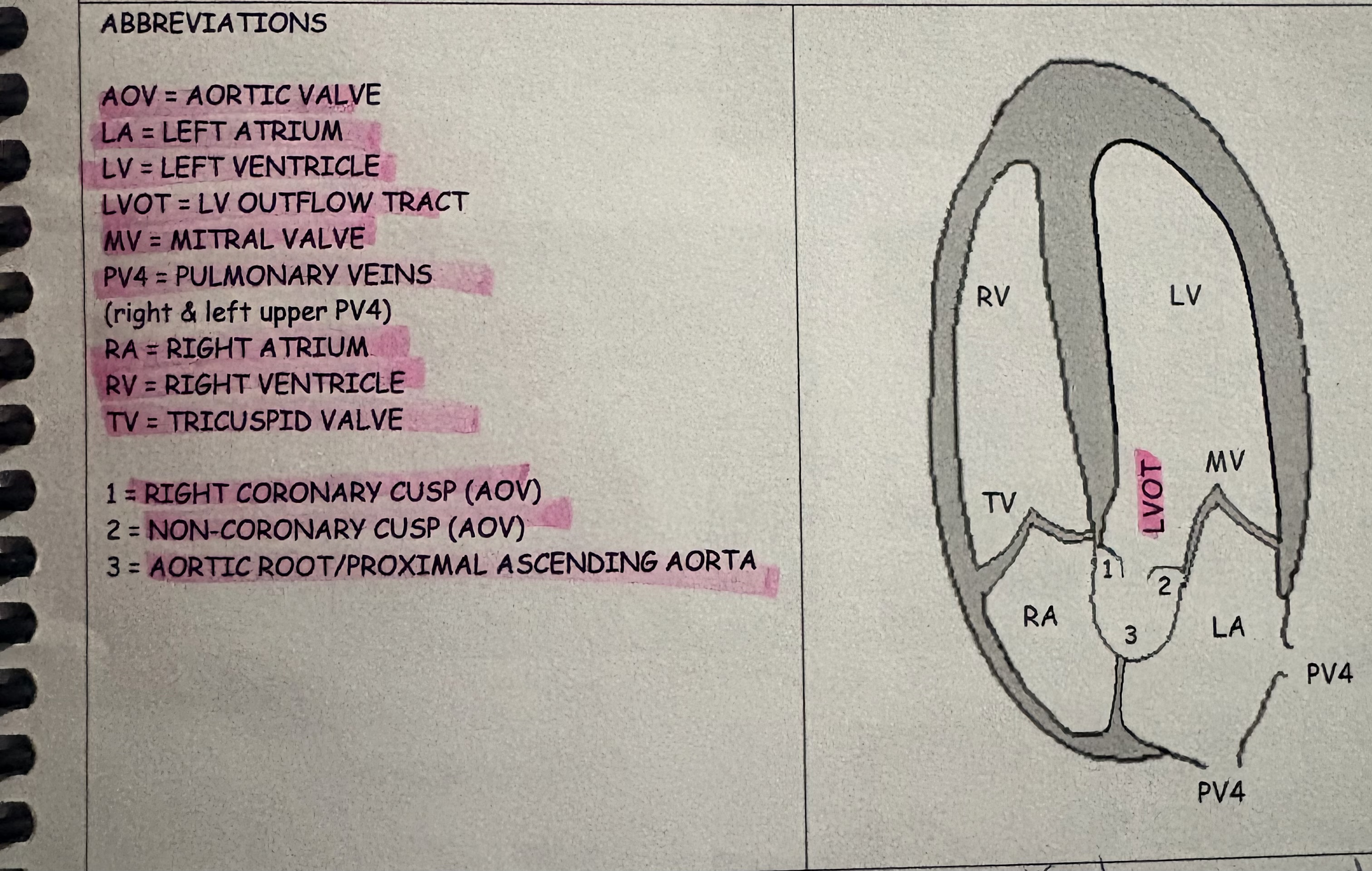
What is the cardiac anatomy visible in 5C?
How should the transducer be placed to view 5C?
From the 4C, angle more anterior. Tail down
AOV in 5C view
RCC and NCC of the AOV are thin, delicate and pliable with unrestricted opening.
Viewed to rule out dilate AOV annulus and flail/prolapse/thickening/calcification of AOV cusps.
LVOT in 5C
Unobstructed and allows laminar or smooth flow from the LV out to the AO. MV apparatus does not interfere with systolic flow through the LVOT. We can rule out SAM (systolic anterior motion) of the MV apparatus without vs with LVOTO (left ventricle outflow tract obstruction)
Important Facts 5C
Normal flow shows below the baseline. The primary color of blood would be blue.
Anything above the baseline would be regurgitation.
Why do we aquire CFD in a 5C view?
To view the LA, MV, LV, LVOT and AOV to rule out abnormalities such as MS, MR, VSD, LVOTO, AS, and AR
Why do we aquire CW of the AOV in a 5C view?
To assess direction and velocity of blood flow. A normal AOV range should be 1 - 1.7 m/s.
Why do we aquire PWD (pulse wave doppler) in a 5C view?
To asses velocity, sound and shape of wave form. A normal LVOR range should be. 0.8 - 1 m/s
How should the transducer be placed for 2C?
Rotated 60-90 degrees counter clockwise from the 4C. Likely toward the patients right ear/shoulder.
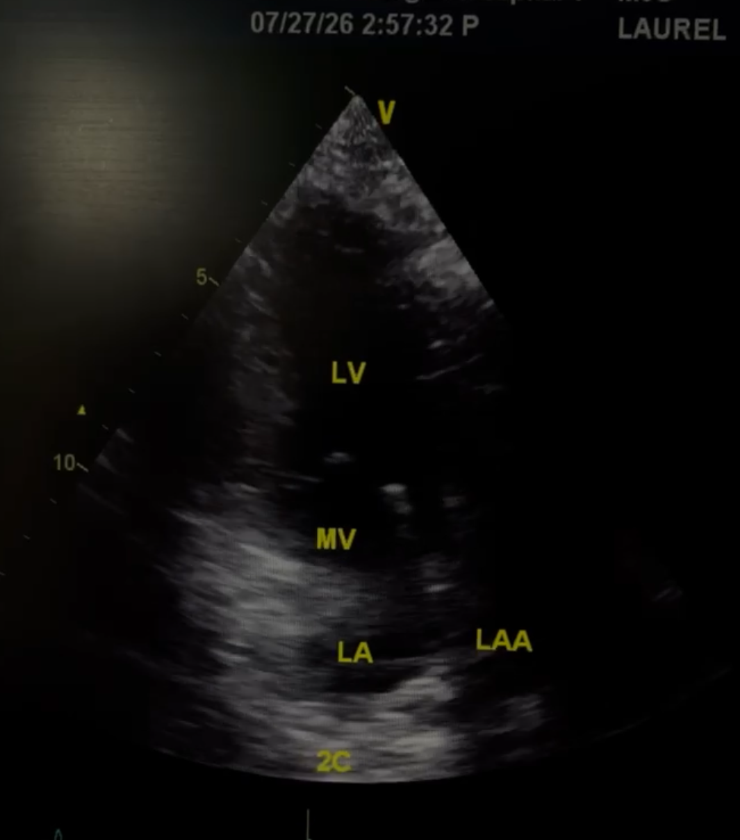
What is the cardiac anatomy for 2C?
The LA in 2C view
Smaller than the LV, it is the most posterior chamber, a reservoir that recieves pulmonary venous return, a passageway to the LV and a contractile pump.
Rule out LA thrombus, LAE, mass
LAA in 2C
(Left Atrial Appendage) a normal embryonic remnanr of the LA. It is long, tubular, hooked appendage that lies within the pericardium. It acts as a decompression chamber that helps decrease LAP whenever elevated or during normal LV systole.
Rule out LAA thrombus
2C View and the MV
Leaflets are thin and pliable, Viewed to rule out abnormalities such as MVP, MAC, dilated annulus etc.
Walls in 2C
Inferior and anterior LV walls squeeze concentrically during systole.
Viewed to rule out LVH (Left Ventricular Hypertrophy)
LV in 2C view
The LV is larger than the LA. Viewed to rule abnormalities such as LV dilatation, decreased LVFX (left ventricular function) WMA, and clot/mass.
LAX of the DOA
A seperate structure, posterior (below) to the LA.
Viewed to rule out AO dilatation, plaque, aneurysm and dissection
Why do we aquire CFD in a 2C view?
To rule out abnormal flow such as MS and MR
Used to asses the LA, MV, LV and DOA
How should the transducer be placed to view 3C?
Rotated 30-60 degrees counter clockwise from the 2C. Likely towards the patients right shoulder
What is the cardiac anatomy for a 3C view?
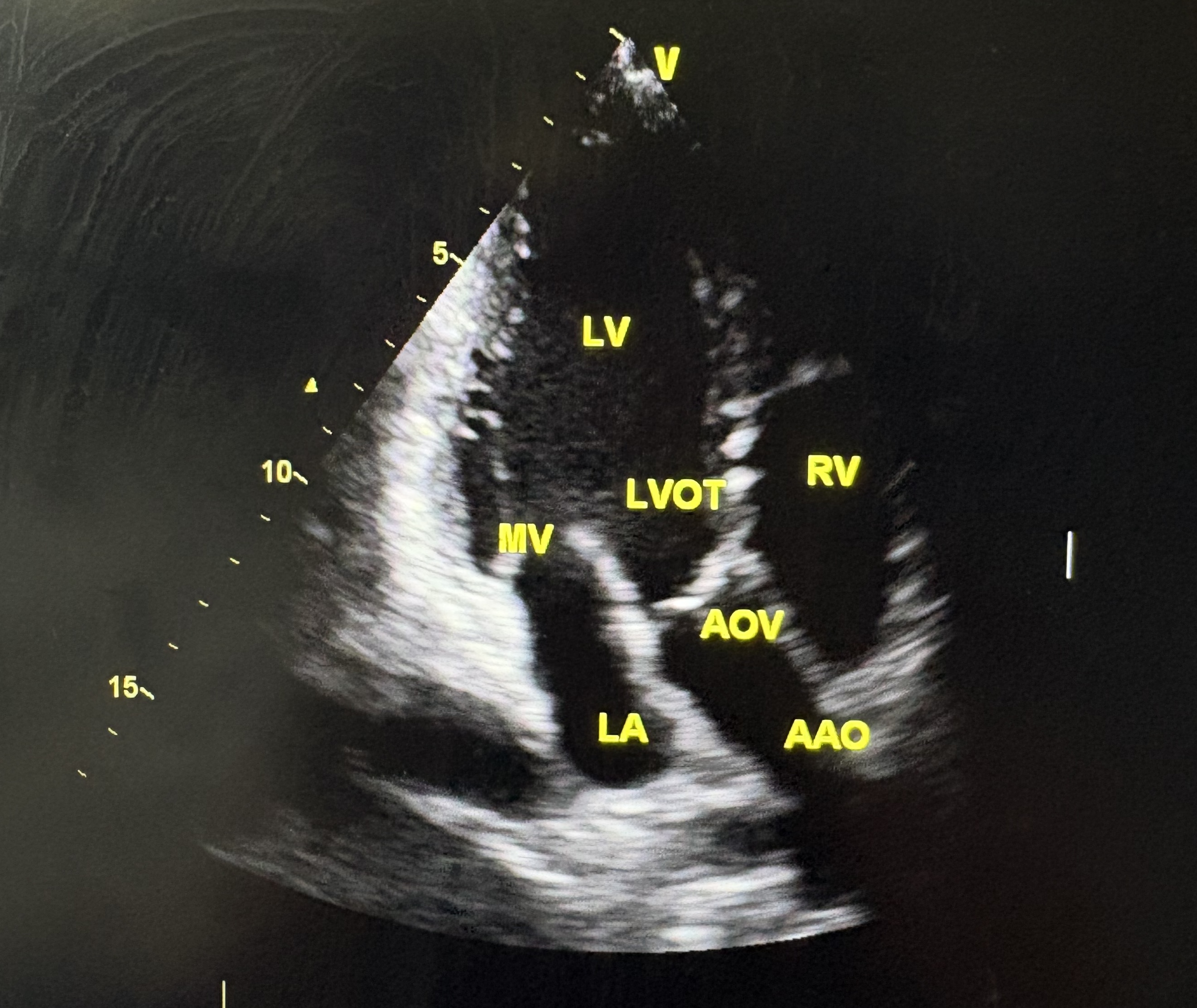
Why do we view a 3C?
To rule out chamber dilatation, decreased LVFX, WMA, LVH, valvular abnormality and AAO aneurysm.
Why do we aquire CFD in a 3C?
To rule out abnormal flow such as MS, MR, LVOTO, VSD, AS, and AR.
How should your transducer be placed for 3C RVIT?
More medial and inferior from the 3C. Often acquired from an off-axis 3C.
What is the cardiac anatomy in a 3C RVIT?
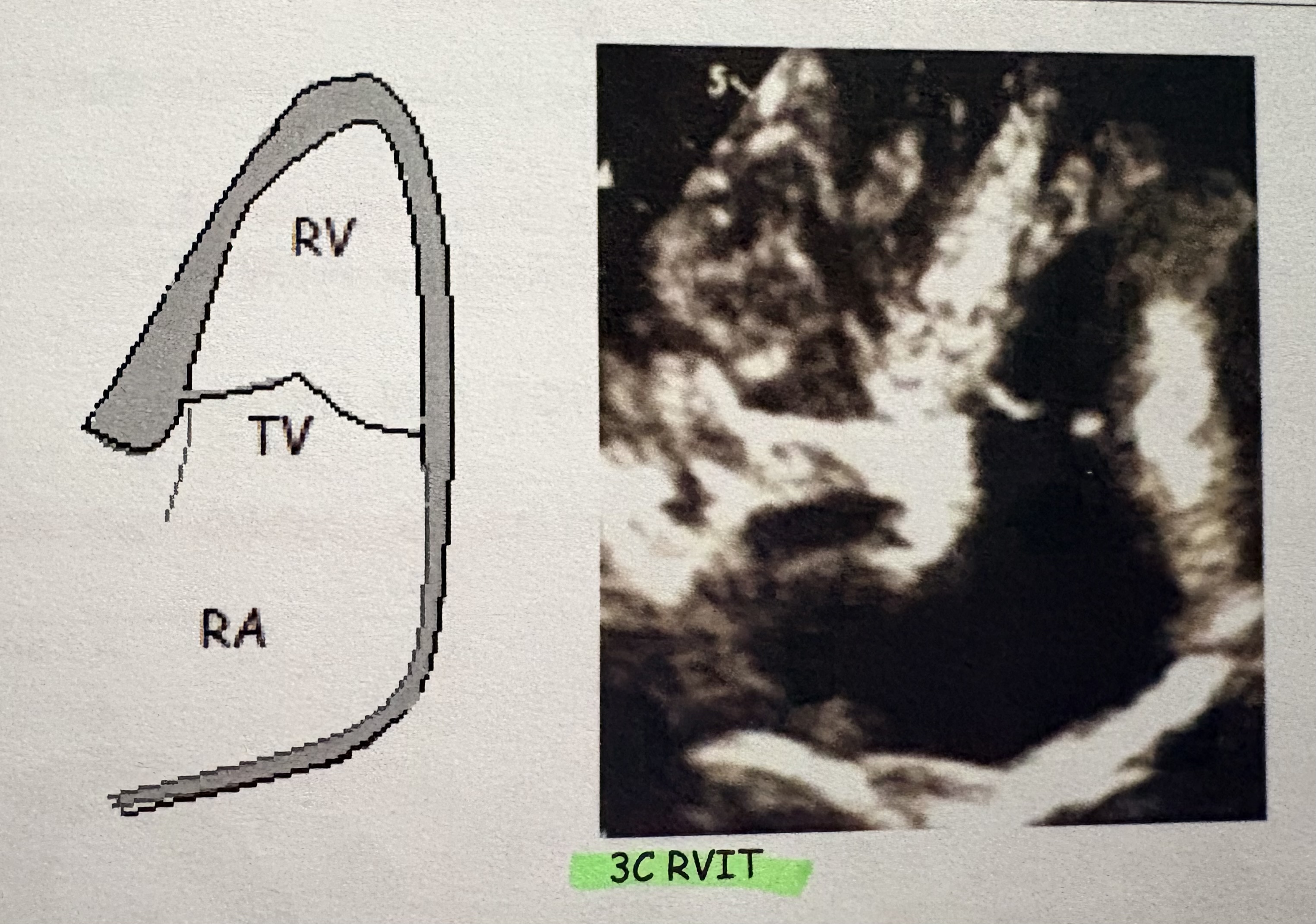
3C RVIT Purpose
To asses 2D, CFD and spectral doppler. Useful for absolute peak TRV and RVSP calculation.
CWD in 3C RVIT
Above the baseline is considered a normal flow. Majority of the blood should be red.
Anything below the baseline would be regurgitation.