BIO 201 Lecture Exam 1
 Call Kai
 Learn
Learn Practice Test
 Spaced Repetition
 Match
Match Flashcards
 Knowt Play
1/156

There's no tags or description
Looks like no tags are added yet.
Name | Mastery | Learn | Test | Matching | Spaced | Call with Kai | Chat |
|---|
No analytics yet
Send a link to your students to track their progress
157 Terms
Functions of the Endocrine System
Regulates growth, development, and metabolism
Maintain homeostasis of blood composition and volume
Controls digestive processes
Controls innume response
Controls reproductive processes
Similarities of Nervous & Endocrine Systems
Both maintain homeostasis
Both communicate via chemicals that bind to receptors
Differences of Nervous & Endocrine Systems
Nervous
Uses neurotransmitters released at synapses
Effects are almost immediate & short-lasting
Endocrine
Uses hormones released into the bloodstream
Effects are in seconds to days & longer-lasting
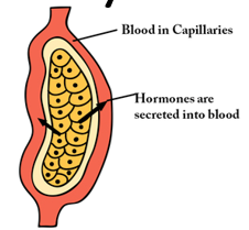
Endocrine Glands
Ductless - Internal Secretion
Hormones act only on target cells with receptors for them
Connective tissue framework
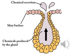
Exocrine Glands
Secrete into ducts or tubes that lead to a body surface
Deliver their products directly to a specific site
Endocrine Organs
glands with a solely endocrine function
Examples of Endocrine Organs
Pituitary Gland
Pineal Gland
Thyroid Gland
Parathyroid Glands
Adrenal Glands
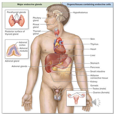
Organs with Endocrine Cell Clusters
Function is not solely endocrine; include:
Hypothalamus
Skin
Thymus
Heart
Liver
Stomach
Pancreas
Small Intestine
Adipose Connective Tissue
Kidneys
Gonads
Hormones
chemical messengers secreted by endocrine glands that regulate functions of other cells.
Have widespread effects throughout the body
Continue circulating the blood until taken up by a target cell or broken down and deactivated
Factors Affecting Levels of Circulating Hormones
Hormone Release
Positively correlated with blood concentration
Hormone Elimination
Negatively correlated with blood concentration
Occurs in multiple ways
Half-Life of Hormone
Water-Soluble = shorter half-life: ~few minutes
Lipid-Soluble = longer half-life b/c carrier proteins protect them
Target Cells
contain specific protein receptors to which specific hormones bind
Receptors
3-D shapes highly specific for hormone or chemical messenger they bind
Can be embedded in plasma membrane
Or
Reside in cytosol or nucleus of target cell
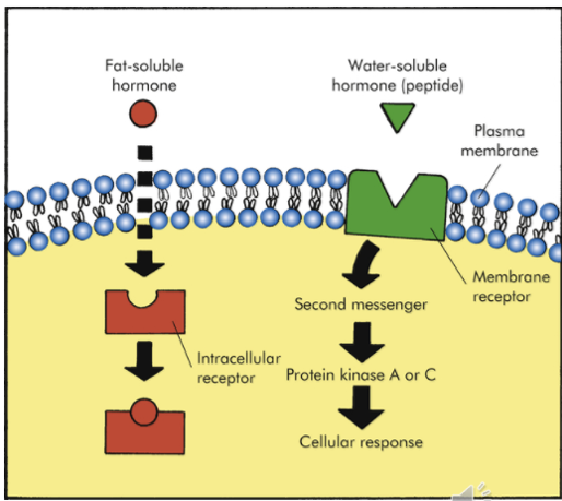
Up-Regulation
process by which target cells produce a greater number of receptors in response to a temporary increase in hormone level of blood
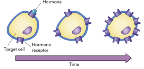
Down-Regulation
process by which target cells decrease number of receptors in response to prolonged exposure to a high level of hormone in the blood
Classes of Hormones
Steroid Hormones
Biogenic Amines
Proteins
Steroid Hormones
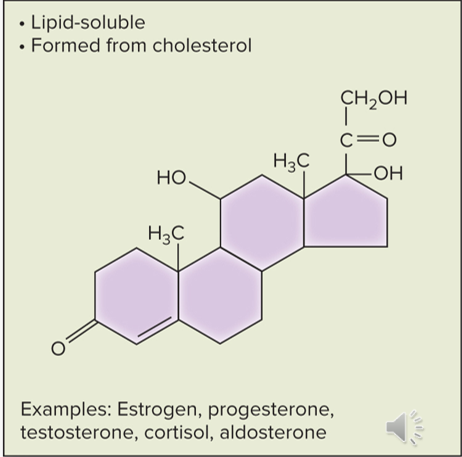
class of hormones that are lipid-soluble and derived from cholesterol
Types of Steroid Hormones
Gonadal Steroids (estrogen, progesterone, testosterone)
Adrenal Cortex Steroids (cortisol)
Calictrol (sometimes classified in this group; more accurately a sterol though)
Biogenic Amines
class of hormones that are modified amino acids and water-soluble
Exception: thyroid hormone is nonpolar and lipid-soluble due to a pair of tyrosines
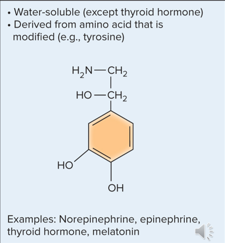
Types of Biogenic Amines
Catecholamines (epinephrine, norepinephrine)
Thyroid Hormone
Melatonin
Proteins
class of most hormones which are water-soluble chains of amino acids; most hormones are a part of this class.
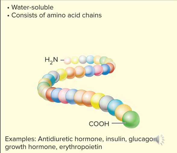
Types of Protein Hormones
Antidiuretic Hormone (ADH)
Insulin
Glucagon
Growth Hormone (GH)
Erythropoietin
Types of cell Signaling
Endocrine
Paracrine
Autocrine
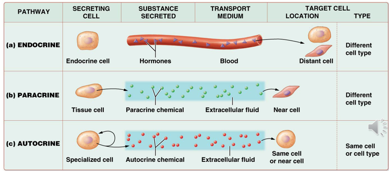
Endocrine Cell Signaling
hormones secreted into blood and taken up by target tissue of a different cell type than the signaling cell

Paracrine Cell Signaling
chemicals secreted into the extracellular space to affect nearby, but different types of cells than the signaling cell

Autocrine Cell Signaling
chemicals secreted into interstitial fluid which elicits effects from the same cell or same cell type as the signaling cell

Local Hormones
hormones not secreted into the bloodstream, but instead into surrounding tissue
Example: Eicosanoids
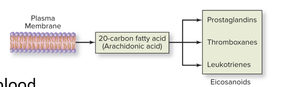
Eicosanoids
local hormone formed from fatty acids within the phospholipid bilayer through an enzymatic cascade
Example: prostaglandins
Function in diverse physiological and pathological systems
Mounting or inhibiting inflammation
Allergy
Fever & other immune responses
Regulating the abortion of pregnancy & normal childbirth
Contribute to the perception of pain
Regulate cell growth
Control blood pressure
Modulate the regional flow of blood to tissues
Prostaglandins
example of an eicosanoid that stimulates pain and inflammatory response
Aspirin and Other Nonsteroidal Anti-Inflammatory Drugs
can be used to block prostaglandin formation reliving pain and inflammation
Ways Hormones are Eliminated from Bloodstream
Enzymatic degradation in liver cells
Uptake by target cells
Excretion by kidneys
Half-Life
time necessary to reduce a hormone’s concentration to half of its original level
Example - testosterone = 12 days
Lipid-Soluble Hormones
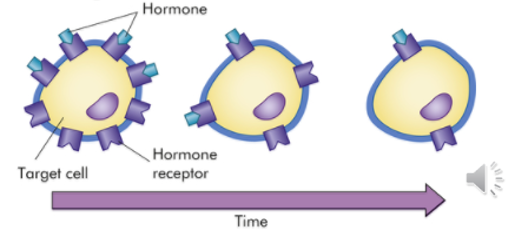
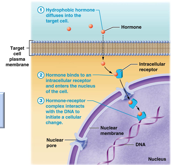
small, nonpolar, and hydrophobic hormones that can diffuse across target cell membranes to reach receptors in the cytosol or nucleus
Hormone-Receptor Complex
complex formed from the binding of a lipid-soluble hormone to a receptor. Complex eventually binds to DNA causing changes in transcription & translation which may have structural or metabolic effects on the cell
Hormone-Response Element (HRE)
point on DNA strand that a hormone-receptor complex is able to bind to influence transcription & translation
Water-Soluble Hormones
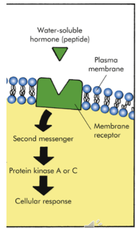
hydrophilic, polar hormones that cannot diffuse across membranes and therefore must attach to receptors in the membrane of target cell to induce a signal transduction pathway to induce change.
First Messenger
hormone that initiates a signal transduction pathway by binding
G-Protein
internal membrane protein that binds a guanine nucleotide; binding of the first messenger turns GDP into GTP, an active form which activates membrane enzymes to catalyze the formation of a second messenger
Second Messenger
a chemical made by membrane enzymes in a signal transduction pathway which modifys cellular activity in someway
Possible Effects of Hormones
Stimulating secretion from endocrine or exocrine cell
Activating or inhibiting enzymes
Stimulating or inhibitng cell division (mitosis or meiosis)
Opening or closing ion channels in plasma membrane —> alters membrane potential (electrical charge)
Activating or inhibiting transcription of genes that code for RNA or proteins (gene expression)
Types of Endocrine Stimulation
Hormonal Stimulation
Humoral Stimulation
Nervous System Stimulation
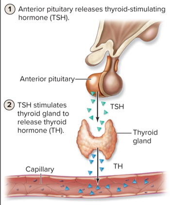
Hormonal Stimulation
release of one hormone causes the release of a different hormone
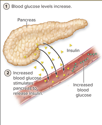
Humoral Stimulation
changes in nutrient or ion levels of the blood causes the release of hormones
Nervous System Stimulation
stimulation of the nervous system causes the release of hormones
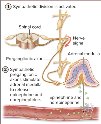
Regulation of Hormone Secretion
Stimulus - regulated physiological variable deviates from its normal range
Receptor - detects the deviation of the variable
Control Center - often the endocrine cell; increases or decreases its secretion of a particular hormone
Effector/Response - hormone triggers a response in its target cells that moves conditions toward the normal range
Homeostatis Range - as the variable returns to normal, feedback to the control center decreases the effector response
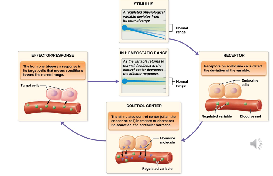
Tropic Hormones
hormones that act on other glands and regulate their hormone secretion
Non-Tropic Hormones
do not cause hormone secretion; instead, control bodily function directly by stimulating or inhibiting target cells
Trophic Hormones
hormones which induce growth in target cells
Example of Trophic Hormone
TSH is both tropic and trophic; causes release of thyroid hormone from the thyroid gland and also causes growth of the thyroid gland. Reason a goiter is a symptom of thyroid issues.
Hypothalamus
Part of the brain, contains neurons
One of the largest endocrine organs
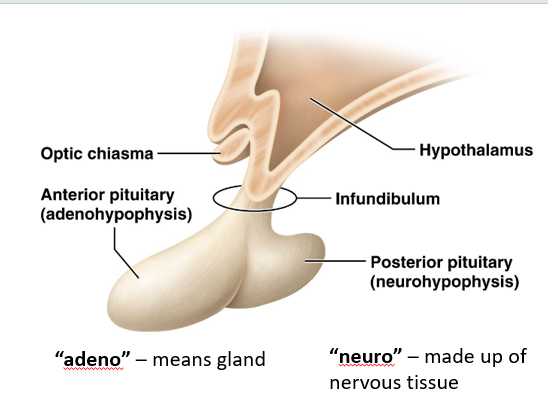
Closely associated with the pituitary gland
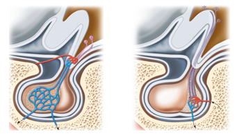
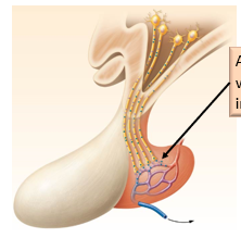
Some neurons enter the posterior pituitary and secrete hormones
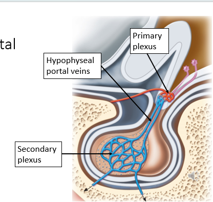
Other neurons secrete hormones into a network of blood vessels that travel into the anterior pituitary, controlling its secretion of hormones
Homeostatic Functions:
Thirst
Hunger
Fluid Balance
Body Temperature
Sleep/Wake Cycle
Certain Reproductive Functions
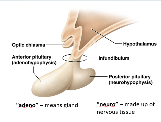
Anterior Pituitary (Adenohypophysis)
“Adeno” meaning gland
Receives blood flow and hormones
Releases its own hormones
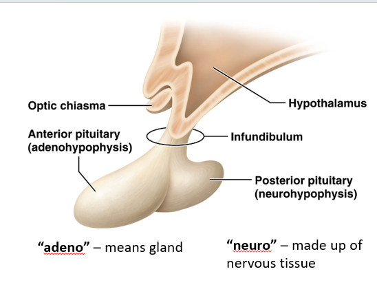
Posterior Pituitary (Neurohypophysis)
“Neuro” since it is made up of nervous tissue
Does not produce or release any of its own hormones; instead hormones of the hypothalamus are released into this area
Hypothalamohypophyseal Portal System
network of blood vessels connecting the hypothalamus and anterior pituitary. Consists of:
Primary Plexus
Hypophyseal Portal Veins
Secondary Plexus
Hormones of the Posterior Pituitary
Not made; just released into this part
Two neurohormones stored in synaptic vesicles in axon terminals
Antidiuretic Hormone (ADH)
Oxytocin
Antidiuretic Hormone (ADH)
Neurohormone made by the hypothalamus and released into the posterior pituitary
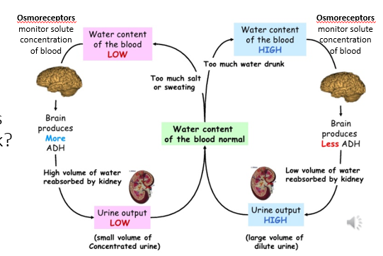
Osmoreceptors send a signal to the brain when the water content of the blood is low or high
Low Water Content —> Increase in ADH —> High volume of water reabsorbed by kidneys —> low urine output & very concentrated urine
High Water Content —> Decrease in ADH —> Low volume of water reabsorbed by kidneys —> high urine output & dilute urine
Diabetes Insipidus
Condition caused by an abnormal lack of ADH secretion or activity
Symptoms
Frequent Urination
Extreme Thirst
Dehydration
Treatment
Synthetic ADH (if secretion is the issue)
Antidiuretics or low-sodium diet (if kidneys are the issue)
Oxytocin
Neurohormone made by the hypothalamus but released into the posterior pituitary
Functions
Muscle contraction of the uterine walls during childbirth
Milk ejection during lactation (Milk let-down reflex)
Target cells
Mammary glands of breast tissue
Smooth muscle of the uterus
Milk Let-Down Reflex
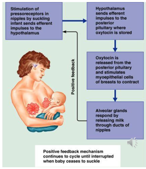
reflex in nursing mothers which exhibits a positive feedback loop
Stimulation of pressoreceptors in nipples by suckling sends impulses to hypothalamus
Hypothalamus sends impulses to posterior pituitary to release oxytocin
Stimulated myoepithelial cells of breasts to contract
Alveolar glands respond by releasing milk through the ducts of the nipple
Continues until the infant stops suckling
Releasing Hormone
hormones produced by hypothalamus capable of accelerating secretion of given hormone by anterior pituitary
Stimulating Hormone
a hormone produced by the anterior pituitary that stimulates another gland to produce another hormone
Inhibiting Hormone
a hormone that inhibits the secretion of another hormone.
Example: Prolactin-Inhibiting Hormone (PIH) of the hypothalamus
Releasing Hormones of the Hypothalamus
Thyrotropin-Releasing Hormone (TRH)
Prolactin-Releasing Hormone (PRH)
Gonadotropin-Releasing Hormone (GnRH)
Corticotropin-Releasing Hormone (CRH)
Growth Hormone-Releasing Hormone (GHRH)
Inhibiting Hormones of the Hypothalamus
Prolactin-Inhibiting Hormone (PIH)
Growth Hormone-Inhibiting Hormone (GHIH)
Tropic Hormones of Anterior Pituitary
Thyroid-Stimulating Hormone (TSH)
Prolactin (PRL)
Follicle-Stimulating Hormone (FSH) & Luteinizing Hormone (LH)
Growth Hormone (GH)
Adenocorticotropic Hormone (ACTH)
Thyrotropin-Releasing Hormone (TRH)
releasing hormone of the hypothalamus that induces the anterior pituitary to release TSH
Prolactin-Releasing Hormone (PRH)
releasing hormone of the hypothalamus that stimulates the anterior pituitary to release PRL
Gonadotropin-Releasing Hormone (GnRH)
releasing hormone of the hypothalamus that stimulates the anterior pituitary to release FSH & LH
Corticotropin-Releasing Hormone
releasing hormone of the hypothalamus that stimulates the anterior pituitary to release ACTH.
Growth Hormone-Releasing Hormone (GHRH)
releasing hormone of the hypothalamus that stimulates the anterior pituitary to release GH.
Amount released depends on person’s age (decreases with age), time of day (increasing during sleeping hours), nutrient levels, stress, and excercise
Prolactin-Inhibiting Hormone (PIH)
inhibiting hormone of the hypothalamus that represses the anterior pituitary from releasing PRL
Growth Hormone-Inhibiting Hormone (GHIH)
inhibiting hormone of the hypothalamus that represses the release of GH from the anterior pituitary
Thyroid-Stimulating Hormone (TSH)
hormone of the anterior pituitary that stimulates growth of thyroid gland and release of thyroid hormone (TH)
Prolactin (PRL)
hormone of the anterior pituitary that stimulates growth of the mammary glands and increases milk production
Follicle-Stimulating Hormone (FSH) & Luteinizing Hormone (LH)
hormones of the anterior pituitary that act on the gonads to stimulate the development of their respective gametes and release sex hormones
Growth Hormone (GH)
hormone of the anterior pituitary that stimulates release of IGF’s from the liver, which synergistically act on all body tissues, especially cartilage bone, muscle, and adipose tissue to stimulate growth
Adenocorticotropic Hormone (ACTH)
hormone of the anterior pituitary that stimulates the adrenal cortex to release glucocorticoids (such as cortisol)
Gigantism
disorder caused by too much growth hormone which causes big people
Acromegaly
growth hormone disorder that affect flat bones of the face, flattening and enlarging the face
Pituitary Dwarfism
lack of growth hormone which causes little people
Anatomy of the Thyroid Gland
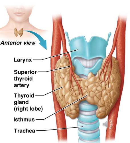
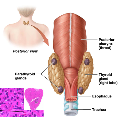
Left & Right Lobes connected at the midline by a narrow isthmus
Highly vascularized
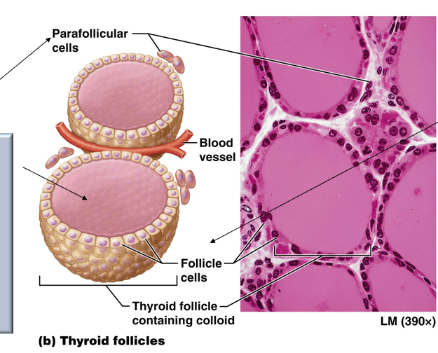
Composed of microscopic follicles
Cellular Structure
Protein-rich, gelatinous colloid in the middle, surrounded by follicle cells
Parafollicular cells around colloid & follicle cells
Parafollicular Cells
cells of the thyroid that release calcitonin hormone leading to a colloid
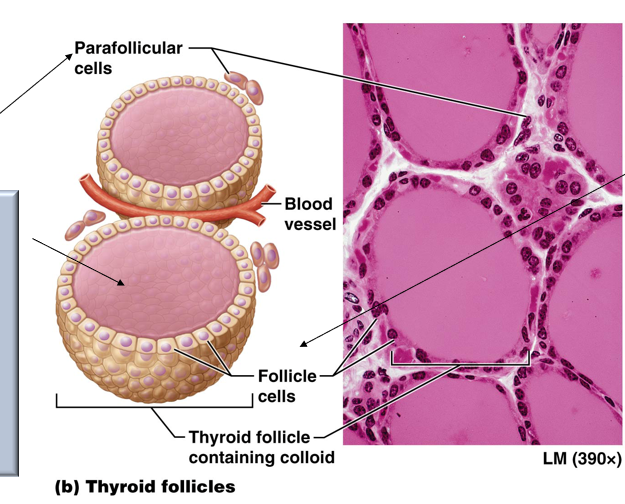
Follicle Cells
cuboidal epithelial cells that surround colloid and produce and release TH
Parathyroid Glands
Typically 3 to 5 separate glands on posterior surface of the thyroid
Secrete parathyroid hormone (PTH) from chief cells
Regulate calcium ion levels
Important for bone health; helps prevent conditions such as osteoporosis
Chemical Structure of Thyroid Hormone
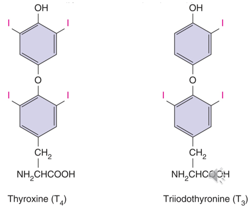
Amino acid core bound to 3 (triiodothyroxine, T3) or 4 (thyroxine, T4) iodine atoms
T4 commonly converted to T3 to target issues. (Both physiologically active, but T3 moreso)
T3 and T4 are more hydrophic than other monoamines, allowing them to enter the target cell’s nucleus
Functions of Thyroid Hormone
Regulation of Metabolic Rate & Thermoregulation
Sets basal metabolic rate by increasing the rate at which cells carry out metabolic reactions
Heat generated through metabolism: critical for core body temp homeostasis
Cold stimulates an increase in metabolic rate, and heat decreases metabolic rate
Promotion of Growth & Development
Required for normal bone growth, muscle growth, and nervous system development
Deficiency leads to birth defects
Synergism with Sympathetic Nervous System
Up-regulates production of receptors for sympathetic neurotransmitters
Helps regulate BP, heart rate, and other sympathetic activities
Regulation of Thyroid Hormones
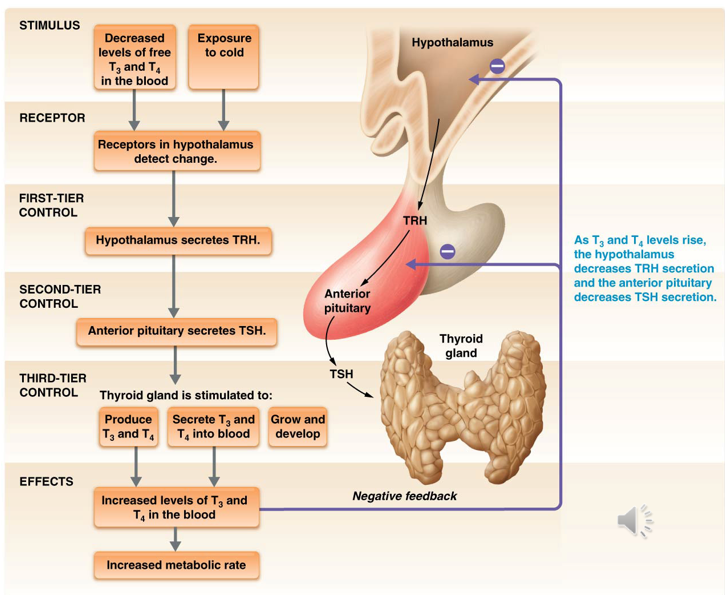
Stimulus: decreased levels of free T3 or T4 or exposure to cold
Receptors in the hypothalamus detect change
First-Tier Control: Hypothalamus releases TRH
Second-Tier Control: Anterior pituitary releases TSH
Third-Tier Control: Thyroid gland produces & secretes T3 and T4 + grows & develops
Effects
Increased levels of T3 & T4 in blood = increased metabolic rate
Parathyroid Hormone (PTH)
Secreted by chief cells of the parathyroid gland
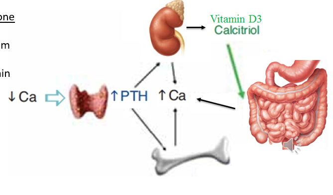
Secreted in response to declining calcium ion levels
Effects:
Increases release of calcium ions from bone by stimulating osteoclast activity
Breaks down bone to free up calcium ions
Acts on kidneys to convert inactive vitamin D into its active form, calcitrol (Vitamin D3)
Increases absorption of calcium from diet
Increases reabsorption of calcium ions in kidneys
Calcitonin
Produced and secreted by parafollicular cells
Released when calcium levels increase above normal (hypercalcemia)
Primary target is osteoclast cells
Inhibits their activity
Allows osteoblast activity
Structure of Adrenal Glands
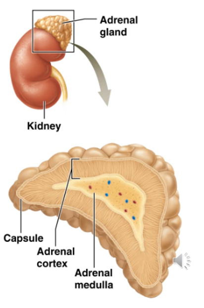
Located on top of the kidneys
Roughly pyramid-shaped
Produces two types of hormones
Steroid Hormones (Aldosterone, Cortisol, & Sex Hormones)
Catecholamines (Epinephrine & Norepinephrine)
Divided into two sections
Adrenal Cortex
Adrenal Medulla
Adrenal Cortex
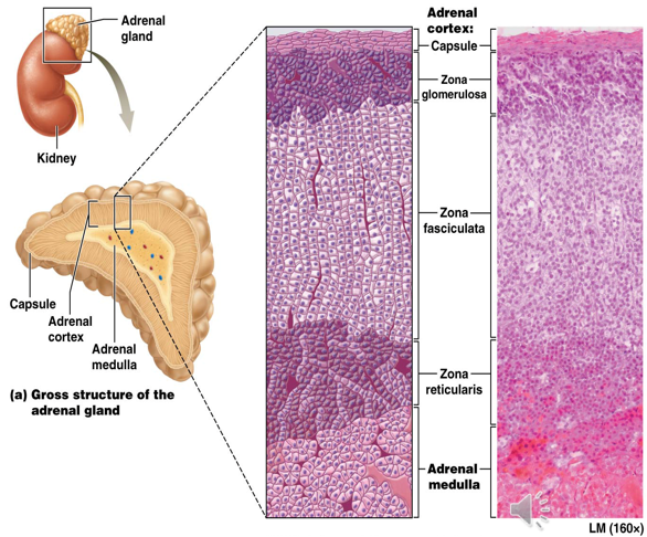
Subdivided into 3 zones
Zone Glomerulosa (Outermost)
Zone Fasciculata (Middle Zone)
Zone Reticularis (Innermost Zone)
Secretes mineralcorticoids, glucocorticoids, & androgenic steroids
Zone Glomerulosa
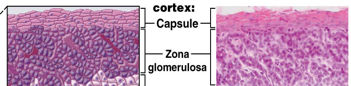
Outermost Zone of Adrenal Cortex
Densely packed cells
Secretes mineralocorticoid hormones (ex. aldosterone)
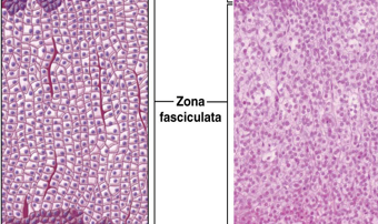
Zone Fasciculata
Middle zone of adrenal cortex
Cells stacked in columns
Secretes glucocorticoids (ex. cortisol)
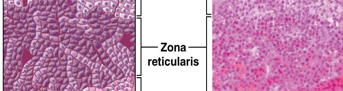
Zone Reticularis
Innermost zone of adrenal cortex
Thin layer of cells; arranged loosely in clusters
Secrete androgenic steroids (gonadocorticoids)
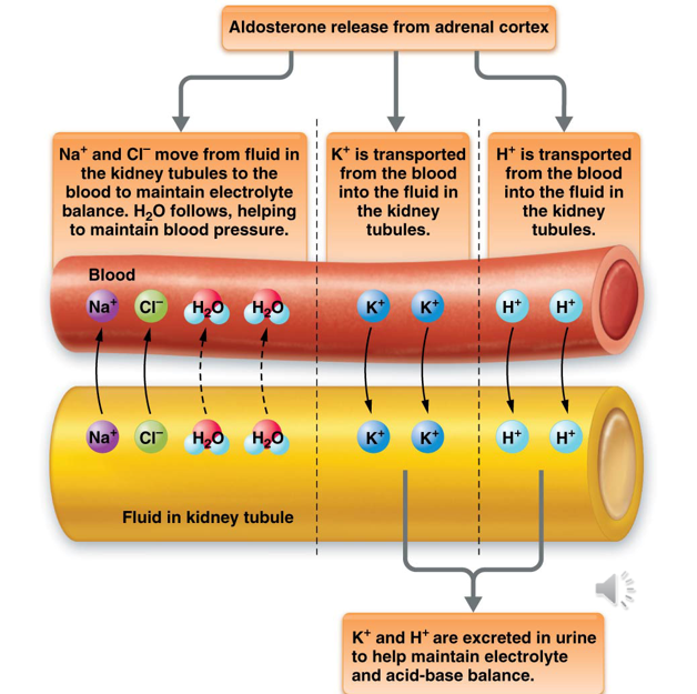
Aldosterone
Regulates the concentration of certain electrolytes (Na+ & K+) in body fluids, blood pH, and blood pressure
Aldosterone released
Na+ & Cl- move from kidneys to blood to maintain electrolyte balance. H2O follows, helping maintain BP
K+ transported from blood to the kidneys
H+ transported from blood to the kidneys
K+ and H+ are both excreted in urine
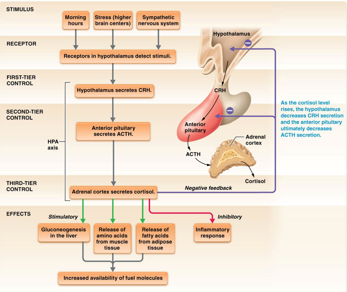
Cortisol (Hydrocortisone)
Most potent glucocorticoid in the body
Receptors located throughout the body = widespread effect
Effects:
Gluconeogenesis in the liver
Release of amino acids from muscle tissue
Release of fatty acids from adipose tissue
Suppresses immune cells & inflammatory response
Regulated by negative feedback loop
Stimulus - morning hours, stress, and sympathetic NS
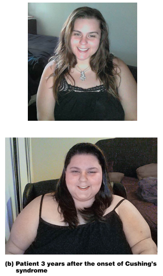
Cushing Syndrome
disorder caused by chronic exposure to excessive glucocorticoid hormones
Symptoms
Obesity
Hypertension
Hirsuitism (excess male-pattern hair growth)
Kidney Stones
Menstrual Irregularities
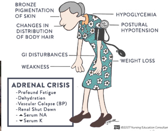
Addison Disease
disorder that develops when the adrenal glands fail
Chronic shortage of glucocorticoids and sometimes mineralocorticoids
May develop from lack of ACTH or lack of response to ACTH
Symptoms
Weight Loss
Fatigue
Weakness
Hypotension
Skin Darkening
Treatment
Therapy with oral corticosteroids
Androgenic Steroids
steroid sex hormones that affect reproductive organs as well as other tissues
Adrenal cortex synthesizes these in small quantities in both genders
Largely byproducts of cortisol synthesis
Can be converted in circulation to the androgen testosterone or the female hormone estrogen
Same general effects as those made by the gonads
Adrenal Medulla
Inner section of an endocrine gland
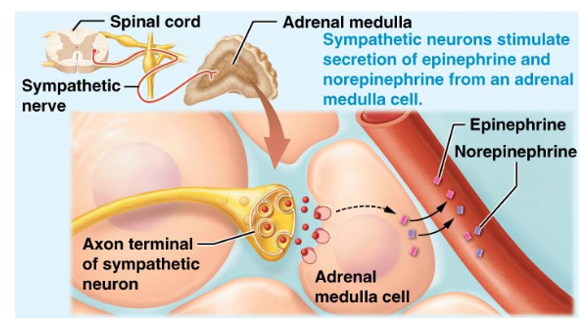
Made up of chromaffin cells
Secretes epinephrine & norepinephrine
Chromaffin Cells
Cells that make up the adrenal medulla
Nervous tissue origins
Secrete mostly epinephrine directly into the bloodstream
Stimulated by
Sympathetic neurons to release
or
By acetylcholine released from preganglionic sympathetic neurons
Epinephrine
Hormone that mediates sympathetic responses to stressful situations (Fight, Flight, or Freeze Response)
Way to reach cells not innervated by the sympathetic NS
Physiological Effects
Increases heart rate and force of heart contraction
Dilates bronchioles
Constricts blood vessels supplying skin, digestive organs, and urinary organs (Increasing blood pressure)
Dilates blood vessels supplying skeletal muscles
Dilates pupils
Decreases digestive & urinary functions