Sheep
 Call Kai
 Learn
Learn Practice Test
 Spaced Repetition
 Match
Match Flashcards
 Knowt Play
1/116

There's no tags or description
Looks like no tags are added yet.
Name | Mastery | Learn | Test | Matching | Spaced | Call with Kai | Chat |
|---|
No analytics yet
Send a link to your students to track their progress
117 Terms
7 Step Process of Herd Investigative Approach
Define the problem
History taking
Environment and distance exam
Not realistic to perform “full farm walk”
Individual exam ± PM exam
± Additional tests (eg. faecal or blood samples)
Must be justified
Data analysis and decision-making
Implement plan and monitor response
ONE: Define The Problem
Farmer Joe asks you to have a look at his flock of sheep as he is “a bit disappointed with the ewe hoggets at the moment”
Question: What additional information do you require to better define the problem?
What does “disappointed” mean?
Death? Poor growth rates?
Number of ewe hoggets and number affected
Signalment of ewe hoggets: Breed, age
Weight of ewe hoggets (when where they last weighed?)
What does “at the moment” mean?
Yesterday? Progressive? Since summer?
Example: Problem = Poor average growth rates of Romney ewe hoggets between December - April (average 27kg and 32kg respectively = ~5kg growth in 5 months) in a herd of 800.
→ Use data where available to define problem clearly
+ve: Helps formulate DDx list to guide investigation
THREE: Distance and Environmental Exam
Importance
11 Features to assess on distance exam
Importance: Usually called out to examine MOB vs. individual → Impossible to conduct individual exam on every animal (unless lifestyle block or prize ram)
Important to assess paddock which animals have been grazing but not always feasible (lack of transport or distance away)
Features:
Signalment and variation in mob
Behaviour, posture and mentation
Swelling or oedema (eg. bottle jaw)
External skin lesions, defects and wool condition (eg. flystrike or FE)
Nasal, ocular or oral discharge
Faeces on ground (consistency and variation)
Sick individuals (depressed, separated from group)
Coughing or dyspnoea
Soft grunting
Bruxism (hunger or pain)
Flystrike or footrot smell
Gut fill
List 3 features to assess on individual examination of a sheep
MM
BCS = Palpate lumbar dorsal and transverse processes
Weight
→ Can perform quickly on large number of animals (50 - 100)
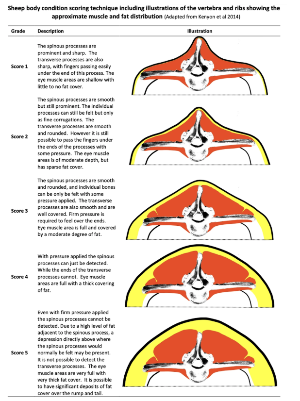
Which sheep should be selected for physical exam?
Disease of entire mob → Assess worst affected and some not so badly affected
Disease of individuals → Examine small number of diseased animals
Random selection is rarely rewarded, but may be justified
7 Differences between sheep and cow PE
MM = 3rd eyelid (vs. vulva of cows) ± gums
Gums in black-faced breeds (eg. Suffolk) are pigmented
Normal: Pale pink
RR and HR = ALWAYS elevated due to stress from handling
Lung Auscultation = Poor correlation between auscultation and pathology
Part fleece so stethoscope contacts skin
Pulse = Femoral artery
GI sounds = GI accidents rare in sheep (pinging uncommonly performed)
Hydration = Pinch upper eyelid
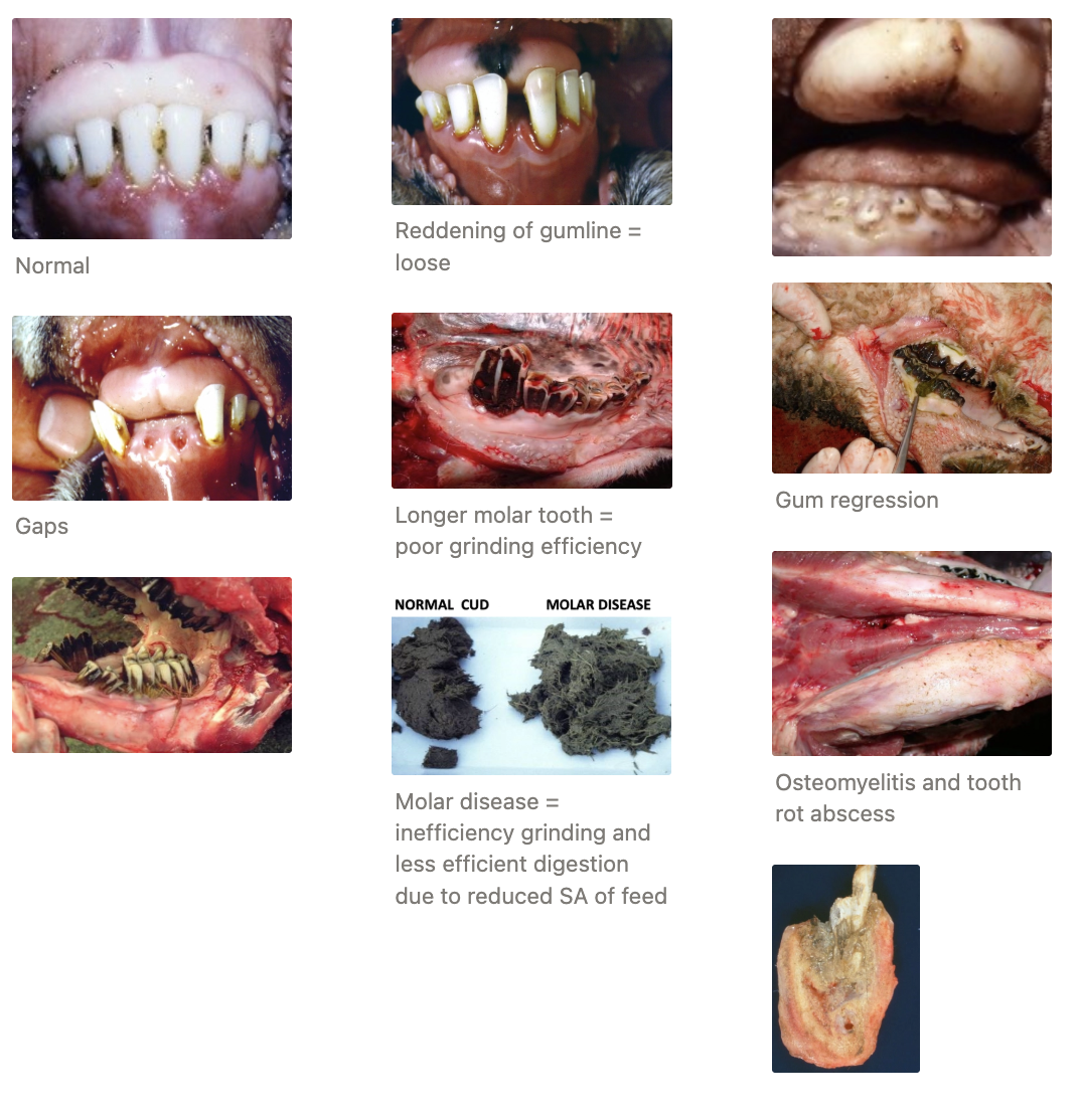
Oral Examination = Tight cheek muscles → Examine incisors and dental pad ONLY
Sharp molars → Do NOT put fingers in mouth
Palpate ventral mandible for osteomyelitis due to tooth root abscess
Age of sheep ≤4yr assessed by number of permanent incisors erupted
1yr = 1 pair of incisors
Normal TPR and rumination rate of sheep
Temperature = 39.5 ± 0.5˚C (sheep are HOT)
HR = 70 - 90bpm
RR = 20 - 30brpm
Rumination Rate = 1 - 3/min
2 Indications for field necropsy
Animals found dead
Alive animals BUT unlikely to recover and/or low value
Necropsy must have high likelihood of helping to achieve diagnosis (i.e. benefit rest of flock to prevent further death)
eg. Ill-thrift ewe
Avoid ill-thrift lambs (treatable disease in high-value stock class)
Ideally ≥3 animals
7 Health and safety recommendations for field necropsy
Care with walking while holding a knife
ONE person cutting at a time
Cut AWAY from yourself and others
Do NOT hand knife to another person (place on ground)
Wear appropriate PPE (overalls, gumboots, waterproofs and gloves)
Hold body part up so skin is cut under tension
Easier to cut UNDER skin vs. on top
Describe the field necropsy technique (+ features to assess)
Field Necropsy = Quick technique assuming limited DDx → Carefully examine few specific organs to rule in/out diseases
More thorough necropsy technique for single animal or many possible DDx
EXTERNAL EXAMINATION
AM: BCS, tag and PE
Assess for paddling (scuff marks in dirt), position of head (opisthotonus), discharge, signs of injury, predation or damage
REFLECT LEGS AND SKIN
Lie in LEFT lateral recumbency (rumen on dependent side)
Reflect cheek tissue to expose molars and assess dentition
Reflect front leg → hindleg (cut ligament to dislocate hip)
Remove skin

OPEN ABDOMEN (+ quick inspection of abdominal organs)
Start caudal to last rib (near spine) and extend cut caudoventrally
ID ileocaecal junction and roll caecum to assess for Johne’s disease
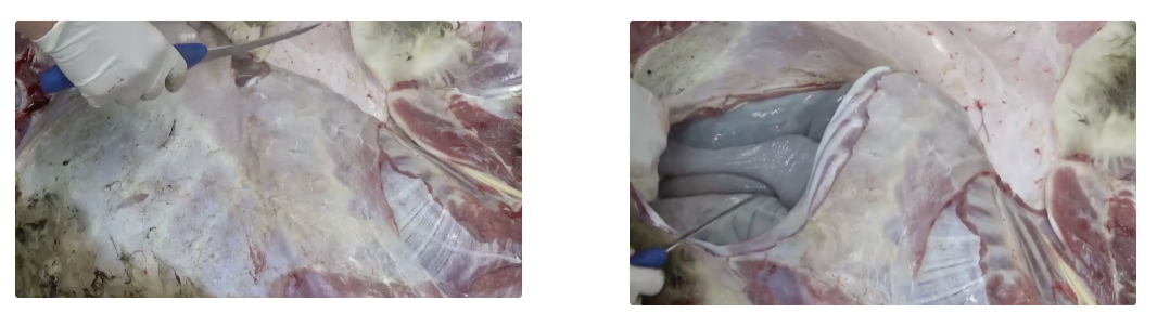
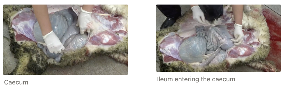
OPEN THORAX
Cut through soft tissue over sternum and ventral to spine → Cut ribs with loppers
Remove pluck by cutting through:
Mediastinum (ventrally)
Oesophagus and CdVC (caudally)
Ao, oesophagus and trachea (cranially)
Care with interpretation:
M. capillaris cysts are NORMAL = Small and gritty lesions over dorsal lung surface
Firm = Abnormal
Cut down trachea into bronchi and bronchioles to ID lungworms
Heart not normally examined in field necropsy unless indicated
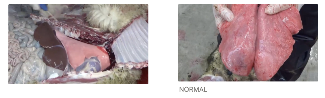
CAREFUL EXAMINATION OF ABDOMINAL ORGANS
Liver: Assess shape, fibrosis and train track lesions for FE and liver fluke
Cut into bile ducts for liver fluke
GI: Normal for slight variation in colour
Intestinal adenocarcinoma
Can cut into abomasum for Haemonchus (not routine) → Tie for worm count or assess top for wriggling worms
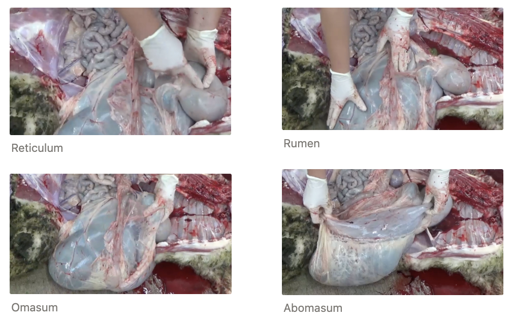
List 5 zoonotic agents of sheep
Leptospirosis → Flu-like symptoms ± severe illness and death
Almost all NZ sheep have serological evidence of exposure
Commonly localised in kidneys and shed in urine
Most dairy herds vaccinated to protect humans but does NOT eliminate shedding
Listeria → Abortion
GI of sheep
Salmonella spp. → GI disease
Normal GI commensal of sheep and cattle
Yersinia spp. → GI disease
Normal GI commensal of sheep and cattle (rare clinical disease)
Campylobacter jejuni → GI disease
Normal GI commensal of sheep and cattle
NOT Zoonotic:
Brucella ovis = ONLY Brucella spp. in NZ AND not zoonotic
BVDV
Rotavirus
Coronavirus
Methods of Sheep Euthanasia
Advantages
Disadvantages
Overdose of Pentobarbitone IV
+ve:
Humane (rapid loss of consciousness)
Minimal distress to owner (eg. lifestyle block)
-ve:
Requires vet (controlled substance)
Residues in carcass (cannot be used in food chain)
$$$
Technically challenging (stick vein)
Throat cut ± Pre-Stunning
+ve:
Simple equipment
Culturally/religiously required in some context
-ve: Animal remains conscious → Pain for some time during exsanguination (major welfare concern)
Stunning with Mallet → Throat Cut
+ve:
Cheap
Superior welfare over non-stunning slaughter
-ve:
Requires skill to ensure effective stun
Aesthetic concerns (cannot use for brain necropsy)
Shoot with Captive Bolt Gun (CBG)
+ve:
Humane (instantaneous unconsciousness)
Does not require firearms license
-ve:
Requires restraint and good aim
Equipment maintenance required
Cannot use brain for necropsy
Shoot with Free Bullet (Rifle)
+ve:
Done from distance
Good for field euthanasia of large animals
-ve:
Safety concerns of free bullet
Requires firearms license and training
Risk of poor aim
Cannot use brain for necropsy
Captive Bolt Gun
Structure and MoA
3 Normal post-CBG responses
Positioning
Sheep (polled)
Sheep and goats (horns)
Cattle
Pigs
Restraint technique
4 Signs of effective shot
5 Signs on ineffective shot
Method of exsanguination
MoA: Steel bolt inside gun powered by small grain bullet
When fired, bolt shoots out to penetrate brain → Concussive forces and damage to brain from bolt (and skull fragments)
No recoil (low-grain cartridge)
→ Immediate and irreversible concussion → Exsanguination or pithing immediately after CBG
Normal Responses:
Sheep to kick after being shot with CBG (after short delay) = Normal post-death neurological reflex
Heart continues to beat (~2 minutes an dup to 10 minutes) after CBG
Occasional agonal gasping (uncommon)
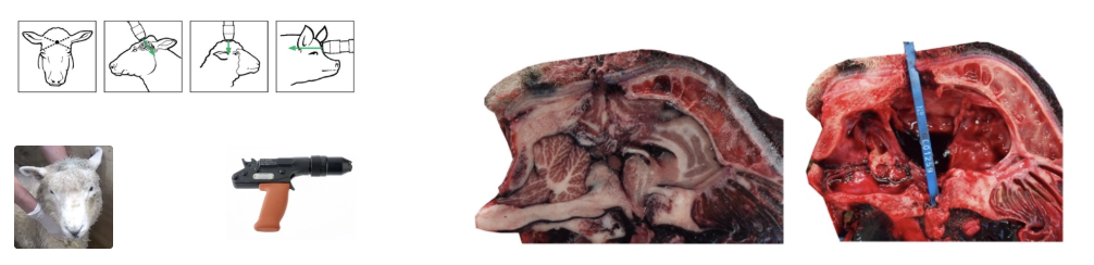
Positioning: Must go through mid-brain down into pons
Different species have different shaped heads → Different species CBG position
Sheep (polled) = Highest point on top of head → Aim directly towards back of throat
Sheep and goats (horns) = Back of head → Aim towards base of tongue (cannot place at highest point of head due to horns)
Cattle = Middle of forehead at crossing point of two imaginary lines between eyes and centre of base of opposite horn
Pigs = 3/4 inches (20mm) above eyes and halfway across forehead → Aim slightly up
Restraint: Adequate restrain sheep ensuring handler out of way (hands down neck and not directly under chin)
Can have 1/both hands on gun (care with hand placement over vent hole)
Effective Ahot:
Dilated pupils
Absent corneal reflex
No jaw tone
No breathing
Exsanguination: Place head around gumboot → Place knife perpendicular just below cervical vertebra (caudal to angle of mandible) and cut outwards/ventrally (check BOTH carotids are cut through)
4 Example ancillary tests
FEC
TE analysis
Liver fluke testing
Serology for specific disease (eg. Johne’s)
CBC, biochemistry and histology rarely indicated
4 Advantages of creating reports for farmers
Clarify thought for you and the farmer
Legal requirement
Allows future monitoring
Builds rapport with farmer
List 7 causes of low reproductive performance (+ 3 common times of callout for the veterinarian)
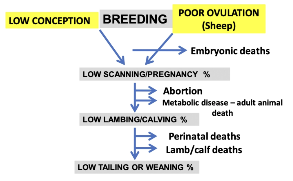
Low conception = Ram factors
Poor ovulation = Ewe factors
Embryonic death
→ Low scanning/pregnancy%
Abortion
Metabolic disease → Adult mortality
→ Low lambing/calving%
Perinatal death
Lamb/calf death
→ Low tailing or weaning%
Scanning important to determine when lamb loss occurred
3 Types of factors to ask the farmer about which contribute to poor reproductive performance (+ examples)
HUSBANDRY FACTORS
Date rams join ewes
Length of mating period/return to service
Lambing period (does it match the length of time rams were with the ewes?)
Ram soundness
Ram:ewe ratio
Teaser ram used?
FARM FACTORS
Geography
South Island → Mate ewes later so lambs miss poor weather events
North Island → Mate ewes earlier so lambs have enough feed when born
Hill country ewes cycle LATER with SHORTER mating period
Age structure of farm
Younger ewes cycle LATER with SHORTER overt oestrus AND lower ovulation rate → Affects management
Vaccination records (eg. Campylobacter, Toxoplasma and Salmonella)
Pregnancy scanning data
Determine where the problem occurred
Make decision about preferential feeding
ANIMAL FACTORS
Nutrition
BWT and BCS
Genetics
Age
Reproductive diseases
Other diseases
Stress
Why are reproductive problems so difficult to investigate? Why are they so important to investigate?
Difficultly: Usually retrospective → Aim to determine cause to prevent problem occurring NEXT YEAR
Importance: Good reproductive performance is fundamental for profitability of a sheep farm
Number of lambs at DOCKING ÷ Number of ewes mated
What is the difference between fertility and fecundity?
Fertility = Ability of animal to get pregnant (affected by ovulation) → Proportion of ewes in lamb
Fecundity = # of offspring produced (number of ovulations per oestrus cycle) → Lambing%
+ Scanning% for timing of loss
Increased lambing% in NZ
5 Reasons why lambing% has increased in NZ
4 Reasons why it is becoming an issue
Causes:
Genetic improvement (incorporated highly fecund breeds as twinning is NOT a highly heritable trait)
Superior nutrition
Superior vaccinations
Hogget mating
Better management (scanning to improve lamb survivability and timing of the breeding season to match weather)
-ve:
Ewe only has two teats
Lower birth weight of individual lambs
Dystocia
Ewe carrying multiples requires more feed
Describe the THREE major ewe factors affecting reproductive performance
NUTRITION = Vital from weaning to mating (3m to get ewe in good BCS for mating)
Flushing/Dynamic Effect = Increase feeding in 3 - 6w pre-mating to increase fecundity
Larger response observed in thinner ewes (BCS 2 - 2.5) but no effect in BCS 3.5 ewes as they already benefit from the static effect
Recommendation: BCS ewes 3 - 6w pre-mating to preferentially feed thinner ewes to utilise flushing effect → Ensure good BCS for mating
BWT and BCS = #1 effect on fecundity
Compromise between reproductive performance and efficiency (higher BWT = higher maintenance requirements)
Reduced fertility if BCS <2 and big -ve impact on fecundity if BCS <2.5
Recommendation:
BCS flock q1m between weaning and mating to preferentially feed
BCS at scanning to preferentially feed
Ensure light ewes meet target weights of ≥55kg AND BCS 3 - 3.5 at mating (effect on fecundity plateaus at 65kg → Decreased ovulation rate when >65kg)
GENETICS = Big impact on fecundity
Introduction of Finnish Landrace = Highly fecund breed into traditional breeds to produce the maternal breed
Ram has less impact on fecundity (lowly heritable trait)
Describe the FIVE minor ewe factors affecting reproductive performance
AGE
Younger sheep = Lower fertility and fecundity
Romney lambs reach puberty at 5 - 8m (~35kg)
Aim ≥40 - 42kg for hoggets at mating → Mate later to ensure they reach these target weights
Commercial ewes culled at 5 - 7yr → Old-age poor fertility is no issue on-farm
REPRODUCTIVE DISEASES
Phytoestrogens = Lucerne and clover (more issue with pure swards or older varieties)
Recommendation: Do NOT graze ewes on lucerne/clover pastures ≥3w pre-mating + during mating
Zearalenone = Fungal mycotoxin that decreases fertility and fecundity
Can test in urine (rare)
Abortive Diseases
AUTUMN DISEASES (breeding season)
Facial eczema → Decreased feed intake
Parasites
Trace element deficiency (eg. selenium)
STRESS → Decreased fecundity and delayed ovulation
eg. Yarding, shearing, transport, dogs, bikes, poor weather events
EXAMPLE: In July, a farmer contacts you because the scanning % of the MA ewe flock is only 142% and they would normally expect ~180%
Based on the scanning data, the issue is reduced fecundity – only 1.6% of the ewes are non-pregnant (normal for this farm) but a higher proportion of ewes are carrying single lambs compared with previous years
Questions:
What ewe factors may have contributed to this problem?
What history questions would you ask?
What ewe factors may have contributed to this problem?
Conception, ovulation rate, embryonic loss
Nutrition prior to mating (BCS)
Stress (events prior to mating)
Age structure of flock (similar effect in 2-tooths?)
Mating length
Phytoestrogens or zearalenone
What history questions would you ask?
Overview of Ovine Abortion
Prevalence
3 Most common DDx
Prevalence: Common (1 - 2% = ~800,000)
Outbreaks most common due to introduction of new disease to unexposed flock
DDx:
Toxoplasmosis
Campylobacter fetus fetus
Salmonella Brandenburg
Toxoplasmosis
Agent
Lifecycle
IP
4 Outcomes of infection
Clinical signs of ewe
PM examination
5 Methods of diagnosis
3 Methods of control/prevention
Agent: Protozoan parasite Toxoplasma gondii
IH = Sheep (ALL mammals, birds and reptiles)
Mouse + cat = Maintenance cycle
ZOONOTIC!!!
DH = Cat (tissue cysts can spillover from GI infection)
Lifecycle:
Cat (DH) infected via:
Bradyzoites within tissue of IH (sheep/mouse)
Sporozoites from sporulated oocysts in environment
Toxoplasma undergoes schizogony and gametogony → Unsporulated oocysts which are shed in faeces
Environment = Unsporulated oocysts undergo sporogony to produce an oocyst with 2 sporocysts each with 4 sporozoites (2 - 3d)
Sporulated oocyst within cat faeces ingested by IH sheep which liberates sporozoites
Sporozoites invade GI cells and transform into tachyzoites which can disseminate to brain, heart, lung, eye, muscle, placenta via monocytes
Clinical signs reflect organs infected by tachyzoites
Rate of division slows due to host immunity (2 - 3w) and switches to bradyzoite formation (100s of bradyzoites within a tissue cyst of ANY tissue)
Tissue cyst ingested by IH/DH which releases tachyzoites
IP: Long (up to 40d from infection to abortion)
Outcomes: Tachyzoites cross placenta to infect foetus
Non-pregnant = Lifelong immunity
Once infected, IH is immune to reinfection as tissue cysts with bradyzoites can periodically breakdown ⇔ tachyzoite which refreshes the parasite population and reinforces immune response
Early pregnancy = Embryonic death → Not pregnant at scanning
Mid - late pregnancy = Abortion
Late pregnancy = Weak lambs (immunity develops)
Stillbirth uncommon
Ewes: Healthy
PM:
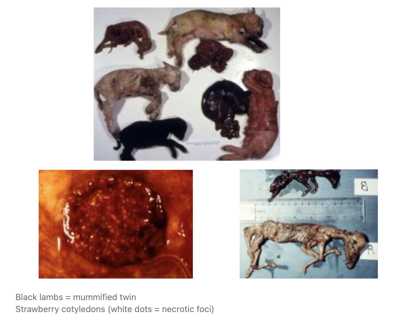
Strawberry cotyledons = Necrotic placental lesions with dystrophic mineralisation
Variable appearance of foetus
One twin may be affected, but other appears okay (both die) as one horn affected more than the other
± Mummified
Diagnosis:
History
Naive ewes (hoggets or 2-tooths) ONLY
Presence of ferrel cats (contamination of hay barns)
Low scanning% (EED)
Clinical signs = Overt abortion with mummified lambs and strawberry cotyledons
Placenta/foetal brain for histopathology → ID tachyzoites
PCR of aborted materials
Foetal heart blood OR serum titres from ewe for Ab
Control/Prevention: Farmer PPE important as zoonotic disease
Dispose of aborted tissue and lambs (consumed by cats to continue lifecycle)
No need to isolate as no sheep-to-sheep transmission
Vaccination with Toxovax = Live attenuated vaccine containing tachyzoites passaged through mice for many years
Single dose ≥4w pre-mating to maiden ewes = Lifelong immunity
May NOT prevent abortion if high environmental challenge with oocysts (20% may still abort)
Control ferrel cats
Campylobacteriosis
Agent
Transmission
3 Sources of infection
IP
Pathogenesis
2 Outcomes of infection
Clinical signs of ewe
PM examination
4 Methods of diagnosis
3 Methods of control/prevention
Agent: Campylobacter fetus fetus = Vibrio bacterium carried in the reproductive tract of sheep
Transmission: Spread via vaginal secretions and aborted materials (unclear what triggers this commensal organism to cause disease)
Sources:
Sheep-to-sheep transmission (high stocking density)
Indirect contact (eg. gumboots)
Carrier birds
IP: 2 - 3 weeks
NOT zoonotic
Pathogenesis:
Ingestion of bacterium
Bacteraemia and spread to the placenta
Necrosuppurative placentitis
Foetal death OR reduced neonatal viability
Lifelong immunity after exposure
Outcomes:
Abortion in last 6w of gestation (abortion storms ≤50% of flock possible)
Late gestation = Stillbirth or weak lambs
More common than toxoplasmosis
Ewe: Clinical healthy ± vaginal discharge
PM:
Foetus with fresh appearance
Necrotic target liver lesions in ¼ of aborted lambs
Placenta oedematous and opaque
Diagnosis:
History
High stocking density
Naive hoggets or 2-tooths affected
Clinical findings
Stillbirth (NOT mummified like Toxoplasmosis)
Necrotic liver of foetus
Foetal liver for histology
Culture of foetal stomach contents
Control/Prevention: NOT zoonotic
Hygiene and reduce stocking density
Antibiotics to manage abortion storm
Vaccination with killed vaccine
Sensitiser + booster 4 - 6w later by mid-pregnancy
Annual booster (most farmers ONLY give to 2-tooth ewes)
Can give early gestation BUT stressful for pregnancy
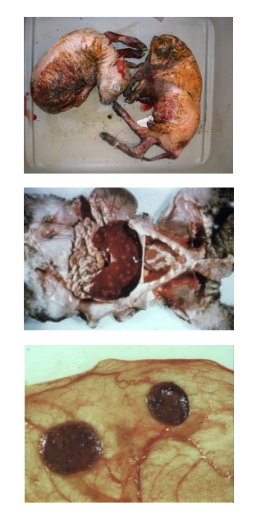
Salmonella Brandenburg
Signalment
3 Sources of infection
Environmental resistance
Outcome of infection
Clinical signs of ewe
PM examination
3 Methods of diagnosis
4 Methods of control/prevention
Signalment: South Island sheep ONLY
Adult ewes more likely than young ewes
Multiples have higher incidence
Can re-abort in subsequent years
Sources:
Carrier sheep in naive flock (excreted in faeces)
Aborted material (risk with high stocking density)
Vectors: Farmer, vet, vehicles, BBG, dust
Resistance: Survives 4 - 6m in environment
Outcome: Abortion storms in last 6w of gestation
3 - 4% of flock over 30d → Up to 50% with no treatment
Ewe: 50% aborted ewes die from necrotising metritis = Ill, dull ± enteritis
PM: Necrotic, macerated or autolysed foetus
Diagnosis:
History = Sick ewe (mature) with septicaemia prior to abortion
Tips of cotyledons to ID organism
Culture of foetal stomach contents
Control/Prevention: Farmer PPE important as zoonotic disease
Antibiotics during outbreak (prevent ewe death NOT abortion)
Vaccination (reduce disease, but small number of abortions still occur)
Salvexin+B in winter
Do NOT vaccinate ewes in the face of outbreak (yarding)
Dispose of aborted material and prevent access to BBG
Lower stocking density and isolate affected ewes to prevent further spread
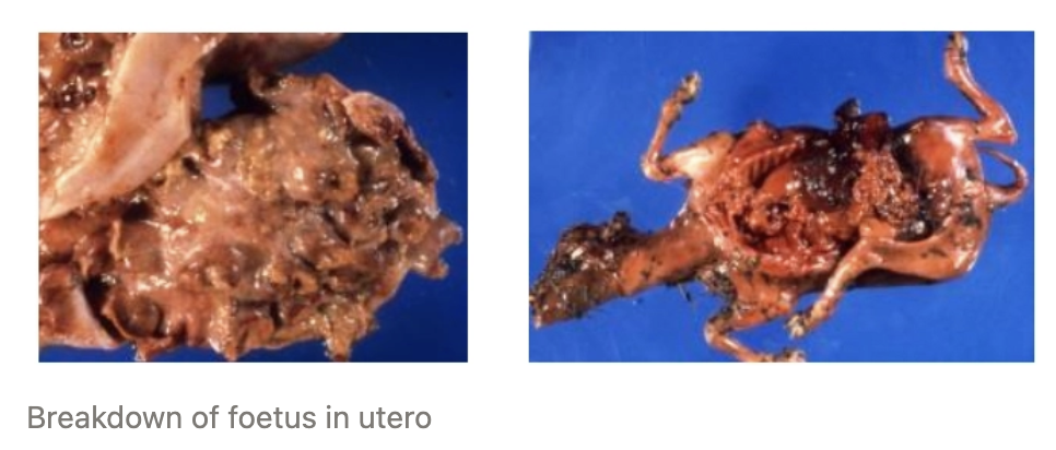
EXAMPLE: In July a sheep farmer rings to say that they have had some abortions occur in the ewe flock
Questions:
What further data could you gather to help define the problem?
What history questions would you ask?
Assuming you visit the farm, what distance and clinical exams would you do?
What would you specifically look for in/on aborted foetuses and placenta?
What samples would you submit to the laboratory for further diagnostic testing?
What recommendations would you make for management of this problem right now (before getting lab results back)?
…
3 Reasons to examine rams for breeding soundness
Ensure healthy rams for sale and fit for breeding
Important in NZ when ram : ewe ratio is ≥1 : 100 → Must be very fit to complete task
Pedigree breeders require it to be accredited-free from Brucella ovis
Excellent opportunity for vet input on farm
3 Times when ram breeding soundness exams should be performed
Pre-Sale (2-tooth rams) = October - November
Pre-Mating = January - February
≥1m pre-mating → Enough time for farmer to replace unsound ram OR make temporarily unsound ram sound (~6w for sperm to develop)
Only stud farms conduct single-sire mating to control genetics (vs. commercial farms with team of rams)
In response to a problem = Low scanning%
List 4 differences between ram vs. bull breeding soundness
Smaller = Examination is easier, faster and safer (more common for vets to check rams)
Main problems = Epididymitis, congenital defects and poor health
Penis/prepuce abnormalities are rare in rams (do NOT need routine checking)
Pedigree rams must be vet-checked pre-sale and accredited free from Brucella ovis
Describe 3 (6) steps of the ram breeding soundness examination
FOUR T’s
TEETH and TOES
Clinical history = Signalment, health, reproductive history and future use
Physical examination = Lameness (ability to mount), BCS and free from disease (eg. flystrike, FE, parasites)
Pre-breeding ram aim BCS 3.5 - 4/5
TESTICLES
Genital examination = Systematic evaluation of scrotum and its contents (scrotum → head of epididymis → testicles → tail of epididymis)
Size
Tone/consistency
Symmetry
Lesions (scrotal skin and wool)
TESTOSTERONE
± Semen evaluation
Not routine for rams as there is goof correlation between appropriately sized, firm testicles, free of defects AND good sperm production
Mating ability (bulls)
Other tests eg. B. ovis
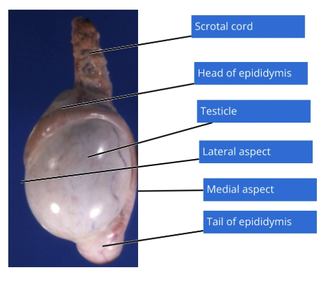
ONE: Testicle Size
Importance
Pre-sale diameter aim
Pre-breeding diameter aim
Importance: Directly proportional to amount of sperm produced (1g testes = 20M sperm)
Judgement call (unsure → recheck several weeks later) as highly variable due to seasonal atrophy
Pre-Sale (15m): ≥28cm diameter
Pre-Breeding (MA): > 32cm
TWO: Testicle Tone
Importance
Normal
4 Causes of soft testes
Importance: Good indicator of sperm production
Sperm takes 6 - 8w to develop and mature → Soft testes in January (unsound) may not recover in the breeding season
Normal: Flexed bicep
Causes of Soft Testes:
Out of season
Over-heating (eg. long wool over scrotum)
Poor health (infection → febrile)
Malnutrition and low BCS (eg. parasites)
FOUR: Lesions
3 DDx for epididymitis
6 Congenital reproductive defects
2 scrotal DDx
Main penis/prepuce DDx
Epididymitis: Enlarged and hardened epididymis (head/tail)
Brucella ovis
G- pleomorphs
Vasectomy (surgical disruption of tubes → sperm leakage)
Congenital: 1 - 5% of rams at pre-sale
Monorchid = ONE testicle develops
Cryptorchid = Partially descended testis
Asymmetrical testis size = Hypoplasia of ONE testicle
Segmental aplasia = Missing bits, usually epididymes)
Micro-orchid = Tiny testis
Hypospadia = Urethral NOT at the tip of the penis
Scrotal:
Scrotal mange (Chorioptes bovis)
Scrotal abscess following shearing cuts
Penis/Prepuce: Balano-posthitis = Pizzle rot (feed → highly alkaline urine → inflammation of penis
Pathogenesis of epididymitis
Disruption of tubular epithelium by pathogen
Inflammatory reaction causes leakage of sperm into interstitium
Sperm outside the testes = Foreign body reaction (haploid cells)
Chronic granulomatous inflammation with fibrosis → Obstructed tubules
2˚ testicular atrophy (orchitis rare)
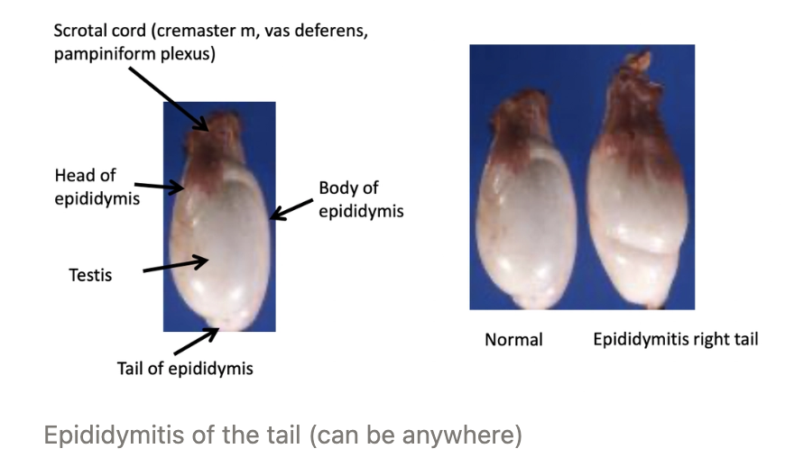
Brucella ovis
Transmission
4 Sources of infection
Diagnosis
Treatment
Transmission: Bacterium shed in semen and spread via direct contact between rams
Ram → Ewe → Ram
Sources: Infected ram
Newly purchase ram
Leased ram contracted to another farm
Neighbouring farms
Transport of rams from different locations → Ram from accredited free farm can still carry brucella ovis)
Diagnosis: Serology to detect Ab (CFT)
Rams with epididymitis require separation and screening with blood test to rule out B. ovis
Pedigree breeders must be accredited free from B. ovis (tested annually) and commercial farms should aim to be free → Reduced ram wastage and superior reproductive performance
Treatment: Cull and eradication programme (ram with B. ovis continues to secrete organism for up to 2yr)
Gram Negative Pleomorphs
2 Agents
Source of infection
Transmission
Diagnosis
Treatment
Agents:
Actinobacillus seminitis
Histophilus ovis
Sources: Widespread in environment
Transmission: Gain entry into urogenital tract by retrograde urethral infection around puberty
Unknown risk factor
Diagnosis: Identified in rams pre-sale (1 - 10%)
Diagnosis of exclusion (must test for B. ovis)
Treatment: Cull, but no eradication programme required
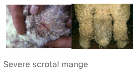
Scrotal Mange
Agent
Pathogenesis
Clinical appearance
Treatment
Agent: Chorioptes bovis mite
Pathogenesis:
ALL rams carry small proportion, but some develop an allergic hypersensitivity reaction
Inflammation and heat → Reduced sperm output
1/3 of testicle must be affected to reduce fertility
→ Temporarily unsound
Clinical Appearance: Scrotum with crusty lesions
Treatment: Ectoparasiticides (OP or ML)
Pizzle Rot
Signalment
Agent
Pathogenesis
4 Sequelae
Species: Balanoposthitis most commonly in wethers (occasionally in rams and bulls)
Agent: Urease-producing Corynebacterium renale
Pathogenesis:
High-protein diet (eg. clover-dominant pasture)
→ High urinary urea is broken down by C. renale to cytotoxic ammonia
Preputial ulceration and 2˚ bacterial infection
Sequelae:
± Extensive balanoposthitis with ulceration and necrosis of head of penis due to obstruction of the preputial orifice
Phimosis
Inability to serve
± Death from urinary obstruction (post-renal azotaemia)
How to classify rams during a breeding soundness exam
NEVER certify as fertile (use sound/temporarily unsound/unsound)
Unsound rams can still be fertile
eg. lesion in ONE testicle with SECOND normal testicle (still get ewe pregnant)
Unsound from commercial perspective (pedigree breeders should never sell and unsound ram)
Classification:
Sound = Ram with no physical or genital defects who is considered sound for breeding or sale
Temporarily Unsound = Ram with physical or genital defects which can be completely resolved BUT currently unsound for breeding or sale
Unsound = Ram with permanent physical or genital defect who is not sound for breeding or sale
Classify the following rams as sound, temporarily unsound or unsound
Scrotum with crusty lesions on bottom half
Soft testes with small scrotal circumference
Right testicle significantly smaller than left testicle
BCS 2/5
Testes with lots of wool on scrotum
Scrotum with crusty lesions on bottom half = Temporarily unsound (scrotal mange)
Soft testes with small scrotal circumference
Temporarily unsound (out of season)
Unsound if at pre-mating (not enough time for recovery)
Right testicle significantly smaller than left testicle = Unsound (but fertile)
BCS 2/5 = Unsound (insufficient time to gain BCS for mating)
Testes with lots of wool on scrotum = Temporarily unsound
Define “ill-thrift” for lambs vs. adults
Lambs = Poor growth rates
Adults = Poor BCS and/or weight loss
6 Benefits of reaching target liveweights for lambs
Reach slaughter weights faster → Fewer days on-farm
Superior schedule prices
Less cost associated with dagging, feeding and treatments
Reduces parasite burden on-farm
More feed partitioned to breeding ewes or other livestock classes
Replacement ewe lambs mature earlier
Opportunity to breed as ewe hoggets (≥42kg)
Superior growth as 2-tooths → Lifelong reproductive performance
Lamb Growth Rates
Time | Ill-Thrift | Target | Maximum |
Pre-weaning | |||
Post-weaning | |||
Average GR throughout life |
Time | Ill-Thrift | Target | Maximum |
Pre-weaning | <250g/d | 300g/d | 400g/d |
Post-weaning | <50g/d | 250g/d on crops (realistic = 150g/d on pasture) | |
Average GR throughout life | 100g/d |
3 Times when a farmer will identify ill-thrift in lambs
Routine weighing (begins at weaning)
Frequency varies between farmers
Observation #1
Slaughter records (eg. % slaughtered at weaning in March)
→ Retrospective and vague data
List 7 DDx for ill-thrift in young livestock
Inadequate nutrition (quality and/or quantity)
Internal parasites
Trace element deficiencies (Co and Se)
Pneumonia
Facial eczema
Flystrike
Ryegrass staggers
*Whole mob issues
3 Methods of diagnosis of undernutrition
History
Pre- and post-grazing covers (sward sticks)
Feed quality by eye or lab testing
Difficult to diagnose → Diagnosis of exclusion
Pneumonia
Seasonality
Geographical distribution
Infectious agents
3 Bacteria
2 Viruses
Pathogenesis
2 Forms
Clinical signs
PM lesions
5 Risk factors
4 Methods of diagnosis
Treatment
8 Methods of prevention
Seasonality: Summer/autumn (December - May)
Distribution: Increasing prevalence in Northern regions
Agents: Complex with many pathogens
Bacteria = Mannheimia haemolytica, Pasteurella multocida and Mycoplasma ovipneumoniae
Viruses = Parainfluenza virus-3 and respiratory syncytial virus
Pathogenesis: Requires respiratory defence mechanisms to be overwhelmed with inciting damage for some pathogens to invade
Young sheep have reduce immunity which is further exacerbated by stressors associated with yarding = Long distance from paddocks → Crowded into dusty yards
Heat stress induces open-mouth breathing and dust inhalation → Air bypasses defence mechanisms (nasal turbinates and mucociliary apparatus)
Viruses and Mycoplasma invade which compromise lung defences
Allows Pasteurella and Mannheimia to colonise lung = Commensal URT species
Cannot diagnose disease with nasal swab
Forms:
Chronic Non-Progressive Pneumonia (CNPP)
Clinical Signs: Slowly progressive and mild
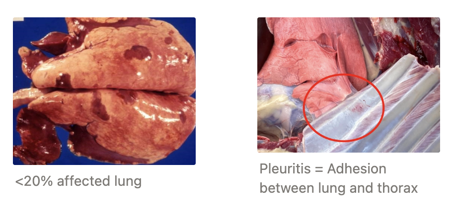
Reduced GR when >20% lung affected
Cough
PM:
Consolidation of cranioventral lung lobes
Pleuritis/pleurisy which can persist as adults = Adhesions between lung and thorax → Reported at meatworks
Occasional chronic lung abscess
Acute Fibrinous
Clinical Signs: Severe and acute
Outbreaks of coughing
SICK animals
Sudden death
PM:
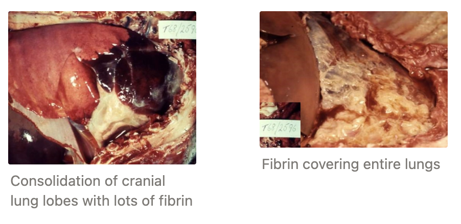
Severe fibrinous pleuritis
Dark red, solid lung lobes
Risks: Stress and yarding
Shearing and weaning on SAME day (running up and down fence lines)
Shearing lambs twice (shed overnight)
Frequent yarding post-weaning (eg. drenching)
Motorised mustering
Increased age of lambs at slaughter
Diagnosis:
History (esp. management)
Clinical signs
Mild
Moderate = Coughing and ill-thrift
Severe = Sudden death
Necropsy (esp. for poor BCS lambs)
Slaughter records for pleurisy
Treatment: NONE for CNPP
Antibiotics useful for early stage acute fibrinous form to prevent lamb death
Prevention: NO vaccination (commensal bacteria)
Allow time post-weaning before shearing
Minimise number of shearing times
Muster in early morning
Slower and calmer yarding (minimise use of dogs)
Minimise time in yards
Keep dogs tied up while in the yards
Sprinkler system to dampen dust
Scrape concrete base of faeces and dust
Define “Wastage”
Natural lifespan of sheep
Age at culling
2 Causes of early death
Wastage = Loss of ewes from flock BEFORE the end of their productive lifetime
Natural Lifespan: 10 - 13yr
Age at Culling: 5 - 7yr
Only 10 - 30% of ewes make it to 6yrs
Causes: Low BCS associated with increased wastage
Premature culling
~7% mortality (most risky period = lambing)
4 Reasons why BCS is superior to BWT
Minimises issues of:
Skeletal size between and within breeds
Physiological status (pregnancy = ewe weight + foetal weight)
Gut fill
Fleece length and wetness
4 Times to BCS sheep
Must encourage farmers to BCS ewes
Post-shearing (accurate by eye at the drafting gate)
Weaning = Lowest BCS
6w Pre-mating (mid-Feb) → Influences fecundity and fertility
Pregnancy scanning (6w pre-lambing)
ID during yarding and may have progressed for some time (4 - 5 yardings/yr)
What does the animal welfare code say about sheep BCS?
Vague = All sheep should be BCS 3 - 4 and any sheep BCS ≤ 1 requires urgent remedial action (euthanasia OR feeding)
Minimum Standard No. 5 on the Code of Welfare for Sheep and Beef Cattle
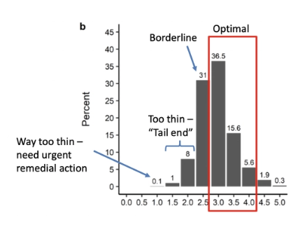
Describe the typical BCS distribution of NZ sheep
Normal distribution
~50% optimal (BCS 3 - 4)
30% borderline (BCS 2.5)
Tail-end = BCS 2
Event well-managed farms will have ewes with low BCS
List 4 times when a vet will intervene with BCS
Farmer concerned about ewes with extremely low BCS
More ewes at tail-end (BCS 2)
Flock average BCS is lower than expected
Investigation of welfare complainy by MPI
DDx for Ill-Thrift in Ewes = Low BCS
4 Treatable DDx
2 Fatal DDx
3 DDx where they do okay if fed well
Treatable:
Undernutrition
Parasitism = Likely symptom of low BCS rather than cause
Liver fluke
Foot disease/lameness
Fatal:
Johne’s disease
Intestinal carcinoma
Okay if Fed:
Dental disorders
Chronic FE?
Lung disease/abscess
Dental Disorders
Importance
Function of incisors vs. molars
Diagnosis
Treatment
Importance: Major reason for culling as important cause of ill-thrift
Function:
Incisors = Cut grass with dental pad
Molars = Flat surface for grinding
Diagnosis: Easy to examine incisors
Impossible to examine molars due to sharp cheek teeth, narrow dental arcade and tight cheek muscles
Severe → Palpable osteomyelitis and tooth root abscess
Treatment: Revise culling policy
Johne’s Disease
Agent
Signalment
Clinical signs
Pathogenesis
PM lesions
Prevention
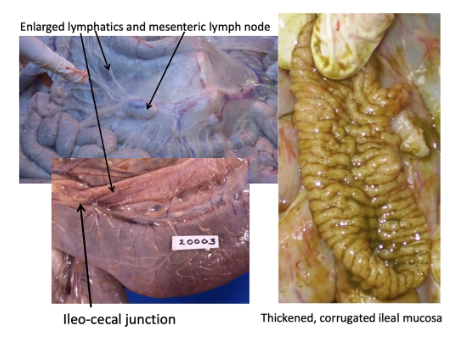
Agent: Mycobacterium paratuberculosis
Signalment: >18m (long IP)
Clinical Signs: Poor BCS and generalised oedema
Otherwise normal temperature, hydration and rumination rate
Usually over winter and spring due to poorer nutrition and added stressors of pregnancy and lactation
Pathogenesis: Chronic granulomatous enteritis → Intestinal malabsorption
PM: Assess ileocaecal junction for thick and corrugated ileal mucosa + enlarged mesenteric lymph nodes
Collect terminal ileum, liver and mesenteric lymph nodes for histology when in doubt
Prevention: Vaccination available to reduce shedding ONLY
DDx?
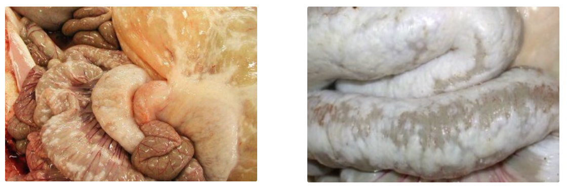
Small intestinal adenocarcinoma (1% of old ewes)
Variable appearance, but usually jejunal area
Fatal
DDx? Significance?
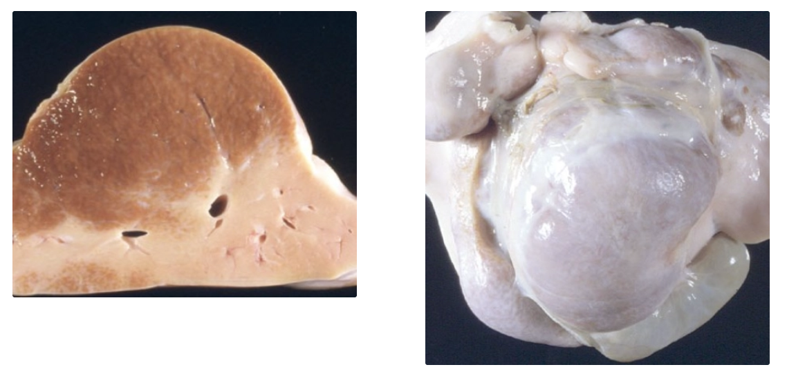
Chronic facial eczema = Fibrosis and end-stage liver disease
May not have a big impact on ill-thrift (often incidental at PM)
Significance of lung abscesses on ill-thrift
Consequence of lamb pneumonia but no data on effects on ill-thrift (presumed)
May rupture → Sudden death
Significance of nematode parasites on ill-thrift
Thin ewes have high FEC likely a CONSEQUENCE of ill-thrift (i.e. reduced immunity) rather than a case
Low BCS → Poor health and immunity → Reduced immunity → Increased worm burden
Recommend drenching thin ewes with high FEC
DDx for Sudden Death
Young sheep (13)
Adult sheep (14)
YOUNG SHEEP
Infectious:
Clostridial
Acute fibrinous pneumonia
Leptospirosis
Haemophilus
Parasites:
Haemonchosis
Other nematodes
Acute liver fluke
Nutritional/Metabolic:
Intestinal torsion (redgut)
Acidosis
PEM
Other:
Toxicity
Climatic/misadventure (eg. hypothermia, lightening strike, drowning, stuck in fence, dog attack)
Anthrax (exotic)
ADULT SHEEP
Infectious:
Salmonellosis
Clostridial
Listeriosis
Parasites:
Haemonchosis
Other nematodes
Acute liver fluke
Nutritional/Metabolic:
Ketosis
Hypocalcaemia
Hypomagnesaemia
Acidosis
PEM
Other:
Toxicity
Climatic/misadventure (eg. hypothermia, lightening strike, drowning, stuck in fence, dog attack)
Anthrax (exotic)
MANY DDx → Requires thorough investigation and PM highly recommended
MUST put short-term plan in place BEFORE leaving farm and often before DDx confirmed
4 Signs indicating a carcass is too autolysed for necropsy
Extremely bloated (bubbles of gas from anus)
Skin sloughing
Blue/green colour (esp. groin)
Smell
Enteric Salmonellosis
Agents
Source of infection
Transmission
Signalment
Seasonality
Prevalence
2 Risk factors
6 Clinical signs
5 PM features
2 Methods of diagnosis
3 Treatments
Prognosis
3 Recommendations when dealing with an outbreak
2 Methods of prevention
Agents: Salmonella Hindmarsh, Typhimurium, Bovismorbificans
Brandenburg → Abortive
Source: Carrier sheep (commensal in intestines)
Transmission: Faecal-oral
Signalment: Adult ewes (often well-fed with good BCS)
Extremely rare in ewe hoggets
Seasonality: December - July (intensive grazing)
Prevalence: 1 - 2%/yr?
Risks:
Stress → Shedding (eg. shearing, yarding, transport)
High stocking density (eg. break-feeding or set-stocking)
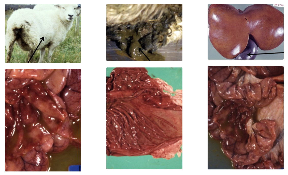
Clinical Signs: Progression over 2 - 3d
Lethargy (lagging behind mob)
± Fever (≥40.2˚C) but may be hypothermic due to recumbency
Khaki green and watery diarrhoea with mucus
Dehydration
Injected MM
PM:
Fatty liver (mobilisation of fat due to anorexia)
Large gallbladder (anorexia → not excreting bile)
Enteritis = Inflammation of intestine and abomasum with watery contents
Petechial haemorrhage of serosal surfaces
Diagnosis:
Culture after 24 - 48hr (growth ≠ diagnosis)
Histopathology up to 1w later
Treatment:
Antimicrobials (if not too sick and well-monitored)
Supportive care (PO fluids)
Euthanasia
Prognosis: 50%
Outbreak Management:
Reduce faecal-oral transmission = Move to new paddock and reduce stocking density
Vaccination = Death stops in 10d
Do NOT vaccinate sick sheep (immunity develops slower than disease progression)
Warn farmer of zoonosis
Prevention:
Reduce risk factors
Vaccinate ewes BEFORE risk period (weaning in November - December)
Sensitiser and booster 4w apart → Annual booster
Enterotoxaemia (“Pulpy Kidney”)
Signalment
Seasonality
Pathogenesis
2 Clinical signs
6 PM lesions
2 Methods of diagnosis
2 Recommendations when dealing with an outbreak
Long-term preventative method
Recommendation if dam is not vaccinated
Signalment: Well-fed lambs ≤6 - 8m (largest of the mob)
Uncommon in cattle and deer
Vaccinated dam → MDA in lambs for 8 - 16w
Most common in unvaccinated weaned lambs (dams ARE vaccinated)
Seasonality: Summer and early autumn (may coincide with weaning or change in feeding for finishing)
Pathogenesis:
Good nutrition/excess CHO → Additional nutrition for bacterial
Proliferation of Cl. perfringens Type D in gut which produces epsilon toxins
Toxins absorbed into bloodstream
Vascular endothelial damage and generalised vasculitis throughout body
Most damage in organs with highest blood flow = Kidneys and brain
Clinical Signs:
Sudden death of unvaccinated lambs on good quality feed
Signs of agonal struggle (pain = Struggle marks from feet, foam from nostrils and head thrown back)
PM:
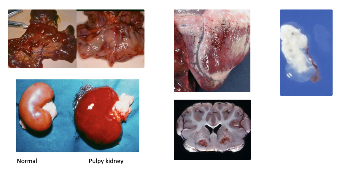
Rapid carcass decomposition and bloating
Excess pericardial fluid and fibrin clots
Petechial and ecchymotic haemorrhage over serosal surfaces of intestine and heart
Pulpy kidney 1 - 2hr PM (NOT pathognomonic)
Glucosuria
Focal symmetrical encephalomalacia = Pathognomonic
Diagnosis: History and clinical signs →
Necropsy of several lambs
± Confirmatory test ($) = Histology of brain and kidney
Detection of epsilon toxin from ileal contents (BUT also found in 20% of healthy animals)
Outbreak Management:
Reduce feed quality short-term
Vaccination in face of outbreak (deaths stop within 2d)
Prevention: Routine vaccination with toxoid (5in1, 7in1 or 10in1)
Ewes: Sensitiser + booster 4 - 6 weeks later
Booster 3 - 4 weeks pre-lambing (peak at colostrogenesis)
Take average of mob due to difference in lambing date
NOT recommend at set-stocking (1 - 2 weeks pre-lambing)
Young lamb: Maternal Ab lasts ~8 - 12 weeks (depends on vaccine) until weaning
Weaned replacement lambs: Sensitiser at docking or weaning then booster 4 - 6 weeks later
Weaned non-replacement lambs: Decide whether to vaccinate, depending on length of time to sale/slaughter
Disease often observed as farmers do not vaccinate lambs destined for slaughter in a few months
ALTERNATIVE if Dam NOT Vaccinated:
Lambs given tetanus anti-toxin and pulpy kidney toxoid at docking (”Lamb vaccine” or “PK-Anti-tet”)
Tetanus #1 risk after docking
Lambs then given 5in1 at weaning and booster 4 - 6 weeks later
List 3 essential vaccines on sheep farms
Toxoplasma
Campylobacter
Clostridia (cheap)
± Scabby mouth (if on farm)
Other vaccines (eg. Salmonella) depends on risk-benefit analysis
Prevalence of metabolic diseases in sheep
High-producing commercial farms (esp. crops)
Lifestyle farmers due to overfeeding
Risk: Underfeeding OR overfeeding
Associated with major physiological challenge to animal (eg. ewes with triplets or high-producing cows pushed to max)
Body good at deal with challenge due to homeostasis UNLESS acute/extreme (livestock at risk but not everyone affected)
Sheep will NOT abort in the face of a metabolic disease (prioritise growth of foetus)
Describe the 2 differences between cattle and sheep metabolic disease
Hypomagnesaemia uncommon in sheep
Ketosis = Pregnancy toxaemia, sleepy sickness or twin lamb disease in sheep
Cattle = At calving or peak lactation
Sheep = Late pregnancy → x2 energy requirements and lower feed intake
BUT further risk factors required to precipitate metabolic disorders (homeostatic mechanisms prevent disease)
List 6 risk factors for metabolic disease in ewes
Old = More foetuses (fecund) and reduced skeletal supply of Ca due to multiple lambings
Multiple foetuses
Sudden feed restriction or diet change (eg. yarding for set-stocking)
eg. Triplet ewes set-stocked on high-quality pasture (eg. Italian ryegrass) from annual ryegrass
Falling plane of nutrition
Concurrent disease (eg. poor dentition or lame)
Inclement weather and inadequate shelter (seek shelter → reduced feeding)
Pathogenesis of ketosis vs. hypocalcaemia
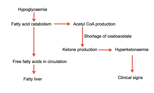
KETOSIS
Hypoglycaemia
Catabolise fat reserves for gluconeogenesis
FFA enters circulation resulting in fatty liver
FFA also used for acetyl-CoA production BUT shortage of oxaloacetate
→ Ketone production and hyperketonaemia
HYPOCALCAEMIA
Ca2+ metabolism complex
Late gestation ewes cannot meet Ca2+ requirements from diet
Up to 20% bone Ca2+ mobilised during pregnancy
When foetal demand exceeds diet and bone Ca2+ → Hypocalcaemia
Clinical signs of ketosis vs. hypocalcaemia
Similar Presentation: Time period = 24 - 48hr depending on severity
Separation from flock, reluctance to move and weak
Paresis and will not running away from farmer
Neurological signs
Depressed and apparent blindness
Recumbent, comatose, die
Differences:
Hypocalcaemia = Ruminal tympany
Ketosis = Wool plucks easily
Biochemistry features of ketosis vs. hypocalcaemia
KETOSIS
Hyperketonaemia = Increased BOH (handheld ketometer)
Ketonuria (dipstick)
Decreased BUN
Increased cortisol
± Hypoglycaemia
± Hypocalcaemia
HYPOCALCAEMIA
Hypocalcaemia (cannot measure on-farm)
± Hyperketonaemia
Often ewes with ketosis ALSO have low Ca2+ and visa-versa
4 Ways to differentiate ketosis from hypocalcaemia
Blood test BEFORE treatment
Ketostix, handheld ketometer and wool pluck → Ketosis
Supplement with calcium regardless
Rapid response = Hypocalcaemia
No response = Ketosis (± euthanasia and necropsy)
Necropsy
Ketosis = Fatty liver and large adrenal glands with dark cortex
Treatment of ketosis (5) vs. hypocalcaemia (2)
KETOSIS
IV dextrose ± PO propylene glycol
PO rehydration (or IV if high-value sheep)
SC Ca2+
Flunixin
± Caesarean section (lifestyle block)
Do NOT place in nursing situation (reduces grazing as no grass and way from flock)
HYPOCALCAEMIA
IV Ca2+ with care ± SC or PO
PO propylene glycol
Prognosis of ketosis vs. hypocalcaemia
KETOSIS
Irreversible if not treated early
Recumbent, neurological signs or renal failure = Poor prognosis
HYPOCALCAEMIA
Excellent prognosis if uncomplicated
List 3 most important sheep skin conditions
Facial eczema (see “Systemic Diseases”)
Scabby mouth
Flystrike (see “Parasitology”)
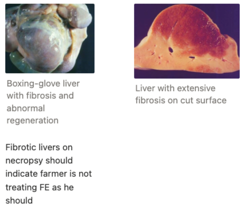
Effects of acute vs. chronic facial eczema
Acute: Major reduction in growth and reproductive performance (eg. ewes in mating season)
Chronic: Unknown effects of liver fibrosis
Scabby Mouth (Orf)
Agent (+ environmental resistance)
Transmission
3 Clinical signs
Treatment
Prevention
Agent: Parapox virus
Highly resistant in environment (persists for 1yr)
ZOONOTIC!
Transmission: Penetrates skin through abrasions
Mainly lambs (soft mouths) eating thistles or other rough feed
Clinical Signs:
Proliferative, solid lesions around mouth ± feet
Reduced GR due to reduced eating
± 2˚ bacterial infection in wet conditions
Treatment: Self-limiting
Prevention: Vaccination with live vaccine scratched into axilla or groin → Cause disease at site that is NOT going to affect production
Recommend ONLY if farmer had disease (otherwise introducing virus onto naive farm)
SINGLE dose to lambs at docking
Check lambs 1w after to assess for scabs
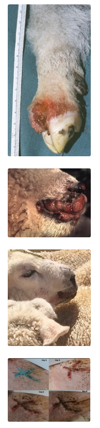
Importance of lameness (2) + 4 times of vet involvement
Importance:
Transport limitations (>10% sheep lame at works → Fine)
$ to treat and control (early treatment important for quick resolution)
Vet Involvement:
Ram soundness exams
Pet sheep and goats
Bad flock problems (10 - 15% flock influenced)
Transport (NO certificates for sheep)
7 DDx for ovine lameness
ALL ovine lameness in the foot:
Ovine interdigital dermatitis (OID)
Footrot
Foot abscess
Toe granuloma
(Arthritis - esp. old pet sheep)
NOT commercial sheep
(White line disease)
(Shelly hoof)
Describe the pathogenesis/progression of ovine foot disease
Moisture/predisposing factors (eg. trauma to skin)
Autumn and spring (wet)
Invasion of interdigital skin by Dichelobacter nodosus
Hoof damage rare as light weight (vs. cattle)
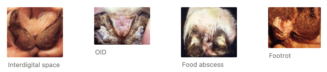
Ovine interdigital dermatitis (OID) = Mild to moderate dermatitis which is easy to treat as a superficial bacterial infection
→
Foot Abscess via infection with Trueperella pyogenes
Initial bacterial infection allows T. pyogenes to invade and penetrate the interdigital skin → JOINTS
Footrot via deeper infection by Dichelobacter nodosus
Bacteria erodes hoof wall
Dichelobacter nodosus
Source
Environmental resistance
3 Risk factors
Source: Anaerobic an present on most NZ farms
Environment: ONLY survives in ruminant feet (environmental survival of 5 - 7d)
Risks:
Virulence of D. nodosus strains (some farms never see footrot as only have benign strains)
Susceptibility of host
Coarse-wool sheep (90%): Usually OID, but footrot uncommon
Fine-wool sheep eg. Merinos (and goats): Footrot can be major problem
Farmers deal with footrot via culling → Indirect selection for high footrot tolerance
Wet conditions
Diagnosis of ovine foot disease
History and clinical findings
Vets rarely involved but should be (improper treatment or ID of cause)
Lab diagnosis NOT useful as culture commensal bacteria
Ovine Interdigital Dermatitis (OID)
2 Clinical signs
Treatment
Clinical Signs: MAY resolve spontaneously in dry conditions
Superficial and mild infection = Erythema and moisture of interdigital space
Mild - moderate lameness
Necessary precursor for foot abscess/footrot
Treatment: Topical antibiotics (blue spray = oxytetracycline) OR footbath
Ideally place in drier environment BUT typically indoors → Reduced grazing
Ideally multiple treatments of blue spray but not feasible on large farm
Footrot
Pathogenesis
4 Clinical signs
3 Treatments
4 Methods of prevention
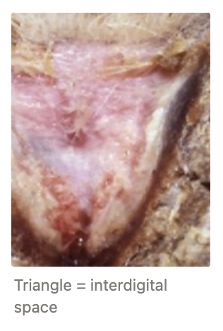
Pathogenesis: Deeper penetration of D. nodosus which produces keratolytic proteases to dissolve the hoof wall → Hoof destruction and under-running
Severity depends on virulent and chronicity (i.e. mild footrot = OID → Continuum of disease)
Clinical Signs:
Moderate - severe lameness
Obvious hoof under-run on examination
Broken skin horn junction which extends under the horn tissue of the heel
Soft horn is separated from underlying sensitive tissue
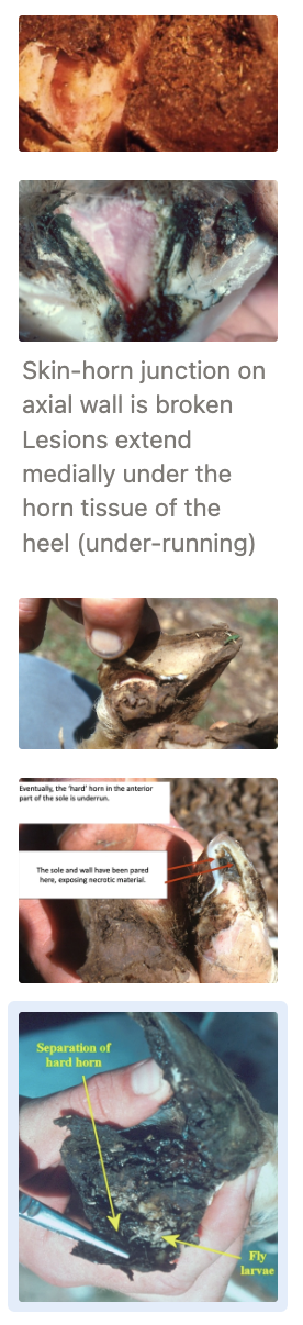
Hard horn in anterior part of sole under-run
Untreated cases can become chronic and severe with deformation of the hoof horn
± Flystrike
Never grows back normally
Small amount of black necrotic tissue (no pus) = Degraded hoof wall
Foul smell
Treatment:
Antibiotics (topical AND injectable) = Injectable penicillin/oxytetracycline and blue spray
Past: Footbath of formalin BUT Lame foot never touches bath
80 -90% recover with SINGLE dose of antibiotics
Trim hoof exposing anaerobic bacteria to air and kill (1 week after antibiotic treatment)
Difficult follow-up on commercial farm
Analgesia
Keep in dry environment
Prevention: Eradication feasible BUT difficult in NZ conditions
Foot bath with zinc sulphate
Useful for superficial infection (OID) → Preventing footrot
Difficult to do well (footrot = hold up lame foot)
“Cure rates” for footrot 30 - 50% with foot bathing ONLY (not effective treatment)
Keep in dry paddock where possible
Vaccine: Footrot ~14w protection
Culling (tolerance for footrot is heritable)
Foot Abscess
Agent
Signalment
3 Treatments
Agent: Trueperella pyogenes infection 2˚ to OID which allows breach of skin integrity of the interdigital space
Signalment: Heavy ewes and rams
Small number of severely lame animals (non-weight bearing)
Treatment: Difficult as most cases are advanced
Cannot insert scalpel to drain as vital structures around foot
Do nothing → recovery in 1 - 2m (ankylosis and bone fusing BUT very painful and welfare concern)
Consider culling as single dose of antibiotics not effective
Amputation of affected digit if appropriate
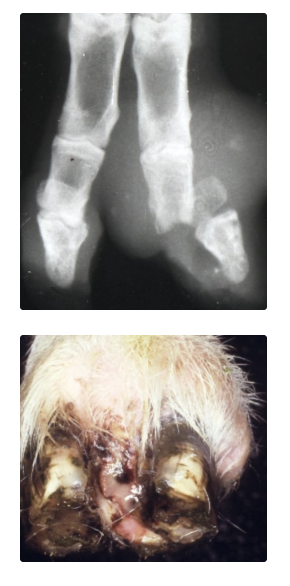
Toe Granuloma
Cause
2 Clinical signs
Treatment
Cause: Toe damage when haemorrhage occurs at trimming (overzealous)
Common for goats and lifestyle sheep
Clinical Signs:
Proud flesh (excessive granulation tissue) with hoof growing around → Pinching
Moderate to severe lameness
Treatment: Difficult
Ideally trim horn, amputate, cauterise, aftercare (regular bandaging)
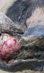
Describe the role of foot trimming
Past: Frequently used to treat footrot BUT inappropriate
Use antibiotics
Over-zealous trimming = Toe granuloma
Useful when:
Hoof is so over-grown it is causing lameness
Hoof shape accumulating debris (white line accumulating pockets of debris)
Common trimming in goats but less common now in sheep farms (culling main treatment option)
9 DDx for neurological disorders of sheep
Listeriosis
Brain and spine abscess
Polioencephalomalacia (PEM)
Tetanus
Ryegrass staggers
Congenital disorders (eg. Border disease)
Metabolic disease (eg. hypocalcaemia, ketosis, hypomagnesaemia)
Scrapie*
Maedi-visna*
*Exotic
List 7 neurological features to assess on distance and physical exam
Symmetrical vs. asymmetrical neurological changes → Drive DDx list
Head carriage (tilting, asymmetrical drooping of lips, ears, nose)
Tremours
Gait, circling, recumbency
Response to humans (lack of awareness of humans = ketosis)
Nystagmus
Cranial nerve reflexes (eg. response to pinching both nostrils and menace response)
Withdrawal reflex (pinch interdigital skin)
History features of:
Listeriosis
Brain abscess
Spinal abscess
PEM
Ryegrass staggers
Scrapie
Signalment | Additional History | Number affected | Clinical Signs | |
|---|---|---|---|---|
Listeriosis | Adult sheep | Fed poor quality silage | Small number | Unilateral CNS deficits |
Brain Abscess | Adult ram | Head-butting wounds | ONE | Unilateral CNS deficits but BAR |
Spinal Abscess | Young pet lamb (2 - 3w) | No colostrum | Small number | Progressive bilateral HL paralysis but BAR |
PEM | Post-weaning | Change in fibre content OR high sulphur | Several | Bilateral and generalise neurological signs |
Ryegrass Staggers | Grazing animals | Hard grazing in summer/autumn | Individuals | Head tremours and staggering with stimulated |
Scrapie | Adult sheep | Exotic | Sporadic | Slow and progressive neurological signs |
Listeriosis
4 Clinical signs
Treatment
Prognosis
Prevention
See “Systemic Disease” for aetiopathogenesis
Clinical Signs: Slowly progressive (BUT often not observed by sheep farmer)
Found dead
Depression and anorexia
Excessive salivation
Unilateral neurological deficits
Facial paralysis (ear drooping and less movement in one nostril)
Circling in ONE direction
Treatment: High dose penicillin EARLY in disease process
Monitor flock to observe early signs of disease (outbreak unlikely)
Prognosis: Poor (micro-abscesses in brain → euthanasia)
Prevention: Prevent remaining flock from eating poor quality silage
Brain Abscess
Pathogenesis
2 Clinical signs
Treatment
Prevention
Pathogenesis: Bacteria gain entry from head wounds via fighting → Large unilateral brain abscess
Clinical Signs:
Unilateral neurological deficits
Circling
Facial paralysis
Otherwise BAR (± depression)
Treatment: Euthanasia
Once showing clinical signs, treatment NOT effective (cannot use antibiotics to clear abscess)
Prevention: Difficult to prevent rams from fighting
Spinal Abscess
Pathogenesis
2 Clinical signs
Treatment
Pathogenesis:
Poor colostrum intake → Failure of passive transfer of immunity
Infection via umbilicus
Bacteraemia
Spinal abscess and compression of spinal cord
Abscesses throughout body (eg. liver, lung, kidneys)
Clinical Signs:
Progressive bilateral HL paralysis ± FL (depending on location of lesion)
No withdrawal reflex of HL
Otherwise BAR
Treatment: Euthanasia (poor prognosis)
PEM
4 Clinical signs
Treatment
For aetiopathogenesis, see “ Systemic Diseases”
Clinical Signs:
INITIAL: Apparent blindness and isolation (no response to humans)
Nystagmus
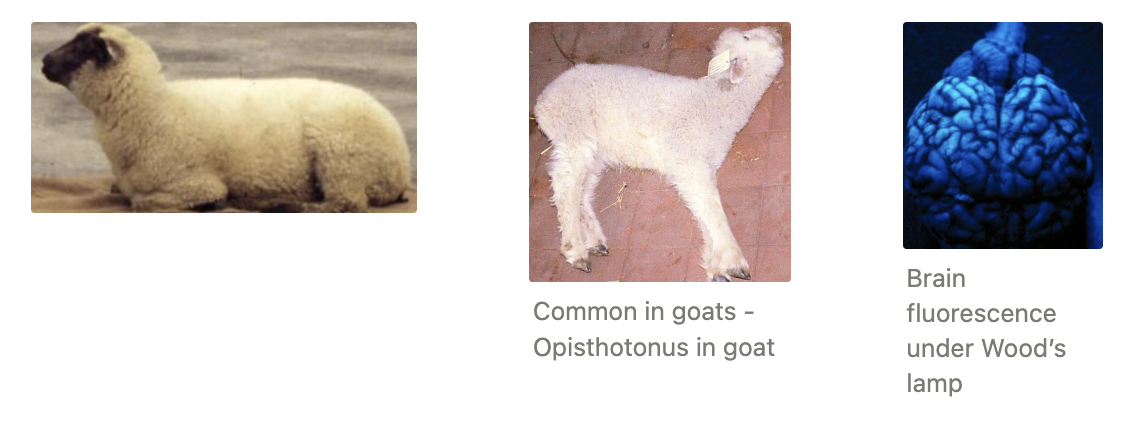
± Dog-sitting with head back (“star gazing”) to relieve pressure from cerebral oedema
→ Lateral recumbency, opisthotonos, seizures and death (3d progression)
Treatment: Rapid response to IV vitB1 → IM
Prognosis: Excellent (if treated before irreversible cerebral necrosis)
Response to treatment aids diagnosis
Transmissible Spongiform Encephalopathy (“Scrapie”)
Agent
2 Clinical signs
2 Methods of surveillance
Agent: Prion protein (exotic to NZ)
Long IP
Clinical Signs: Slowly progressive
Pruritic and nibbling form
Incoordination form
Surveillance: Prove to trading partners we are free from scrapie
Active = Random histology of brains at meatworks
Passive = Vet submits brain to lab when suspicious of disease
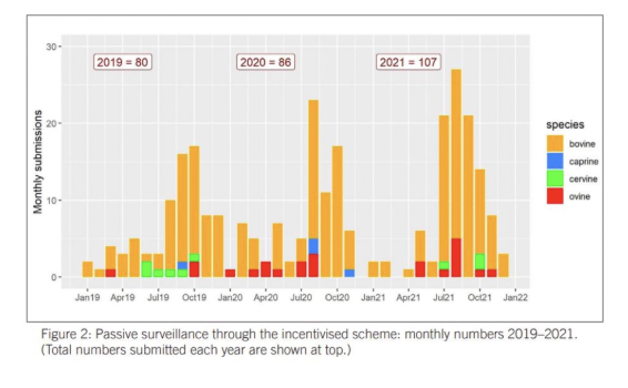
Monetary incentives for vet and farmer
Ovine Infectious Keratoconjunctivitis (“Pinkeye”)
2 Agents
Severity
Source
Morbidity
4 Progressive clinical signs
3 Treatments
Prevention
Agent: NO cell wall → Different AB requires to cattle
Chlamydia = Self-resolving when mild
Source: Dust, wind and flies → Irritation to eye
More common in summer/autumn (yarding and dry/stalky grass)
Mycoplasma = More severe ± recurrence
Source: Carrier sheep, transmitted by direct contact and fomites (dry/stalky grass)
Morbidity: 10 - 20% (highly contagious)
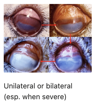
Clinical Signs: Unilateral OR bilateral
Red sclera with mild tear-staining
→ Spontaneous resolution OR
Progression to corneal oedema and severely red sclera
Apparent blindness
Pus in eye → Rupture
Treatment: Cattle penicillin-based pinkeye treatments are NOT effective in sheep
Spontaneous resolution (eg. mild Chlamydia)
Oxytetracycline diluted in spray bottle (Chlamydia and Mycoplasma)
Oxytetracycline SC/IM (Mycoplasma)
Prevention: Isolate affected animals (yarding increases transmission)
Entropion
Signalment
Clinical signs
2 Treatments
Prevention
Signalment: Recently born lambs
Clinical Signs: Eyelashes rub against cornea = Corneal ulcer
Risk of misadventure and mismothering
Treatments: Do NOT prescribe eye ointment to pet lambs without seeing them
Manually evert eyelid for 30s (mild cases)
SC injection of AB into lower eyelid (bleb evert eyelid)
Prevention: Cull carrier ram (inherited)
Vaginal Prolapse (“Bearings”)
Signalment
Prevalence
4 Risk factors
3 Protective factors
6 Treatments
Signalment: Late-gestation, multiple-bearing ewes
Prevalence: 1% (#1 welfare concern to farmer at lambing)
Risks are poorly understood as prevalence is highly variable between farms and years
Risks: Increased intra-abdominal pressure
High fecundity
Hill country (ewes sit with rump down to relieve weight from diaphragm)
Gain in weight pre-tup to scanning → Increased weight of placenta and conceptus
Feeding swede late pregnancy (high water content and bulky feed in rumen)
Protective:
Culling policy of bearing ewes and their offspring
Mid-pregnancy shearing
VitD injections during pregnancy? (Increase Ca2+ metabolism and muscle strength)
Treatment:
Assess viability of ewe and prolapse
Fresh prolapse vs. damaged/gangrenous
Herniation of intestines = Bad prognosis
Reduce vaginal prolapse
Epidural (1.8mL local + 0.2mL 2% xylazine) → Reduce straining and pain
Lift prolapse to allow urination (avoid bladder rupture)
Clean prolapse
Lubricate prolapse and use flats of hands to reduce
Place retaining suture/device (recurrence of prolapse possible as pre-lambing vs. post-calving)
Umbilical tape with purse-string suture around vulva = Vet insertion and farmer removal
Bearing harness #1 for farmer (reduce hunching during straining and enables lambing through harness)
Some farmers use ear tags and safety pins
Antibiotics (penicillin)
Analgesia (NSAIDs)
Monitor for lambing to remove retaining device