Unit 4
 Call Kai
 Learn
Learn Practice Test
 Spaced Repetition
 Match
Match Flashcards
 Knowt Play
1/139

There's no tags or description
Looks like no tags are added yet.
Name | Mastery | Learn | Test | Matching | Spaced | Call with Kai | Chat |
|---|
No analytics yet
Send a link to your students to track their progress
140 Terms
what part of the heart is MC injury in trauma
the Right ventricle due to anterior position 43%
penterating truama catagories
low velocity- ex knife stab
medium -
high - ex rifle or shrapnel injury
primary presentation of cardiac truama
tamponade and hemorrhage, tamponade decreases survival rate
tamponade occurs
when a clot and surrounding pericardial fat partially seals the defect
what if the diagnoist gold standard
subxiphoid window
tx of penetrating cardiac trauma
depends to the stability of the pt, most wounds are repaired through a left thoracotomy
when there is a blunt trauma
might not be able to tell about cardiac injury get and ECG, cardiac markers, holter, CT, or MRI
all pt who have significant mechanism on injury should have a
screening ECG
why are in v tachy and fib uncommon in hospital
cause most do not survive
how to treat trama
treat underlying cause, arrythmias are treated for rate control and suppression of ectopy, hemodynamic instability a definitive echo should be obtained. when there is severe ventricular dysfunction and low cardiac output, inotropic support to aviod a primary ischemic event
pericarditis
inflammation of the pericardium may be due to systemic dx, neoplasm, radiation, drug toxicity, hemopericardium, post cardiac surgery
types of pericarditis
acute less than 4 weeks
incessant less than 3 months
recurrent: after sx free interval
chronic: greater than three months
acute pericarditis
can be infecious and non infecious
infectious pericarditis
viral is MC- enterovirus, coxsackie and echo virus
purulent- most common S. aures, s. pneumoniae, other streptococci caused by hematogenous spread or from a close organ or complication from penetrating injury
TB
non infectious acute pericarditis
uremic, complication of CKD
neoplastic
early post MI typically occurs within several days of a transmural MI due to inflammation adjacent to myocardial necrosis
dressler syndrome: delayed immune-medicated pericarditis occurs weeks to months after MI
radiation, autoimmune, drug induced, injury
know the different between dressler and early post MI
sx of pericarditis
substernal cp: usually sharp and pleuritic that improves with sitting upright, worsen supine and can radiate to neck, shoulder, back, or epigastrium
friction rub if there is no increase pericardial fluid
what does pericarditis sx require
two of the following:
sharp pleuritic pain
pericardial friction rub
consistent changed on ECG
new or worsening pericardial effusion
what sx occurs pericardial effusion develops in pericardidits
dyspnea, rapid respiratory rate
what sx occurs pericardial temponade develops in pericardidits
pulsus paradoxus, JVD
pericarditis TX
viral or idiopathic is self limiting used NSAID and colchicine (for 3 months) can sue PPI protection for the high dose NSAID
if it purulent do an emergency surgical drainage and IV abx
if TB do regimen
make sure they rest from physical activity
pericardial effusions what should you look for
determine to size
small less than 10mm
mod: 10-20mm
severe: greater than 20mm/ 2cm much greater risk for developing tamponade
pericardial effusion to tamponade
is not dependent on size the rate of fluid accumulation determines hemodynamically consequences more than the size alone a rapidly developing smaller effusion can still cause a tamponade
what can echo differentiate on pericardial effusion
differentiate circumferential from loculated effusion
sx for effusion/tamponade
dyspnea and tachy
pleuritic CP
cough/ hiccups
hoarseness
syncope
sx are most common associated with rapidly accumulating effusion
PE findings for pleural effusion and tamponade
Normal physical exam is most common unless cardiac tamponade is present
• Tachycardia can be present without tamponade
• Hypotension
• Muffled heart sounds
• Elevated JVP
• Pulsus paradoxus
•Electrical alternans
what sx is most concerning for tamponade
syncope
Beck Triad
hypotension
JVD
muffled heart tones
may also have pulsus paradoxus and electical alternans
diagnostics for effusion/ tamponade
slide 29
hallmark finding for effusion on cxr
water bottle configuration
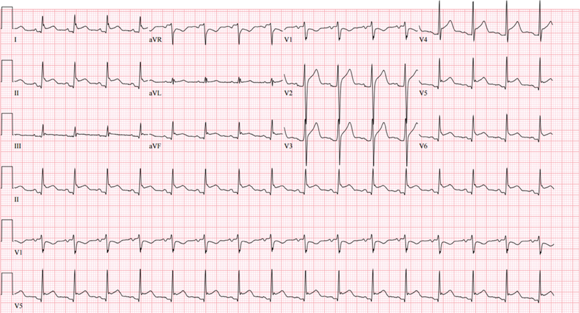
pericarditis
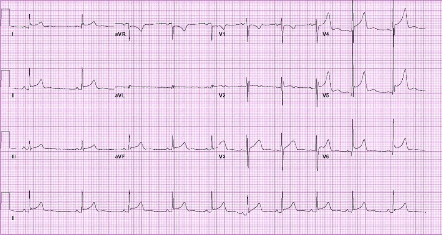
pericarditis
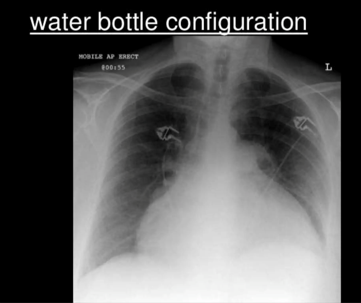
plerual effusion
pericardial treatment
Pain control with:
NSAIDS and colchicine
Treat underlying cause
Routine diagnostic pericardiocentesis and/or pericardial biopsy are not indicated unless high index of suspicion for a bacterial, fungal, or protozoal infectious etiology or suspicion of cancer.
Pericardiocentesis should be performed:
Cardiac tamponade
Large pericardial effusions
constrictive pericarditis
when a thickened fibrotic pericardium, impedes normal diastolic filling and produces chronically elevated venous pressure
what is MC od constrictive pericarditis
TB in undeveloped countries
echo distinctive finding for constrictive pericarditis
septal bounce that reflects the early filling
sx of con peri
•Slowly progressive dyspnea
•Fatigue, and weakness.
•Chronic edema
•Hepatic congestion ascites
oAscites often seems out of proportion to the degree of peripheral edema.
•Elevated jugular venous pressure
•Kussmaul sign—a failure of the JVP to fall with inspiration
•The apex may actually retract with systole and a pericardial “knock” may be heard in early diastole.
•Atrial fibrillation is common.
what imaging can you do for constrictive pericarditis
cxr: can show norm or cardiomegaly
echo shows septal bounce
cardiac CT or MRI
• Invasive hemodynamics are particularly useful when the diagnosis remains uncertain or when distinguishing constriction from restriction..
•Cath will help differentiate constrictive pericarditis from a restrictive cardiomyopathy
tx
§If inflammatory treat with anti-inflammatory medications
§ Diuretics for venous congestion and edema
§ Diuretics for symptomatic venous congestion — use cautiously to avoid excessive preload reduction., use loop diuretics (oral torsemide or bumetanide, if bowel edema is suspected or intravenous furosemide), thiazides, and aldosterone antagonists
§ If diuretics are no longer working: Surgical pericardiectomy.
oPericardiectomy removes only the pericardium between the phrenic nerve pathways
§ Morbidity and mortality after pericardiectomy are high (up to 15%)
Poor prognostic predictors include prior radiation, kidney dysfunction, higher pulmonary systolic pressures, abnormal LV systolic function, a lower serum sodium level, liver dysfunction, and older age
how to differentiate cardiomyopathy and constructive pericarditis
Restrictive Cardiomyopathy:
◦Diastolic dysfunction → ventricle can’t relax → high filling pressures.
◦Systolic function often preserved until late.
Pericarditis:
◦Thickened pericardium → abrupt stop in diastolic filling (“square root sign” on catheterization).
◦Myocardial contractility usually normal.
Presentation
Both can present with:
•Right heart failure signs (JVD, peripheral edema, ascites)
•Kussmaul’s sign (↑ JVP with inspiration)
•Pericardial knock (in pericarditis)
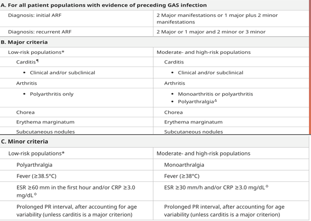
jones criteria
to determine rheumatic fever
we used revised jones criteria
JONES= joint pain, oh heart (carditis), nodules, erythema marginatum, sydham chorea
infective endocarditis
usually bacterial that cause inflammation of the endocardium and the valves.
I endocarditis usually involves
mitral valve and aortic valve MC
TV is most common with IV drug use
usually involves a valve
acute v subacute endocarditis
•Acute = S. aureus + aggressive + can attack normal valve
Subacute = viridans strep + indolent + usually abnormal valve
acute IE
Infection of previously normal heart valve by highly virulent organism (staphylococcus aureus) that rapidly produces necrotizing and destructive lesions
Death can occur within days to weeks despite appropriate treatment with antibiotics and surgery
subacute infective endocarditis
Infection by organisms of lower virulence (Streptococci viridans) that causes insidious infections of damaged/deformed valves (rheumatic heart disease, congenital bicuspid aortic valve, calcified/degenerative changes) with over all less destruction
Disease has course of weeks to months and cure can be achieved with antibiotics
IV drug user usually affects
the tricuspid valve
what destroys the cardiac tissue in infecious endo
the formation of vegetations
IE pathogenesis
1. Endocardial Injury
•Turbulent blood flow from an abnormal valve
•Injury from an intracardiac device/catheter
2. Platelets + Fibrin Deposit
•A small sterile platelet-fibrin deposit forms on the injured surface
3. Transient Bacteremia
•Bacteria enter the bloodstream
•Organisms adhere to the platelet-fibrin surface
4. Vegetation Forms
•Bacteria multiply within the platelet-fibrin material
•Vegetation can cause valve destruction, embolization, and persistent infection
•
Important Exception
•S. aureus is highly virulent and can infect a previously normal valve.
Valve injury → Platelet/Fibrin → Bacteremia → Vegetation
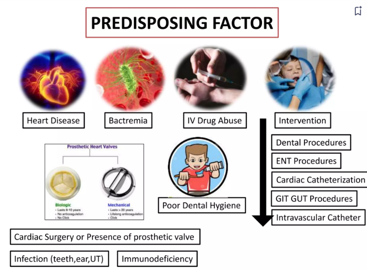
why are the predisposing factor
causes of the IE the bugs for native
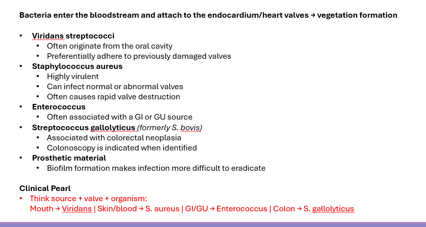
•S. aureus
•Streptococci (S. viridans), enterococcus (S. faecalis)
A more rare cause is the gram negative HACEK group
causes of the IE the bugs for IV
•S. aureus (MCC)
•Enterococci, streptococci
•Other organisms: Gram negative (Pseudomonas), Fungi (Candida)
causes of the IE the bugs for prosthetic valve
•Early (<12 months): nosocomial infections: Staphylococci – S. aureus, S. epidermidis, gram-negative and fungi (Candida).
•Late (>12 months postop)- similar to NVE (SE for early surgery)
figure out
fungal IE
common with heroin that is black tarr cause they have to dissolve in lemon acid
what are risk factors for native valve endocarditis
Mitral valve
•Mitral valve prolapse, mitral regurgitation
•Rheumatic heart disease (now less common)
Aortic valve
•Calcific aortic stenosis (60-70 year old)
•Bicuspid aortic valve (50-60 year old)
•Rheumatic heart disease (now less common)
Tricuspid valve (IV drug use)
Ventricular septal defects (and other congenital heart defects)
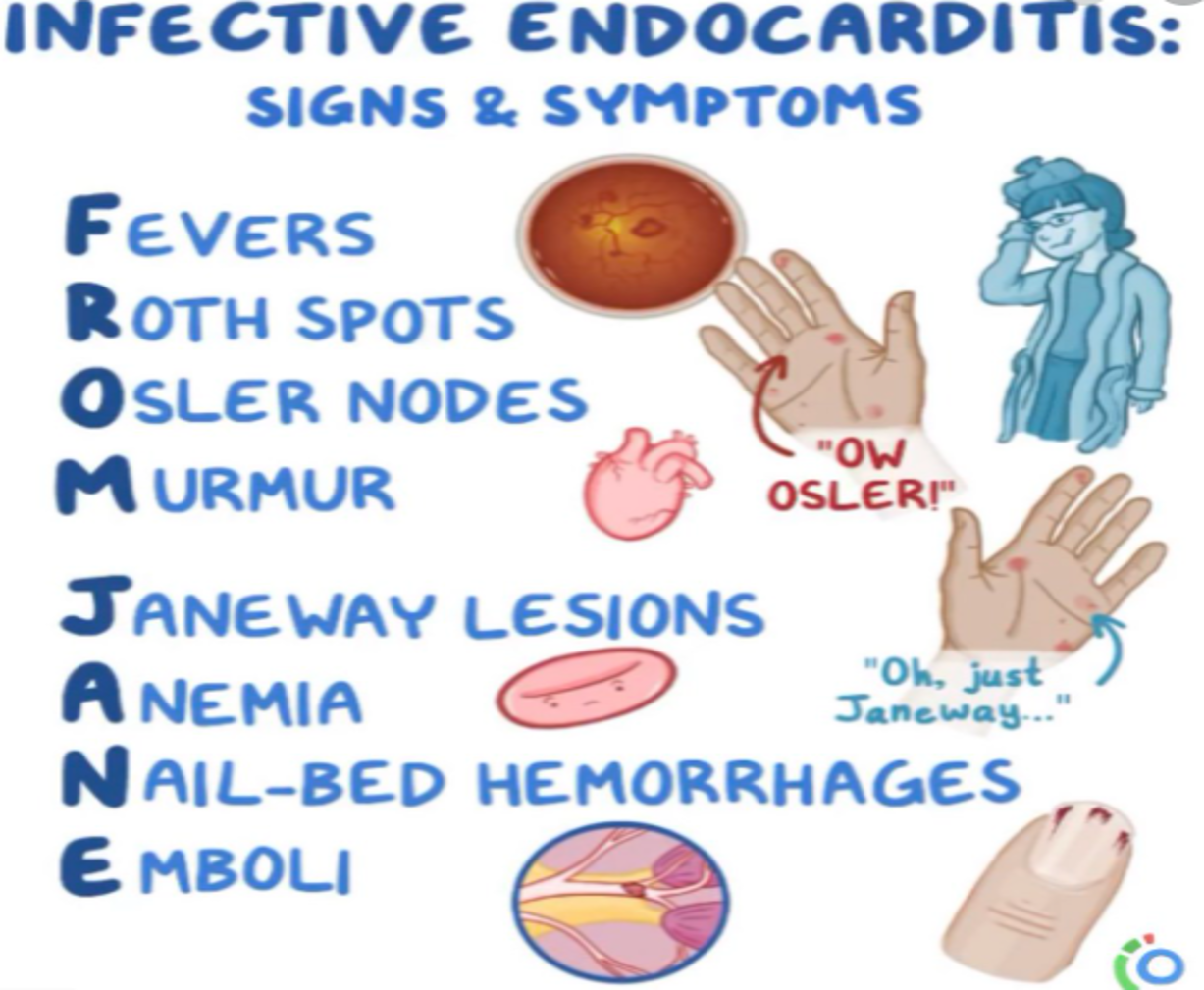
CM of IE
fever MC sx with possible chills, rigors, night
heart murmur hear in nearly all but might not be heard in right sides (IV drugs)
fatigue, arthralgia
anemia
left sided valve lesions can lead to regurg and new onset of left sided heart failure
stroke pt with fever
ENDOCARDITIS until proven otherwise the cause of the embolism can be broken off parts of the vegetation
new or acute aortic regurg is
IE until proven otherwise
CM of IE
How to dx IE
blood culture: obtain 3 from separate venipuncture site before abx when the pt is stable should be more than 10mL
echo (TTE is first line can to TEE when need investigate more
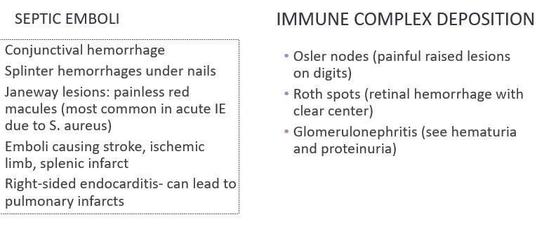
can looks for septic emboli
testing for immune complex deposition
other labs 1.may show anemia, leukocytosis, elevated ESR/CRP
what criteria is used of IE
the Duke must know
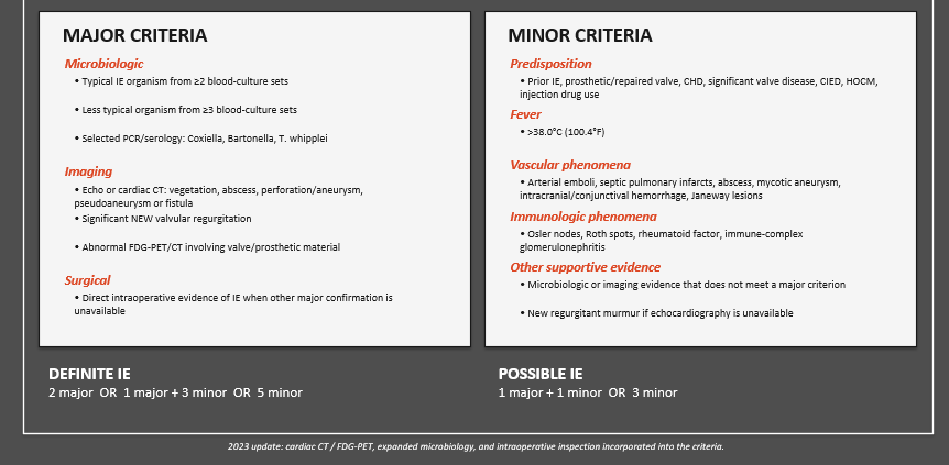
tx of IE
1.Multidisciplinary team: Infectious Disease, Cardiology, Cardiothoracic surgery
2.Draw blood culture before giving any antibiotics!!
3.Start empiric IV therapy promptly in acutely ill patients.
4.Tailor therapy to organism, susceptibilities, native vs prosthetic valve and local resistance.
5.Treatment is usually prolonged IV antimicrobial therapy.
6.IV antibiotics:
•2-6 weeks for native valve
• ≥6 weeks for prosthetic valve
7.Consider early surgery when indicated (usually 7-10 days if not better on abx)
what imaging to look for septic emboli
: Chest Xray or chest CTA, MRI of brain, heart, spine, abdominal CT or ultrasound
how to test for immune complex deposition
urinalysis, BMP, rheumatoid factor
empiric tx for IE
Empiric treatment with antibiotics Only if severe/acute. You want to cover for most common organisms (staph, strep, enterococcus)
Best to consult ID before starting empiric
•NVE(native valve): Vancomycin + ceftriaxone (Cipro if severe PCN allergy)
•PVE: vancomycin + cefepime or piperacillin-tazobactam with antipseudomonal activity
These will change frequently
do you need to use anticoagulation for IE
not need for native
CAN NOT use if septic can cause intracerebral hemorrhage
if they have mechanical valve they will usually get anticoag
when should prophylaxis tx be given
1. Prosthetic cardiac valve (includes bioprosthetic and mechanical)
2. Previous infective endocarditis
3. Congenital heart disease (CHD)
◦Unrepaired cyanotic CHD, including palliative shunts and conduits
◦Completely repaired CHDs with prosthetic material or device during the first 6 months
◦Repaired CHD with residual defects at the site or adjacent to the site of the prosthetic patch or device
4. Cardiac transplantation recipients in whom cardiac valvulopathy develops
treatment for surgery prophylaxis (like what kind of procedure needs prophylaxis)
•Dental procedures involving manipulation of gingival tissue, periapical area, or perforation of the oral mucosa. (Not for minor procedures)
•Respiratory tract procedures with incision/biopsy of mucosa (e.g., tonsillectomy, adenoidectomy).
•Procedures treating infected skin or musculoskeletal tissue (e.g., abscess drainage), using appropriate antibiotics against staph and β-hemolytic strep.
when is surgery needed for IE
•Acute heart failure unresponsive to medical therapy
•No response to antimicrobial therapy after 7-10 days (persistent fevers, + blood cultures)
•>1 systemic embolus
•Perivalvular leak or myocardial abscess
•Severe valve regurgitation and mobile vegetation > 10 mm
You are doing
great
ambulatory monitoring depends on
frequency of sx
when to do holter
sx that are hours to weeks just records will not be able to react
when to get event/ mobile cardiac telemetry
intermittent sx that are over weeks (they can press a button to get immediate help?)
when to use implantable cardiac monitor (loop recorder)
infrequent or unexplained events - long term monitoring - does not do anything about the rhythm unless put with pacemaker
what can wearable devices detect
possible a fib but cannot establish dx, abnormal findings needs ECG confirmation, performance varies by device and population
what is an EP study
an invasive catheter-based test that maps the heart electrical activity
electrode catheters are advanced through vein into the heart and records electrical signals from inside the heart
What does an EP study mmonitor
•Location and mechanism of an arrhythmia
•SA and AV node/conduction system function when clinically indicated
•Presence of an accessory pathway (e.g., WPW)
•Ability to induce an arrhythmia under controlled conditions
when to use EP study
•Recurrent or unexplained SVT
•Suspected AVNRT or AVRT/WPW
•Selected wide-complex tachycardias
•When catheter ablation is being considered
what is catheter ablation
•Minimally invasive procedure used to eliminate abnormal electrical pathways or arrhythmia-producing tissue
•An EP study first maps/localizes the abnormal electrical activity
•Radiofrequency energy, cryotherapy, or other energy sources are then used to ablate the target tissue
common use of cather ablation
•AVNRT → ablate the slow pathway
•AVRT/WPW → ablate the accessory pathway
•Typical atrial flutter → ablate the reentry circuit
•Focal atrial tachycardia → ablate the ectopic focus
•Atrial fibrillation → usually involves pulmonary vein isolation (PVI)
•Ventricular tachycardia → may be used in selected/recurrent VT
what can catheter ablation do for AVNRT
ablate the slow pathway
catheter ablation for AVRT/WPW
ablate the accessory pathway
what will catheter ablation for focal atrial tachy
ablate the ectopic focus
what will catheter ablation do for ventricular tachy
may be used in selected/ recurrent VT
what testing can fing the electrical problem
EP study finds and maps the electrical problem
what can treat electrical problems
catheter ablation
what are the mechanism of arrhythmias
abnormal automaticity
abnorm impulse conduction
reentry
triggered activity
why do arrhythmias happen
the impulse starts abnormally, travels abnormally, gets caught in a loop or triggers an extra beat
abnormal automaticity
cardiac cells fire when they should not
creates and abnormal/ ectopic pacemakers
ex are PA, PCV, focal tachycardia
abnormal impulse conduction
•Electrical impulse is delayed or blocked
•Examples: AV blocks, bundle branch blocks
reentry
•Electrical impulse travels in a continuous loop, repeatedly reactivating tissue
•Major mechanism of many tachyarrhythmias
•Examples: AVNRT, AVRT/WPW, atrial flutter
triggered activity
•An extra impulse occurs after the normal action potential
•Often associated with electrolyte abnormalities, medications, or prolonged QT
•Example: torsades de pointes
No clear P waves + irregularly irregular
AF
sawtooth flutter waves
a flutter
Regular narrow-complex tachycardia
think SVT a type of AVNRT
Wide-complex tachycardia
VT first, with structural heart dx
Progressively longer PR → dropped QRS
mobitz 1
Fixed PR + sudden dropped QRSs
MObitz II
P waves and QRS march independently
complete heart block