TOP 1 HISTOLOPATHOLOGIC TECHNIQUES EXAM
 Call Kai
 Learn
Learn Practice Test
 Spaced Repetition
 Match
Match Flashcards
 Knowt Play
1/70

Earn XP
Description and Tags
TOP 1 EXAM
Name | Mastery | Learn | Test | Matching | Spaced | Call with Kai | Chat |
|---|
No analytics yet
Send a link to your students to track their progress
71 Terms
Fixation
One of the most critical step in histotechnology
Process that preserves tissues from decay, thereby preventing autolysis and putrefaction
The process should be carried out as soon as possible after removal of tissue from the body
Autolysis and putrefaction
What does fixation prevent?
Goal of fixation
Preserve the morphologic and chemical integrity of the cell in as life - like manner as possible.
Harden and protect the tissue from the trauma of further handling.
Objectives of fixation
To preserve the tissue (stops all cellular activities)
To prevent breakdown of cellular elements (prevent autolysis and putrefaction)
To coagulate or precipitate protoplasmic substances (renders tissue components insoluble)
Hydrogen ion concentration
Temperature
thickness of the section (block)
Osmolality
Concentration of solutions
Duration of fixation
Main factors involved in fixation
Speed
Penetration
Volume
Duration of fixation
Practical considerations of fixation
Hydrogen ion Concentration
(pH 6 to 8)
for routine surgical specimens, room temperature.
Temperature
Thickness of the section (block):
Electron microscopy: 1 to 2 mm²
Light microscopy: 2 mm² (recommended / 4-10mm)
Measurements should not be compromised to obtain full penetration and satisfactory fixation.
Large tissues like the uterus should be opened or sliced thinly.
Hypertonic
causes cell shrinkage.
Isotonic and Hypotonic
causes cell swelling and poor fixation.
For best results,
use slightly hypertonic solutions with an osmolality of 400 - 450 mOsm (isotonic solutions are 340 mOsm).
Concentration of solutions
Formaldehyde is most commonly used as 10% solution while glutaraldehyde is used as 3% solution. Low concentration (0.25%) of glutaraldehyde is ideal for immunoelectron microscopy.
Prolonged fixation:
may cause cell shrinkage and hardening of tissues.
Incomplete fixation:
softening of tissues, very hard to cut during section - cutting.
Speed
specimens are placed in fixative as soon as it is removed from the body. This is done to prevent autolysis and putrefaction.
Penetration
formalin diffuses into tissues at a rate of approximately 1 mm per hour and slows down as it goes deeper. However, this varies with different types of tissues.
Volume
the ideal fixative to tissue ratio is 20:1
Duration of fixation
the usual length of time for fixation is 24 hours but some tissues take longer times to fix than others.
Simple Fixatives:
made up of only one component substance
Compound Fixatives:
made up of two or more fixatives
Microanatomical Fixatives
Permit general microscopic study of tissue structures and normal intercellular relationship of tissues
- for general microscopic study of tissue structures.
Cytological Fixatives:
preserve specific cellular components
Nuclear fixatives
preserves nuclear structures of the cell. They usually contains glacial acetic acid (pH 4.6 or less) which fixes nucleoprotein.
Cytoplasmic fixatives
Useful for preservation of cytoplasmic details or structures (pH greater than 4.6). This must never contain glacial acetic acid because it destroys mitochondria and Golgi bodies.
Histochemical fixative
Preserve chemical constituents of the cell
Lipids:
mercuric chloride or potassium dichromate
Phospholipids:
Baker's formol-calcium
Cholesterol:
digitonin
Carbohydrates:
alcoholic fixatives
Glycogen:
Rossman's fluid or cold absolute alcohol
Proteins:
neutral buffered formol saline or formaldehyde vapor
Electron microscopy:
double fixation
Electron Cytochemistry:
Karnovsky's paraformaldehyde_glutaraldehyde solution
Acrolein:
mixture of glutaraldehyde or formaldehyde
rapid penetration, preserves morphology and enzyme activity at low concentrations v immersion fixation of surgical biopsies.
Aldehyde fixatives
for routine paraffin sections, electron microscopy, histochemistry and enzyme studies
Formaldehyde (Formalin)
gas produced by the oxidation of methyl alcohol
buffered atph 7 to 8
— hypoxia in tissues leads to acidity which favors the
formation of Formalin heme pigments (black, polarizable
deposits)
Pure Stock: 40% formalin
Dilution: 1:10 (10% solution); 1:20 (5% solution)
Paraformaldehyde
white precipitate due to prolonged storage
may be removed by filtration or addition of 10% methanol
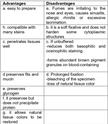
10% Formol-Saline
central nervous tissues and general post-mortem tissues for histochemical
examination
ideal with most stains including silver impregnation
duration of fixation: more than 24 hours (slow fixative)
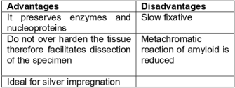
10% Neutral Buffered Formalin
MOST WIDELY USED FIXATIVE IN ROUTINE
HISTOLOGY
preservation and storage of surgical, post-mortem and research specimens
best fixative for frozen sections
best fixative for iron pigments and elastic fibers
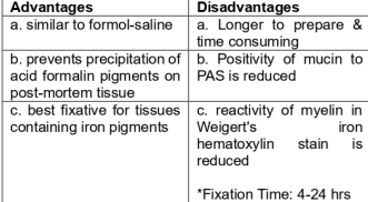
Formol-Corrosive
routine post-mortem tissues
for lipids, especially neutral fats and phospholipids
no need for washing-out
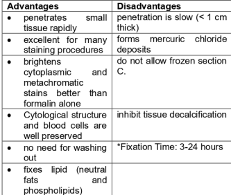
Glutaraldehyde
* made up of two formaldehyde residues linked by three
carbon chains
used in conjunction with osmium tetroxide
» Fixation time: ½ hour to 2 hours
ADVANTAGES
a. stable effect on tissues
b. preserves plasma
c. less tissue shrinkage
d. recommended for enzyme histochemistry & EM
e. it does not cause dermatitis and less irritating to the nose
DISADVANTAGE
| b. penetrates tissues more slowly
| c. tends to make tissue proteins better more brittle
Alcoholic Formalin
= used to fix sputum
« for the demonstration of immunoperoxidase activity
Mercuric Chloride
most common metallic fixative
V tissue photography
V tissues contain black precipitates of mercury (except Susa)
Zenker’s Fluid
fixing small pieces of liver, spleen, connective tissues fibers and nuclei
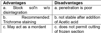
De-zenkerization:
removal of mercuric deposits in tissues
Zenker-formol (Hellys solution)
pituitary gland, bone marrow and blood containing organs (spleen and liver)
*Fixation time: 4-24 hours
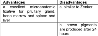
Heidenhain's Susa Solution
v for tumor biopsies
v excellent cytologic fixative
*after using This, transferred the tissue to a high grade alcohol to avoid undue swelling.
Fixation Time: 3-12 hours
B-5 Fixative
for bone marrow biopsies
Osmium Tetroxide(Osmic acid; Os04)
v used in EM both as fixative and a heavy metal
stain.
v excellent stain for lipids in membranous structure
and vesicles.
Chromic acid
preserves carbohydrates
Potassium Dichromate
preserves lipids and mitochondria
Regauds Fluid (Mullers Fluid)
v demonstration of chromatin, mitochondria, mitotic figures, Golgi bodies, RBC and colloid-containing tissue.
*Fixation time: 12-48 hours
Orth's Fluid
v study of early degenerative processes and tissue
necrosis
v demonstrates Rickettsia and other bacteria
*Fixation Time: 36-72 hours
LEAD FIXATIVES
v demonstration of acid mucopolysaccharides
PICRIC ACID FIXATIVES
are soluble in water, therefore, tissues should not be washed in running tap water (use 70% alcohol instead)
Bouin’s Solution
v fixation of embryos and pituitary biopsies.
Brasil's Alcoholic Picroformol Fixative
v fixative for glycogen
GLACIAL ACETIC ACID
v fixation of nucleoproteins
Ethanol (95%)
- Preserves but does not “fix” glycogen granule
Methanol
- for dry and wet smears, blood and bone marrow samples
Isopropyl Alcohol (95%)
- for touch preparation
Carnoy’s fluid
- most rapid fixative
- for chromosomes, lymph glands, urgent biopsies and brain tissue for the diagnosis of rabies
.Newcomer’s
- demonstration of mucopolysaccharides and nucleoprotein
Gendre’s fixative
alcoholic form of bouins solution
Alcoholic Formalin
used for fixation or post-fixation of large fattyspecimens(particularly breast).
Flemming's (chrome-osmium HAc)
- Permanently fixes fats and recommended for fixing nuclear structures
. Flemming's w/o acetic acid
- For cytoplasmic structure
TRICHLOROACETIC ACID
v may also be used as a weak decalcifying agent
ACETONE
v use at cold temperature (-5 to 4 °C)
v fixation of brain tissues for rabies diagnosis
WASHING OUT
- The process of removing excess fixative from the
tissue after fixation in order to improve staining
and remove artefacts from the tissues.
- Several solutions may be used: tap water, 50 -
70% alcohol and alcoholic iodine.