PREMALIGNANT & MALIGNANT LESIONS
 Call Kai
 Learn
Learn Practice Test
 Spaced Repetition
 Match
Match Flashcards
 Knowt Play
1/55

There's no tags or description
Looks like no tags are added yet.
Name | Mastery | Learn | Test | Matching | Spaced | Call with Kai | Chat |
|---|
No analytics yet
Send a link to your students to track their progress
56 Terms
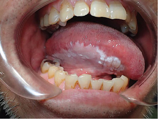
CASE: A 58-year-old patient has a persistent white plaque on the lateral tongue. It does not wipe off, and no specific traumatic, infectious, or other white-plaque diagnosis explains it. What is the best clinical term for this lesion, and what does that term imply?
ANSWER: Leukoplakia. It is a clinical description for a white plaque that cannot be characterized clinically or pathologically as another disease. It does not name a specific histopathologic alteration, but its use usually implies concern for a premalignant process. A biopsy is required for a tissue diagnosis and management planning.

CASE: An oral biopsy shows irregular epithelial stratification, loss of basal cell polarity, drop-shaped rete ridges, increased and abnormally superficial mitoses, dyskeratosis, anisonucleosis, nuclear and cellular pleomorphism, increased nuclear-to-cytoplasmic ratio, atypical mitoses, enlarged nucleoli, and hyperchromasia. What process is the pathologist describing?
ANSWER: Epithelial dysplasia. The lecture divides dysplastic changes into architectural abnormalities and cytologic abnormalities. These features support a dysplastic epithelial process rather than a purely clinical label such as leukoplakia.

CASE: Two leukoplakic lesions are biopsied. Lesion A shows moderate epithelial dysplasia; lesion B shows severe epithelial dysplasia. Which lesion has the greater malignant transformation potential, and what approximate ranges were emphasized in lecture?
ANSWER: Severe dysplasia has the greater risk. Lecture values: moderate dysplasia ≈ 4–11% malignant potential; severe dysplasia ≈ 20–43% malignant transformation potential.

CASE: You are comparing three persistent oral white lesions: one is uniformly smooth and white, one is rough/nonhomogeneous, and one has mixed red-and-white speckling. Rank them from least to most concerning for a premalignant/malignant process.
ANSWER: Homogeneous → nonhomogeneous → speckled/erythroleukoplakia. General rule: the less homogeneous, less uniformly white, and less smooth a lesion is, the more worrisome it becomes.

CASE: A large heterogeneous leukoplakic lesion contains a smooth white area and a rough red-white area. You plan an incisional biopsy. From which area should you sample? If biopsy shows moderate dysplasia or worse, what management principle was emphasized?
ANSWER: Biopsy the most clinically severe/concerning area. Moderate dysplasia or worse warrants complete removal if feasible. Mild dysplasia may be excised if practical; otherwise close monitoring is appropriate. Follow-up is extremely important.
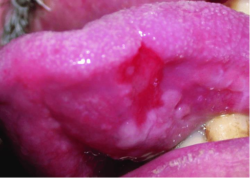
CASE: A previously white oral plaque develops scattered red areas. The red regions appear thin and nonkeratinized compared with the white portions. What is the clinical diagnosis, and why are the red areas concerning?
ANSWER: Erythroleukoplakia (speckled leukoplakia). The red areas reflect epithelium that is too immature or atrophic to produce keratin. These lesions frequently show advanced dysplasia on biopsy. Lecture pearl: red lesions are worse than white lesions.

CASE: A patient with mild epithelial dysplasia has no clinical change over several years of surveillance. The patient asks whether every dysplastic lesion eventually becomes cancer. How should you answer based on the lecture?
ANSWER: No. Not all dysplasia or leukoplakia progresses. Some mild dysplasia can remain mild indefinitely, which is why risk-based follow-up rather than assuming inevitable cancer is important.

CASE: A long-term moist snuff user places tobacco in the mandibular vestibule. The mucosa at that exact site is thin, rippled, almost translucent, soft/velvety, and has an indistinct border. There is local gingival recession and staining, but no pain, ulceration, or induration. What is the most likely diagnosis?
ANSWER: Tobacco pouch keratosis (smokeless tobacco keratosis). Histology is nonspecific, commonly hyperkeratosis and acanthosis. Epithelial dysplasia is uncommon and, when present, is typically mild.

CASE: A patient has classic tobacco pouch keratosis. What is the first management step, and when is biopsy indicated?
ANSWER: Recommend stopping all smokeless tobacco for about 2–4 weeks, then re-evaluate. If the lesion persists, biopsy is indicated. If the patient will not stop, encourage switching placement sides and re-evaluate. Even after no or mild dysplasia, monitor regularly and re-biopsy if size, texture, color, or symptoms change.

CASE: A patient with a long history of betel quid use reports progressive difficulty opening the mouth, generalized oral burning, and intolerance to spicy foods. Exam shows blotchy marble-like mucosal pallor, stiffness, reduced tongue mobility, and palpable fibrous bands in the buccal and labial mucosa. What is the diagnosis?
ANSWER: Oral submucous fibrosis. It is a high-risk precancerous condition characterized by chronic progressive scarring of oral mucosa and is linked to betel quid/areca nut exposure.

CASE: A patient with submucous fibrosis has a maximum interincisal opening of 16 mm. How severe is this finding, and what cancer-risk facts and follow-up principle were emphasized?
ANSWER: Severe trismus; <20 mm is considered severe. Dysplasia is found in about 10–15% of biopsies, SCC in at least 6%, and patients are about 19 times more likely to develop oral cancer. Close clinical follow-up is required. Unlike standard tobacco pouch keratosis, the condition does not regress simply with habit cessation.
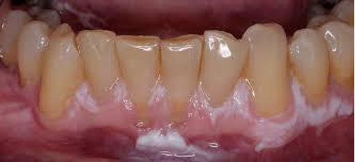
CASE: An older woman who does not use tobacco develops multiple rough, spreading keratotic plaques involving the gingiva and other oral sites. The plaques recur and continue to enlarge despite several biopsies and excisions. What is the most likely diagnosis?
ANSWER: Proliferative verrucous leukoplakia (PVL). It is a special high-risk form of leukoplakia, has a strong female predilection, has minimal association with tobacco, and is a clinical rather than histologic diagnosis. SCC often develops within about 8 years of the initial PVL diagnosis.
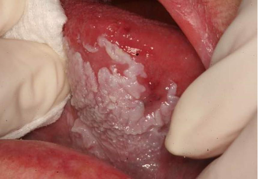
CASE: A patient with established PVL develops a new keratotic plaque while older plaques are changing in surface texture. What is the appropriate long-term strategy?
ANSWER: Indefinite regular monitoring with frequent biopsies. New leukoplakias should be biopsied, and existing plaques should be re-biopsied when size, shape, or surface texture changes.
CASE: Three malignant tumors arise from different tissues: one from oral epithelium, one from connective tissue, and one from melanocytes. What broad cancer term applies to each?
ANSWER: Epithelial malignancy = carcinoma. Connective-tissue malignancy = sarcoma. Melanocyte malignancy = melanoma.
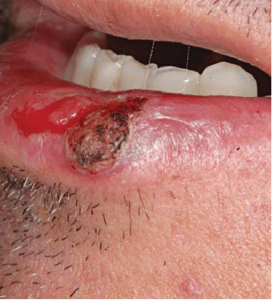
CASE: You are counseling patients about site-specific oral cancer risks. Which extrinsic exposure is especially relevant to vermilion lip cancer, which infectious risk is especially relevant to oropharyngeal cancer, and which two classic exposures are major oral-cavity risks?
ANSWER: Vermilion lip: sunlight. Oropharynx: HPV. Oral cavity: tobacco smoke and alcohol are major extrinsic risks. The lecture also lists intrinsic/generalized factors such as malnutrition and iron-deficiency anemia.
CASE: A patient with known Plummer-Vinson syndrome asks why oral cancer screening matters. Which cancer sites were specifically noted as having increased risk in the lecture?
ANSWER: Oral, oropharyngeal, and esophageal cancers.

CASE: A 67-year-old man with a long history of heavy smoking and alcohol use reports a lateral tongue lesion present for 6 months. It is painless, firm/indurated, and ulcerated. What is the most likely diagnosis?
ANSWER: Conventional oral squamous cell carcinoma (traditional SCC presentation). The classic lecture pattern is an older man with a persistent lateral-tongue lesion for 4–8 months plus heavy smoking and alcohol history.

CASE: A patient has a persistent painless indurated ulcer on the posterolateral tongue. Which oral malignancy should be considered first?
ANSWER: Squamous cell carcinoma. The posterolateral/ventral tongue is one of the most common intraoral SCC sites, and tongue SCC commonly presents as a painless indurated mass or ulcer.

CASE: An older outdoor worker develops a slowly enlarging destructive ulcer on the lower lip vermilion. The lesion has been neglected for months. What diagnosis is most consistent with the lecture, and how does it differ from typical intraoral SCC?
ANSWER: Squamous cell carcinoma of the lip vermilion. Its pathophysiology is more like SCC of sun-exposed skin. It is typically slow growing, and fewer than 10% of patients have lymph-node metastasis, although neglected lesions can cause major local destruction.

CASE: A patient with a long-standing red-white precursor lesion on the floor of the mouth develops a persistent exophytic/ulcerative mass. Which malignancy is most likely, and what precursor relationship is especially important at this site?
ANSWER: Floor-of-mouth squamous cell carcinoma. Among intraoral carcinomas, floor-of-mouth lesions are the most likely to arise from a pre-existing leukoplakia or erythroplakia/erythroleukoplakia.
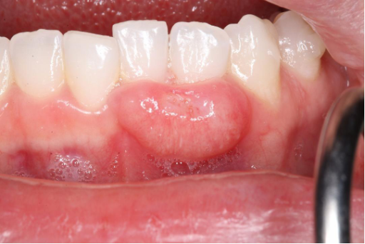
CASE: An older woman with little to no tobacco history has a persistent irregular gingival mass/ulcer that has not responded to routine dental care. Which malignancy should remain high on the differential despite the absent smoking history?
ANSWER: Gingival squamous cell carcinoma. Gingival SCC is the oral SCC site least associated with tobacco smoking and has the greatest female predilection.
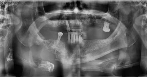
CASE: A panoramic radiograph shows a destructive intrabony jaw lesion. Biopsy demonstrates squamous cell carcinoma, with the dominant disease process centered within bone rather than presenting primarily as a surface mucosal lesion. What lecture entity does this represent?
ANSWER: Intrabony squamous cell carcinoma.
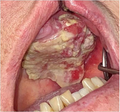
CASE: A patient has a persistent ulcerative, destructive lesion of the palate. Biopsy shows squamous cell carcinoma. Which site-specific lecture entity is this?
ANSWER: Palatal squamous cell carcinoma.
CASE: A patient has newly diagnosed oral SCC. The oncology team documents the primary tumor, cervical lymph-node status, and whether distant spread is present. What staging framework is being used, and what primary-tumor features were emphasized in the lecture?
ANSWER: TNM staging: Tumor, Nodes, Metastases. The lecture emphasizes tumor depth of invasion and tumor size, plus lymph-node and distant metastases. These factors affect treatment and prognosis.

CASE: A biopsy is clearly epithelial and shows abundant keratinization. Invasion is present but subtle enough that it takes careful examination to identify. What grade of conventional SCC best fits?
ANSWER: Well-differentiated SCC. Well-differentiated tumors have abundant keratinization and retain obvious epithelial differentiation.
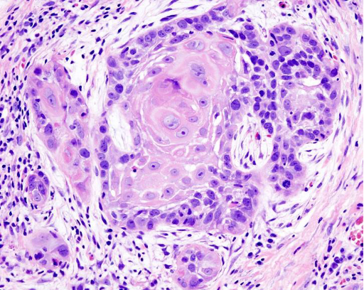
CASE: A biopsy is unmistakably epithelial and also unmistakably invasive. Keratinization and atypia are intermediate between the well- and poorly-differentiated ends of the spectrum. What grade is most likely?
ANSWER: Moderately differentiated SCC. The lecture notes this is the most common grade and gives the practical clue: you are sure it is epithelial and sure it is invasive cancer.

CASE: A highly atypical invasive malignant tumor has minimal keratinization and marked cellular/nuclear pleomorphism. It is obviously malignant and invasive, but the epithelial origin is difficult to recognize and the cells may appear spindled. What grade is most likely?
ANSWER: Poorly differentiated SCC. Poorly differentiated tumors may have little to no keratinization and can resemble a sarcoma.

CASE: Two epithelial biopsies show severe atypia. In specimen A the abnormal epithelium remains confined to the epithelial compartment; in specimen B malignant cells extend into the underlying connective tissue. Which one is invasive SCC?
ANSWER: Specimen B is invasive SCC. The slide diagram contrasts carcinoma in situ with invasive SCC by whether malignant epithelium has extended into the underlying tissue.
CASE: A 43-year-old woman who has never smoked has a persistent tongue lesion. A clinician initially considers cancer unlikely because she does not fit the traditional demographic pattern. What lecture point argues against dismissing SCC on that basis?
ANSWER: Tongue cancer has been increasing in patients who do not fit traditional demographics, particularly younger patients in their 40s who are nonsmoking females. The lecture also notes a study association with ≥1 sugar-sweetened beverage/day (4.87× higher likelihood), while emphasizing that the overall likelihood remains low.

CASE: A smokeless-tobacco user develops a diffuse, painless, thick white plaque with a verruciform surface in the mandibular vestibule. It is locally extensive but there is no evidence of nodal metastasis. What is the most likely diagnosis?
ANSWER: Verrucous carcinoma, a low-grade variant of SCC. Common sites include mandibular vestibule, buccal mucosa, gingiva, tongue, and hard palate. Metastases are very uncommon, but local destruction can be substantial.
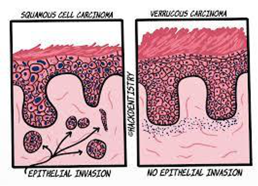
CASE: A broad verruciform oral tumor has architecture consistent with verrucous carcinoma. Why is distinguishing it from conventional SCC clinically important?
ANSWER: Verrucous carcinoma is diagnosed more by epithelial architecture than cytologic dysplasia and, unlike conventional SCC, does not show the same invasive pattern emphasized in the lecture. Treatment is usually more conservative surgery and generally does not include lymph-node dissection.
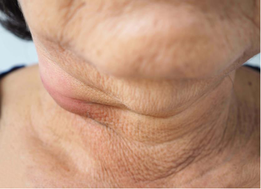
CASE: A 48-year-old patient with little smoking or drinking history presents with an asymptomatic enlarged cervical lymph node and mild dysphagia. Examination identifies a lesion in the tonsillar/base-of-tongue region. Which cancer subtype should be strongly considered?
ANSWER: HPV-positive oropharyngeal squamous cell carcinoma. Oropharyngeal sites include soft palate, tonsils, base of tongue, and pharyngeal wall. HPV16 is the most common strain emphasized, and an asymptomatic cervical node is a common presenting sign.

CASE: A tonsillar SCC biopsy is reported as p16 positive, and cervical lymph-node metastasis is already present. Does nodal spread automatically imply a worse prognosis than conventional SCC, and what prevention point was emphasized?
ANSWER: No. HPV-positive SCC commonly has nodal spread by diagnosis yet has a better prognosis than conventional SCC. The biopsy report may identify it as p16+ SCC. Gardasil protects against the high-risk HPV strains discussed in the lecture.
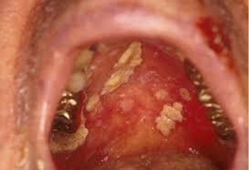
CASE: A patient receiving anticancer therapy develops painful oral ulcerations and easy oral bleeding. Which two oral complications were emphasized as the most likely complications of cancer treatment?
ANSWER: Mucositis and hemorrhage. Other possible outcomes listed include xerostomia, loss of taste, osteoradionecrosis, trismus, dermatitis, and developmental abnormalities.
CASE: A chemotherapy patient develops spontaneous gingival/oral bleeding. Laboratory testing shows a markedly reduced platelet count from marrow suppression. What is the mechanism of the oral hemorrhage?
ANSWER: Thrombocytopenia secondary to bone-marrow suppression.

CASE: Patient A develops painful mucositis within a few days of systemic chemotherapy, mainly on unattached mucosa. Patient B develops mucositis during the second week of head-and-neck radiation, involving tissues throughout the radiation field. Which treatment pattern matches each patient?
ANSWER: Patient A = chemotherapy mucositis; Patient B = radiation mucositis. Both can produce very painful ulcerations with removable yellowish fibrinous surfaces. Mucositis generally resolves slowly over about 2–3 weeks after treatment cessation.
CASE: A patient receiving high-dose chemotherapy asks whether ice-chip cryotherapy can reduce oral mucositis. What protocol and limitations were emphasized in the lecture?
ANSWER: Place ice chips in the mouth about 5 minutes before chemotherapy and continue for about 30 minutes; cold may work through vasoconstriction. The lecture notes no evidence for effectiveness in radiation therapy. Palifermin is more associated with hematologic cancers and is not approved for use in carcinoma.

CASE: One year after head-and-neck radiation, a patient has exposed nonvital mandibular bone that has persisted for more than 3 months. There is no evidence of recurrent neoplastic disease. What is the diagnosis?
ANSWER: Osteoradionecrosis (ORN): exposed, nonvital irradiated bone persisting >3 months in the absence of neoplastic disease. It most often occurs about 4 months to 3 years after radiation.
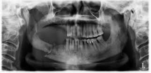
CASE: A patient is scheduled for head-and-neck radiation and has several nonrestorable, periodontally involved teeth. What dental principle best reduces future ORN risk, and why is the mandible particularly important?
ANSWER: Prevention is the best strategy. Extract nonrestorable and periodontally involved teeth and maintain meticulous oral hygiene; ideally allow at least 3 weeks after extractions before radiotherapy. ORN is thought to relate to hypovascularity and hypoxia, and the mandible is involved far more often than the maxilla (about 24× in the lecture).
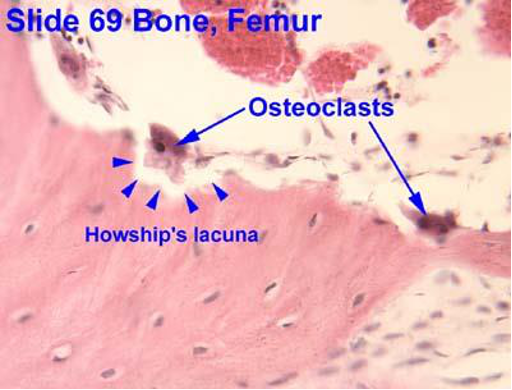
CASE: A patient taking a bisphosphonate asks how the drug increases bone density. Which bone cell is primarily inhibited, and why can the drug remain clinically relevant long after the medication is stopped?
ANSWER: Bisphosphonates slow bone breakdown by inhibiting osteoclast activity. The lecture notes that osteoclasts and osteoblasts retain the medication, osteoblasts incorporate it into bone matrix, and the resulting half-life is longer than 10 years, so drug activity can persist long after therapy stops.
CASE: A patient receiving therapy for cancer is being assessed for jaw osteonecrosis risk. Which medication groups and specific agents were emphasized as being associated with MRONJ?
ANSWER: Antiresorptive and antiangiogenic therapies. The lecture especially identifies nitrogen-containing bisphosphonates, denosumab, and romosozumab as currently important implicated medications.


CASE: A patient previously treated with an antiresorptive drug has exposed mandibular bone for 10 weeks. The patient has never received radiation to the jaws and has no metastatic jaw disease. Does this meet the lecture criteria for MRONJ?
ANSWER: Yes, if the exposed bone is in the maxillofacial region. Required criteria: current or previous antiresorptive/antiangiogenic therapy; exposed bone for >8 weeks; and no history of radiation therapy or metastatic disease to the jaws.

CASE: A cancer patient on IV antiresorptive therapy develops exposed mandibular bone after an extraction. Which details make MRONJ especially concerning in this case?
ANSWER: MRONJ is much more common in patients taking bisphosphonates for cancer than for osteoporosis, often follows trauma such as extraction (67% of cases in the lecture), and involves the mandible more often. Other listed risks include advanced age, corticosteroids, diabetes, chemotherapy, smoking/alcohol, poor oral hygiene, and drug use >3 years.
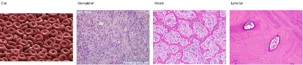
CASE: After an extraction, normal socket healing is disrupted because necrotic tissue prevents formation of healthy granulation tissue. Put the normal healing sequence in order and explain why necrosis interferes with closure.
ANSWER: Extraction → clot → granulation tissue → woven (immature) bone → lamellar (mature) bone. Without proper granulation tissue, re-epithelialization cannot occur. The lecture notes the overall maturation process takes about 2–8 months.
CASE: A patient is about to begin antiresorptive or antiangiogenic therapy. What preventive dental steps should be prioritized before treatment starts?
ANSWER: Emphasize excellent oral hygiene, more frequent prophylaxis, fluoride treatment/toothpaste, extract nonrestorable or periodontally questionable teeth when appropriate, and emphasize tobacco cessation. Dental optimization before higher-risk therapy is a major prevention strategy.
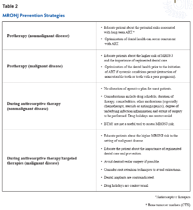

CASE: A patient receiving antiresorptive/targeted therapy for malignant disease has a tooth that would normally be extracted, but dentoalveolar surgery carries a higher MRONJ risk. What alternative principle may be considered?
ANSWER: Consider root-retention techniques to avoid extraction when appropriate. The lecture prevention table also emphasizes avoiding dentoalveolar surgery if possible in malignant-disease patients on these therapies and notes that dental implants are contraindicated in that setting.

CASE: A patient with established MRONJ has rough exposed bone with a loose sequestrum and local inflammation. What treatment options were listed, recognizing that choice depends on severity?
ANSWER: Smooth/remove rough edges, remove sequestra, use chlorhexidine rinse, antibiotics when indicated, and for severe disease consider segmental resection. Management depends on severity.
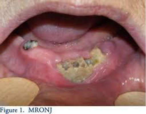
CASE: A patient has exposed jaw bone (or a fistula that probes to bone) but has no pain, erythema, or infection. Which MRONJ stage is this?
ANSWER: Stage 1.

CASE: A patient has exposed jaw bone with pain, infection, and surrounding erythema, but no pathologic fracture, extraoral fistula, or lytic extension to the mandibular border/sinus floor. Which MRONJ stage is this?
ANSWER: Stage 2.
CASE: A patient with painful infected MRONJ develops a pathologic mandibular fracture. Which MRONJ stage is this, and what other findings can qualify a lesion for the same stage?
ANSWER: Stage 3. Other qualifying findings include an extraoral fistula or lytic changes extending to the inferior border of the mandible or to the sinus floor.
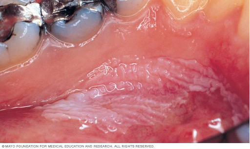
CASE: A 1.8-cm heterogeneous oral lesion is clinically suspicious for premalignancy or malignancy. You do not want to remove the entire lesion before diagnosis. What biopsy type is indicated?
ANSWER: Incisional biopsy: remove a representative sample rather than the entire lesion. Lecture indications include suspected premalignant/malignant lesions and lesions >1 cm. Incisional approaches include scalpel, laser, and punch.

CASE: A large red-white lesion contains a bland smooth region and a focally ulcerated, irregular region. You can sample only one area initially. Which area should be biopsied, and what error are you trying to avoid?
ANSWER: Biopsy the most clinically concerning area. Including some normal-looking tissue can be helpful, but it is more important to capture the worst area. Sampling error can underdiagnose a lesion—for example, reporting moderate dysplasia when SCC is present elsewhere. Multiple samples can be taken when needed.
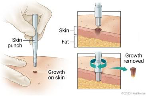
CASE: You are deciding between a punch and scalpel incisional biopsy. The lesion is a broad epithelial plaque where dysplasia is the major concern. Why might a punch be a less ideal choice?
ANSWER: Punch biopsy gives substantial depth but relatively little surface epithelium, making it less helpful for evaluating epithelial dysplasia. It is better suited to skin, soft-tissue, and salivary lesions. The lecture notes that oral pathologists generally prefer scalpel biopsies.
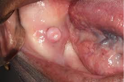
CASE: A 6-mm pedunculated oral nodule is strongly suspected to be benign and can be removed completely with little morbidity. Which biopsy type is most appropriate?
ANSWER: Excisional biopsy—remove the entire lesion. Lecture/INBDE indications include a lesion strongly suspected to be benign, <1 cm, or pedunculated and easily removed in total.
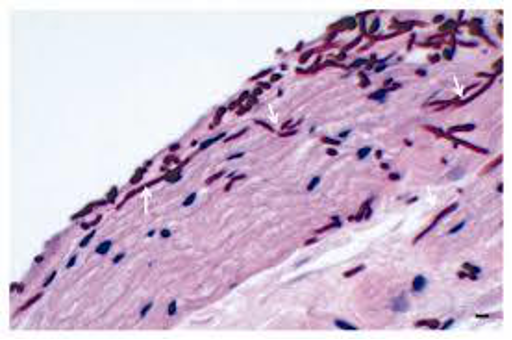
CASE: A 21-year-old with asthma uses an inhaled corticosteroid and develops a wipeable white plaque on the tongue. Which simple biopsy/sampling method discussed in the lecture can help detect the likely process?
ANSWER: Cytology (surface scraping), used in oral pathology primarily to detect Candida and HSV. In this case, the wipeable white plaque in an inhaled-steroid user points to Candida.

CASE: A patient has a mucosal lesion clinically suspicious for herpes simplex virus. You want to sample surface cells rather than remove a tissue wedge. Which diagnostic sampling method from the lecture is appropriate?
ANSWER: Cytology—scrape cells from the surface of the lesion. The lecture emphasizes cytology primarily for detecting Candida and herpes simplex virus.