Med I: Intro to Pathphysiology
 Call Kai
 Learn
Learn Practice Test
 Spaced Repetition
 Match
Match Flashcards
 Knowt Play
1/81

There's no tags or description
Looks like no tags are added yet.
Name | Mastery | Learn | Test | Matching | Spaced | Call with Kai | Chat |
|---|
No analytics yet
Send a link to your students to track their progress
82 Terms
Physiology
study of how it normally works (physio = nature/func)
Pathology
study of strucutal alternatives in cells, tissues, & organs that help ID cause of diease
what changes occured to cells/tissues/organs
Pathophysiology
disease caused what clinical effects?
study of functional change sin cells, tissues, & organs altered by disease and/or injury
etiology
study of cause(s) of disease &/or injury
Idiopathic
disease w/ no identifiable cause
Nosocomial
disease acquired as consequence of being in the hospital/ originate in healthcare
iatrogenic
disease and/or injury as result of medical intervention/treatment
clinical manifestation
Sx/s
signs
objective finding observed/ measured on physical examination
(temperature/pos. straight leg raise/ decrease visual acuity
symptoms
subjective findings pt experience/ reports
ex: chills/sore throat/ pain/ blurry vision
complications
additional unexpected problems as a result of disease/ treatmet
sequelae
residual effect of previous disease/injury
Nucleus
Control center! (DNA)
Contains nucleolus
Function:
“instructions”
cell division and control of genetic info
replication and repair of DNA and transcription of info stored in DNA → RNA
Cytoplasm
aqueous solution that fills cytoplasmic matrix (1/2 volume of euk cell)
crowded w/ ribosome making proteins
Ribosomes
RNA- protein complexes → loca of RNA transcription (carries DNA’s message)
info from DNA (mRNA) to build proteins and provide sites for cellular protein synthesis
Endoplasmic Reticulum
Transportation syst of proteins and enzymes (make and process)(outer nuclear membrane)
RER: synthesis folding and transport of protein components
move to memb…?
SER: synthesis of steroid hormones and ?? of variety of reactions to remove toxic compounds
start processing proteins/lipids
Golgi Apparatus
networks of flattened smooth membrane and vesicles located near nucleus
packages and processes proteins into secretory vesicles that break away from golgi to intra- and extra- cellular destinations
packing system (UPS - stick label and ship out)
Lysosomes
sac-like; contain digestive enzymes to breakdown cellular components, bacteria, and foreign substances (into → amino acids, fatty acids, carbs)
Mitochondria
Power plant of cell~
extract energy from organic compounds (most ATP generated here!)
Plasma Membrane Function
Control composition of space/ components they enclose
via selective permeability - enable exclusion of molecules
Plasma Membrane Component: Functions of Plasma Membrane Proteins
It itself is a lipid bilayer! (lipid inside w/ -charged hydrophilic heads!)
Transport Channel: use for charged ions! (can’t go thru charged heads and nonpolarized tails)
Enzyme: catalyst
Cell Surface Receptor: ***drugs typically act here!!
Cell Surface Markers
Cell Adhesion
Attachment of cytoskeleton: keep shape/form of cell
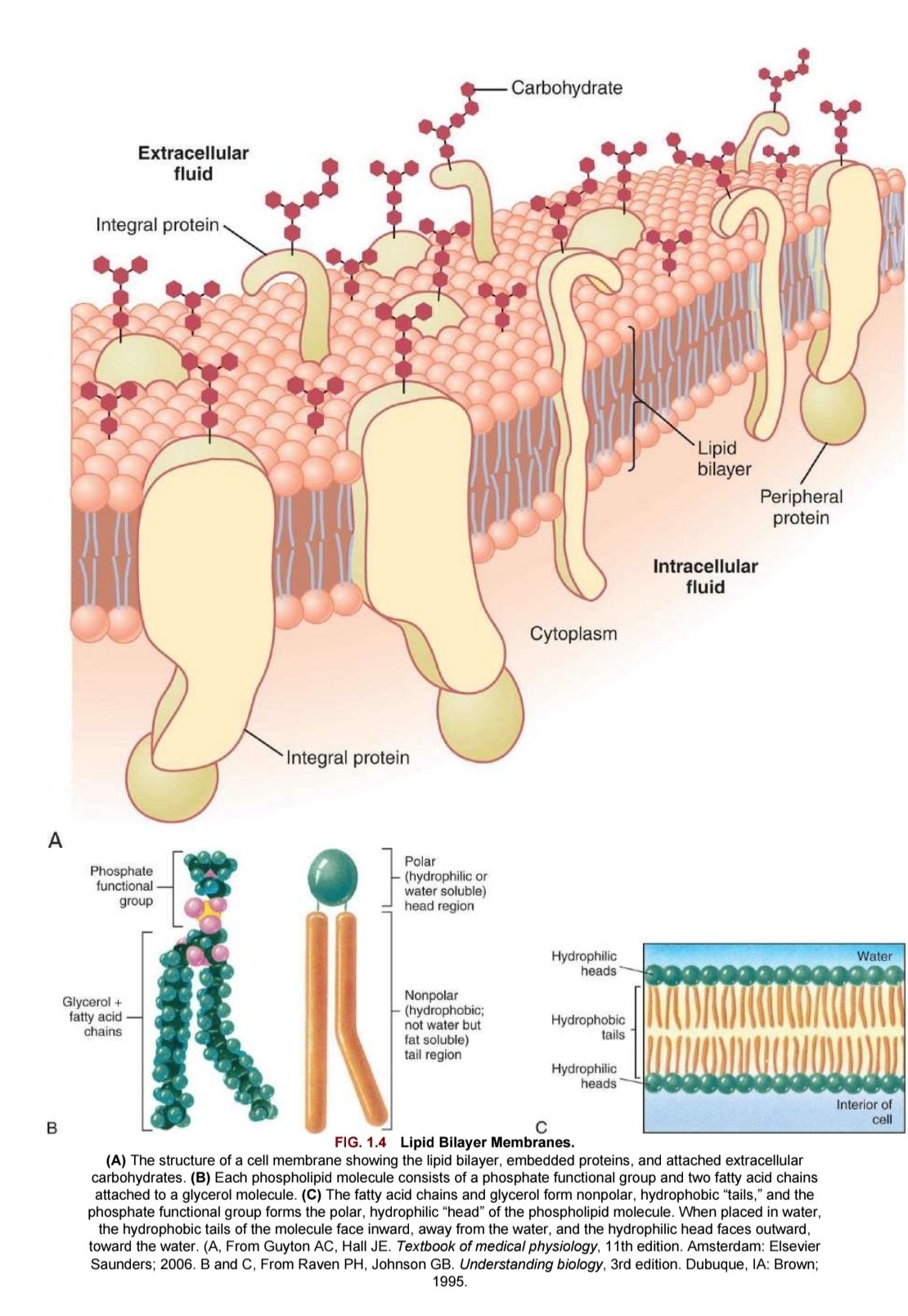
Lipid Bilayer: embedded proteins and attached extracellular carbohydrates
Lipid: all lipids amphipathic (1pt hydrophobic other hydrophilic = 2layers)
func: barrier to diffusion of water and hydrophilic substances while allowing lipid-soluble molecules (eg o2 co2) to diffuse readily
molecular glue (structural integrity)
mains: phospholipids, cholesterol, glycolipids (w/ carb - cell recog)
Proteins: most of plasma membrane’s tasks; major workhorses of cell
lots of membrane proteins
Func:
recog and binding units (recptors) for substance in/out of cell
pores/transport channel (ions & specific carriers fo AA and monosacc)
cell surface markers (glycoproteins on carbs (cell recog)
cell adhedison molecules (CAM)
catalysts
Carbohydrates:short chains of sugars/carbs w/in plasma membrane’s tasks (mostly bound to glycoproteins and glycolipids)
glycocalyx: all carbo on outside of plasma membrane + carb coating
help protect cell for mechanical damage
Func:
cell-cell recog
adhesion
slimy surface → mobility of other cells thru narrow surfaces
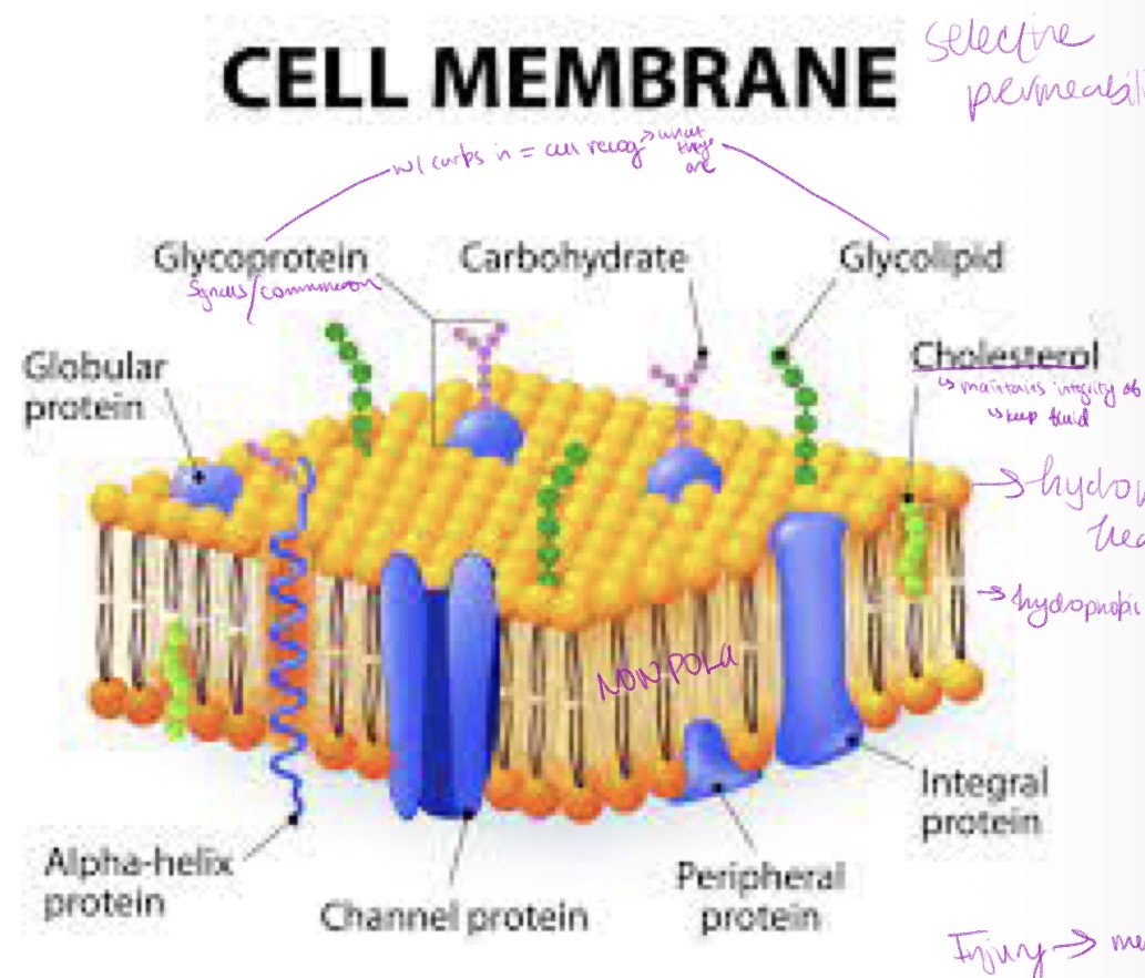
Lipid Bilayer in Physiologic Conditions
1: Solid Gel Phase - L beta (only phospholipid bilayer)
2: Liquid-ordered Phase: L o (cholesterol and sphingolipid glycosphingolipid lipid)
3: Fluid liquid crystalline/ Liquid-disordered Phase: L alpha (disordered phospholipids w/ cholsterols)

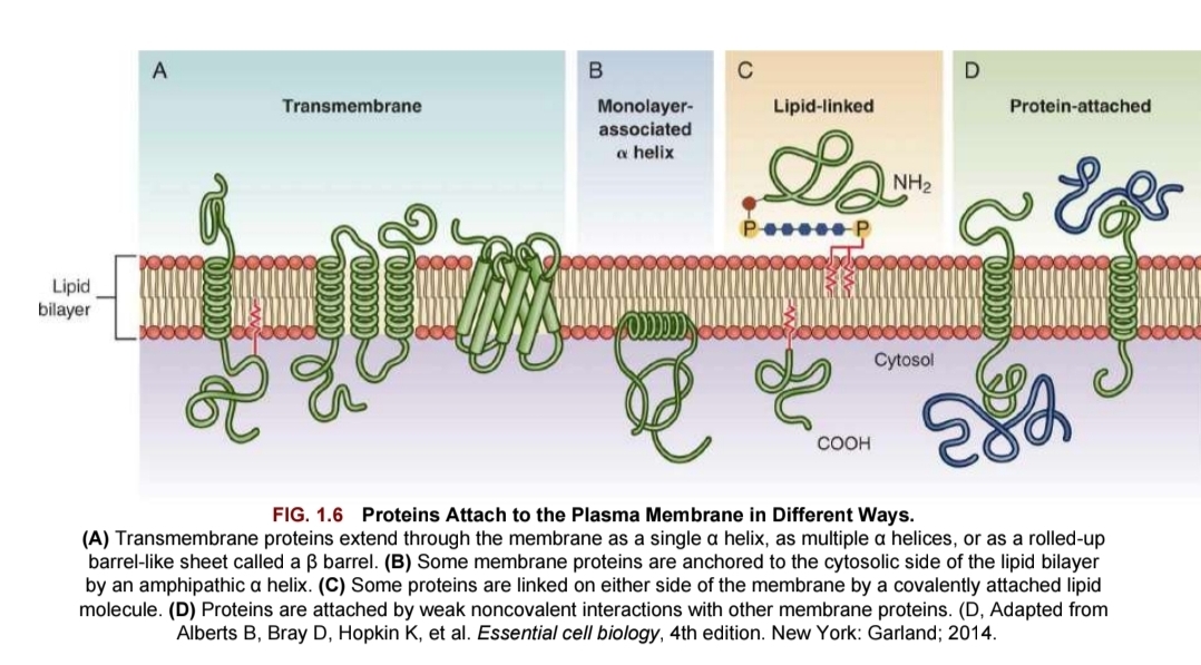
Cell-to-Cell Communcation
Contact Signaling: via plasma membrane bound receptor
signaling molecule from signaling cell accepted at receptor site on target cell
ex: fetal development
Remote signaling: via secreted molecules
carrier protein transports small hydrophobic signal molecule which accepted by intracellular receptor protein in nucleus of target cell
Contact signaling: via gap junctions
touch and move
typ of cardiac and neurons requiring electrical current communication and coordination
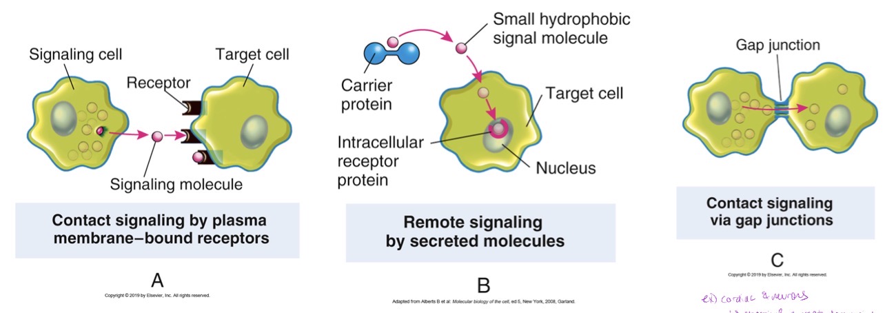
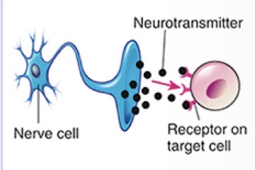
Cell Signaling
Neurotransmitter: nerve cell secretes neurotransmitters that are accepted by receptors on the target cell

Cell Signaling
Neurohomone secretion: neuron secretes neurohormones thru bloodstream and to target cell
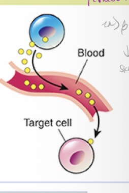
Cell Signaling
Hormonal/Endocrine: source cell secretes receptors to target cell via bloodstream and stream
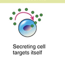
Cell Signaling
Autocrine: cell secretes receptors that bine to same cell (to itself)
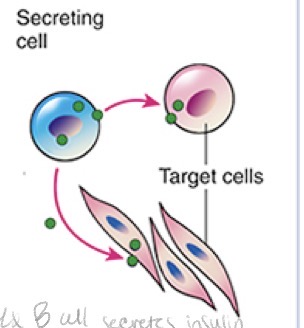
Cell Signaling
Paracrine:source cell secretes receptors for target cells

Cell Signaling
Contact-Dependent: membrane signal molecule links the source cell and target cell
Plasma Membrane Receptors: Channel Linked Type
AKA ligand-gated channels: rapid synaptic signaling b/t electrically excitable cells
channels open/close briefly (neurotransmitters) → change ion permeability of plasma memb of post synaptic cell
Plasma Membrane Receptors: Catalytic Type
Activated by ligands; func as enzymes
Compostion: transmemb proteins
Plasma Membrane Receptors: G-Protein Linked Type
Indirectly activate/inactive plasma membrane enzyme/ion channel
Mediated by guanosine triphosphate (GTP) binding regu protein (G protein)
Activation: chain of reactions to alter concentration of intracellular messengers (cAMP/Ca/signaling molecules )
Active Cellular Intake
1: Coupled Pump: uphill transport of 1 solute to downhill transport of another solute
2: ATP-Driven Pump: drive uphill transport from hydrolysis of ATP
3: Light-Driven Pump: bacteria energy from sunlight to drive uphill tansport

Passive Transport: Diffusion
Solute molecules move from HIGH → LOW concentration

Passive Transport: Osmosis
Solvent molecules move from LOW → HIGH solute concentration

Type of Passive Diffsuion
Simple Diffusion: ready diffusion down concentration gradient (O2, N, urea, H2O, CO2, glycerol)
Channel-Mediated
Transporter-Mediated
macromolecules too large to diffuse
ions (can be repelled if line w/ same charge. no necessarily prevent diffusion but can slow down)
Filtration (Passive Diffusion)
Movement of water AND solutes thru membrane b/c of a greater pushing pressure/force on 1 side of the membrane (hydrostatic pressure)
ex vasc system
Osmolality
measure of number of milliosmoles/kilogram (mOsm/kg) of H2O
AKA concentration of molecules per weight of water
Osmolarity
measure number os milliosmoles/liter (mOsm/L) of solution
AKA concentration of molecules per volume of solution
Osmotic Pressure
amount hydrostatic pressure req to oppose osmotic mvmt of water
factors: type and thickness of plasma memb, size of macromolecules, conc of molecules/cont gradient, solubility of molecules
Oncotic Pressure
overall osmotic effect of colloids (plasma proteins)
Tonicity
effective osmolatity of solution
(isotonic/hypertonic/hypotonic)
Isotonic
same osmolality/conc of particles
(~285 mOsm/kg for intra/extracellular fluid)
Hypertonic
COnc of >285-294mOsm/kg
Cell will shrink (more solute outside)
Hypotonic
Lower concentration (more dilute than body fluids <285mOsm/kg)
Cell will bloat (more solute inside)
Transport of Glucose
Transport Syst:
passive: protein channel
active: symport w/ Na+
Tissues: MOST
Transport of Amino Acids
Transport Systems:
AA specific transporters: couple channels (intestines, kidney, liver)
AA (except proline): Active symport w/ Na+ (liver)
Specific AA: Active group translocation (small intestine)
Transport of Na+
Transport Systems: simple passive transport
Tissues: distal renal tubular cells
Transport of Na/H+
Transport Systems: active antiport; proton pump
Tissues: proximal renal tubular cells and small intestines
Transport of Na+/K+
Transport Systems: Active ATP driven protein channel
Tissues: plasma membrane!! (most)
Transport Ca2+
Transport Systems: ATP driven antiport w/ Na+
Tissues: ALL cells (antiport in RBC)
Transport of H+/K+
Transport Systems: Active
Tissues: Parietal cells of gastric cells secreting H+
Atrophy
response to DECR in work load/adverse envrionmental factor
→ smaller size and more efficient func
Hypertrophy
Incr in work load
→larger cell size and incr in functioning tissue mass
Hyperplasia
incr in number of cells in tissue/organ (only w/ cell capable of mitotic division)
Metaplasia
reversible change in which one cell type→replaces
usually response to chronic irritation/inflammation (may be precancerous)
cont/persistent stress likely → cancer
Dysplasia
deranged cell growth (may be reversible)
→varied cell sizes, shapes, and organization
STRONG precursor to cancer
require surgical removal
Cellular Injury (genetic)
alter cells nucleus and plasma membrane’s structure, shape, receptors, or transport mechanisms
Cellular Injury (Epigenetic)
Induction of mitotically heritable alterations in gene expression w/o changing DNA
Cellular Injury (Nutritional imbalances) SAME
Pathophysiologic cellular effects develop when nutrients are not consumed in diet and transported to body’s cells
OR when excessive amts of nutrients consumed and transported
Cellular Injury (Temp extremes) PHYSCIAL
Hypothermic: high intracellular sodium conc
abrupt drops: vasoconstrictions, incr viscosity of blood → ischemic injury, infarction, necrosis
Hyperthermic: varies based on severity of extent/nature/intensity
Compressive waves of air/fluid followed by decr pressure: may collapse thorax, rupture internal solid organs, and cause widespread hemorrhage
co2 and n normally dissolved in blood can precipitate from solution and form small bubbles (gas emboli) → hypoxic injury and pain
Cellular Injury (Illumination) RADIATION
Fluorescent/halogen lamps: harmful oxidative stress
UV light: skin cancer
Blue light: Induces ROS and s-opsin aggregation
Cellular Injury (mechanical stresses) PHYSICAL
Injury via physical impact/irritation:
compression: perpendicular force
tension: stretching force
torsion: twisting force
fluid shear forces: layers rubbing against each other
Cellular Injury (Noise)
Acute loud noise OR cumulative effects of var intensities/frequencies/duration
Cellular Injury (Chemical Injury)
drugs (overdose), lead, mercury
Cellular Injury (Biological Agents)
Viruses, Parasites, Bacteria
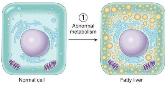
Mechanism of Intracellular Accumulations - abnormal metabolism
normal cell converts to fatty liver cell w/ accumulation of fat molecules
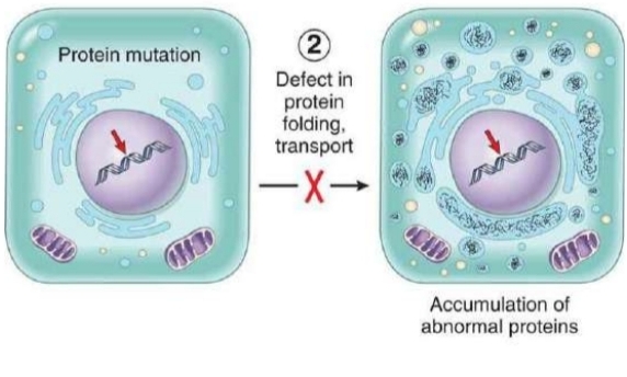
Mechanism of Intracellular Accumulations - Defect in protein folding, transport
protein mutation in normal cell results in accumulation of abnormal proteins w/in cell
Mechanism of Intracellular Accumulations - lack of enzymes
enzymes in normal cell balances w/ complex substate proportional to soluble products: no soluble products? endogenous material accum → lysosomal storage disease

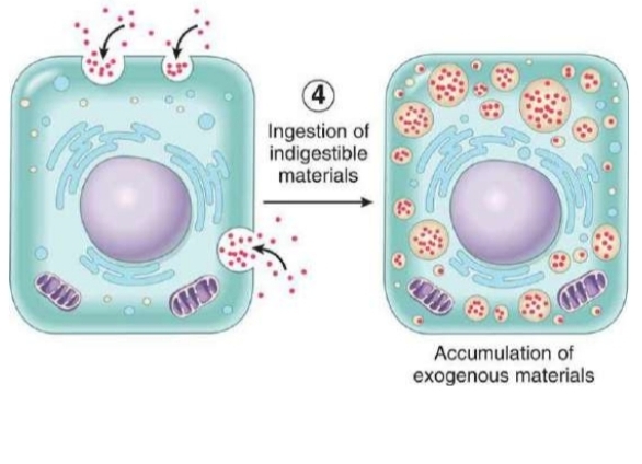
Mechanism of Intracellular Accumulations - ingestion of indigestible materials
normal cell surrounded by indigestible materials. INGESTION → exogenous material accumulates w/in call
Physiologic Adaptive Changes
Normal response to internal/external stimuli help maintain homeostasis and support bodily function.
Pathophysiologic Adaptive Change
Abnormal changes in response to disease, injury, harmful conditions
Apoptosis
Normal cell death as a result of aging “programmed cell death”
little-no inflammation; only few cells involved
disregulated: patholgic! either excessive/insufficent
leades to cancer, autoimmune, neurodegen, ischemic injury
Necrosis
Pathological cell death - autolysis after death of entire organism
cell death in organ/tissue of still living person
Unregulated enzymatic destruction → loss of cell memb integrity → inflammatory response
induced by injury
large areas of contiguous cells involved
Manifestations of Cellular Injury - Fever
release of endogenous pyrogens (IL-1, TNF-α, prostaglandins) from bacteria or macrophages
acute inflammatory response
Manifestations of Cellular Injury - Increased HR
Incr in oxidative metabolic process FROM FEVER
Manifestations of Cellular Injury - Incr in leukocytes
aka leukocytosis
incr in total #WBC b/c infections (5k-9k/mm³ is NORMAL)
Manifestations of Cellular Injury - pain
various mech (release of bradykinins obstruction, pressure)
Manifestations of Cellular Injury - Lactate dehydrogenase
Released from RBC, liver, kidney, skeletal muscle
Manifestations of Cellular Injury - Creatinine kinase
Release for skeletal muscle, brain, heart