3 - Development
 Call Kai
 Learn
Learn Practice Test
 Spaced Repetition
 Match
Match Flashcards
 Knowt Play
1/115

There's no tags or description
Looks like no tags are added yet.
Name | Mastery | Learn | Test | Matching | Spaced | Call with Kai | Chat |
|---|
No analytics yet
Send a link to your students to track their progress
116 Terms
Development
gradual modification of anatomical structures and physiological characteristics from fertilization to maturity
Development is how anatomical structures ______
change
Why to cells differentiate?
to create the diverse tissues of the adult
Cells differentiate due to changes in gene expression
some genes are “turned off” while others are “turned on”

Developmental History of a Human
Pre-embryonic period
Embryonic period
Fetal period
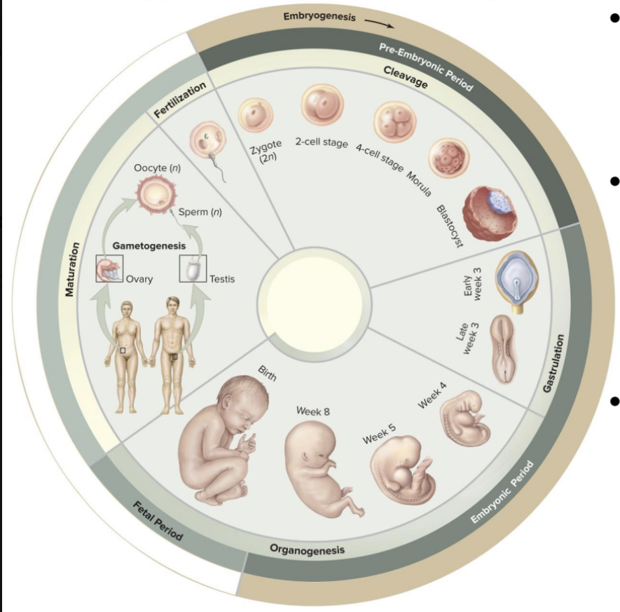
Pre-embryonic period
First two weeks after fertilization of egg
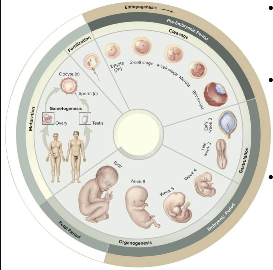
Embryonic period
Third through eighth weeks
The embryonic period is where all major organ systems:
begin to develop
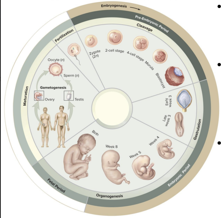
Fetal period
Ninth through thirty-eighth weeks
The fetal period is where growth and development:
continues and ends at birth
Fertilization
fusion of two haploid gametes, secondary oocyte and sperm, 23 chromosomes
zygote
diploid cell with 46 chromosomes
Oocyte is _______ times larger than sperm
2000
What provides organelles and nourishment for ~1 week?
oocyte
Fertilization occurs:
in uterine tube within a day of ovulation

Spermatozoon Head
contains nucleus and chromosomes
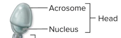
Acrosome
a compartment at tip of spermatozoon head

Acrosome contains
enzymes essential to fertilization

Spermatozoon Neck
contains centrioles

Spermatozoon Middle piece
mitochondria and microtubules

Spermatozoon Tail
only flagellum in the human body

The secondary oocyte leaving the follicle is in
metaphase of meiosis II

Cells of the corona radiata
surround the oocyte
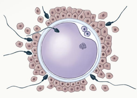
For fertilization to occur,
sperm must penetrate the corona radiata

The sperm binds receptors in
the zona pellucida beneath corona radiata

enzymes from the acrosome
digest a path to surface of oocyte

when the sperm contacts the surface,
the sperm and oocyte membranes fuse
Oocyte activation
Fusion between sperm and oocyte membranes
Enzymes inactivate sperm receptors and harden the zona pellucida
only one sperm fertilizes
Dad’s contribution = 23 chromosomes, 1 centriole, and
determines sex
The oocyte completes
meiosis II
Cellular metabolism increases as
many proteins necessary for development are rapidly produced

Amphimixis
the male and female pronuclei (1n+1n) fuse

2n Cell =
zygote has 46 chromosomes

The zygote begins
cleavage, a series of cell divisions

A single cell ultimately develops into
a mature adult

Differentiation
changes in genetic activity of specific cells

As the zygote divides, cells have
differing cytoplasmic compositions

differing cytoplasmic compositions
alters the genetic activity of each cell
facilitate the development of diverse cell lines
The time spent in prenatal development
gestation
Gestation is divided into
three trimesters (each 3 months long)
First Trimester
Period of embryological and early fetal development
Rudiments of all major organ systems appear
Second Trimester
Development of organs and organ systems
Body shape and proportions change
Third Trimester
Rapid fetal growth and deposition of adipose tissue
Most major organ systems are fully functional
First Trimester includes four major stages
Cleavage
Implantation
Placentation
Embryogenesis
Cleavage
a sequence of cell divisions that transforms a zygote into a multicellular blastocyst
Implantation
begins when the blastocyst attaches to the endometrium and continues as the blastocyst invades maternal tissues
Placentation
occurs as blood vessels form around the blastocyst and the placenta develops
Embryogenesis
the formation of a viable embryo with foundations for all the major organ systems
The First Trimester
Most dangerous period in prenatal life
~40% of conceptions survive first trimester
Pregnant women should avoid drugs and other disruptive stresses

The cells produced by cleavage divisions
blastomeres
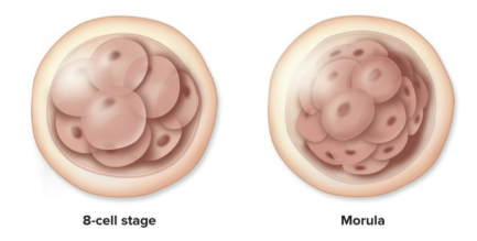
3 days of cleavage, the pre-embryo is a solid ball of cells
morula

The cells of the morula reorganize into a hollow ball
blastocyst
blastocoele

inner cavity
blastocoele

blastomeres then form
trophoblast
inner cell mass
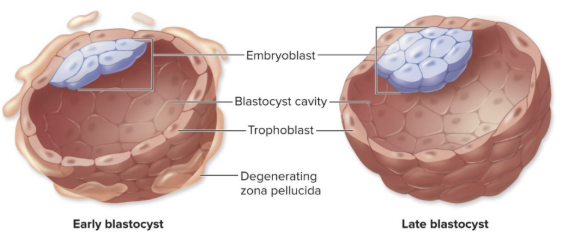
trophoblast
outer layer facilitates nutrient exchange
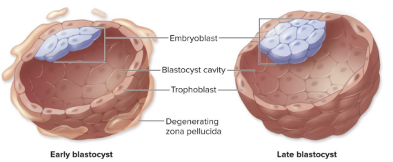
inner cell mass
cells that will form the embryo
Implantation occurs
~7 days after fertilization
Blastocyst reaches
uterine lining
Trophoblast
Erodes into uterine epithelium
Divides into 2 layers
secretes human Chorionic gonadotropin
Formation of the Bilaminar Germinal Disc and Extraembryonic Membranes
~ 8 days, bilaminar germinal disc
bilaminar germinal disc
two layers of differentiated cells
bilaminar germinal disc layers
Hypoblast
Epiblast
3 extraembryonic membranes:
1. Yolk sac
2. Amnion
3. Chorion
1. Yolk sac:
early blood cell and vessel formation
2. Amnion:
contains fluid-filled amniotic cavity
3. Chorion:
Outermost membrane forms part of placenta
Main functions of the placenta:
Metabolic
Immune
Endocrine
Metabolic function of the placenta:
share resources with mom
Immune function of the placenta:
isolate baby from mom
Endocrine function of the placenta:
produces hormones itself; takes over the ovaries
connecting stalk
develops into the umbilical cord
contains umbilical arteries and vein
Placenta is both embryo and mother
Embryo – chorion/chorionic villi contain branches of umbilical vessels
Mom - functional layer of the endometrium
Gas, waste, nutrient, and some immunity exchanged
the placenta also acts as
an endocrine organ
Human chorionic gonadotropin (hCG), human placental lactogen (hPL)
hormones produced by the placenta
placental prolactin
helps produce milk
relaxin
targets connective tissue
softens collagen in CT
collagen connections between bone
Land R colaxal bone
hips widen
- opens up birth canal
progesterone, estrogens
sex hormone production
The Endocrine Placenta maintains
uterine lining
The Endocrine Placenta prepares
mom for delivery and childcare
At ~12 days, what stage is development at?
gastrulation
~12 days, Gastrulation
Form all three primary germ layers from epiblast cells
Ectoderm, mesoderm, endoderm
Once all three primary germ layers are present =
embryo
Starts with epiblast cells migrating through
primitive streak
All three germ layers are derived from
the epiblast
Cells between the epiblast and hypoblast
mesoderm
Some displace the hypoblast cells
endoderm
Cells remaining in the epiblast
ectoderm
deuterostome
animals with a second mouth and anus
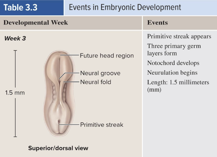
Embryonic Period begins with
establishment of three primary germ layers

By end of embryonic period,
main organ systems are developed

At 1 month, the embryo is
about 0.2 in long and weighs 0.02 g

At 2 months, the embryo is
about 1 inch long and weighs about 2.7 g

At 3 months, the embryo is
about 3 inches long and weighs about 26 g

During Second Trimester
Fetus grows faster than surrounding placenta
Growth in length
Mom’s survival probability lowers as
baby’s weight increases

During Third Trimester
Most of the organ systems become ready
Growth rate starts to slow
Largest weight gain
Fetus and enlarged uterus displace many of the mother’s abdominal organs

At 4 months, the embryo is
about 5 inches long and weighs about 150 g

Pregnancy and Maternal Systems
Developing fetus is dependent on maternal organ systems

Maternal adaptations include increases in:
Respiratory rate and tidal volume
Blood volume
Nutrient and vitamin intake
Glomerular filtration rate
Size of uterus and mammary glands
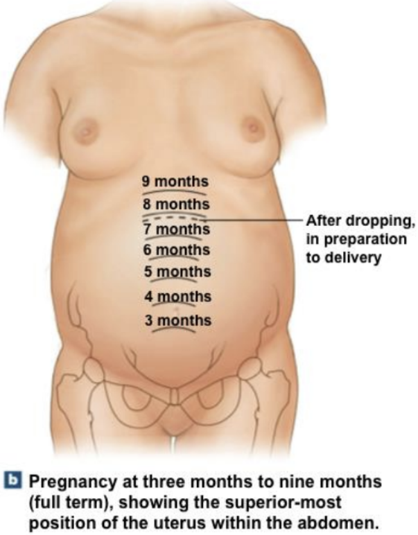
Third Trimester involves
Structural and Functional Changes in the Uterus