The Lymphatic System and Lymphoid Organs and Tissues
1/70
There's no tags or description
Looks like no tags are added yet.
Name | Mastery | Learn | Test | Matching | Spaced |
|---|
No study sessions yet.
71 Terms
What are three components of the lymphatic system?
- Lymphatic vessels
- Lymph
- Lymph nodes
Explain Bulk Flow across Capillary Walls?
The hydrostatic and colloid osmotic pressures operating at capillary beds force fluid out of the blood at the arterial ends of the beds ("upstream") and cause most of it to be reabsorbed at the venous ends ("downstream").
As much as ____ of fluid becomes part of the interstitial fluid, daily.
3 L
Once interstitial fluid enters the lymphatic vessels, it is called
lymph (lymph = clear water).
The lymphatic vessels form a _______ ______ system in which lymph flows only toward the heart.
one-way
Where does the transport of lymph begin?
blind-ended lymphatic capillaries
Lymphatic capillaries are widespread, but they are absent from...
bones (including bone marrow) and teeth.
In the brain, __________ form a system of channels that act like lymphatic vessels.
astrocytes
Astrocyte channels in the brain connect to the true lymphatics found in the _________ and together form the __________ ___________.
meninges, glymphatic system.
What is the purpose of the glymphatic system?
Drains extracellular fluids and waste products from the brain during sleep.
What impact could the malfunction of the glymphatic system have?
May have a role in the development of degenerative diseases such as Alzheimer's.
What are the two unique structural modification lymphatic vessels have to in increase their permeability.
- The endothelial cells forming the walls of lymphatic capillaries are not tightly joined. (contain minivalves)
- Collagen filaments anchor the endothelial cells to surrounding structures so that any increase in interstitial fluid volume opens the minivalves
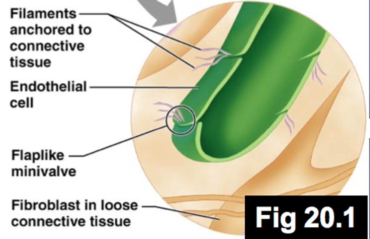
Explain the one-way swinging doors (minivalves) in the lymphatic capillary wall.
When fluid pressure in the interstitial space is greater than the pressure in the lymphatic capillary, the minivalve flaps open, allowing fluid to enter the lymphatic capillary.
However, when the pressure is greater inside the lymphatic capillary, it forces the endothelial minivalve flaps shut, preventing lymph from leaking back out as the pressure moves it along the vessel.
T/F: Proteins in the interstitial space are unable to enter blood capillaries, but they enter lymphatic capillaries easily
True
T/F: When tissues become inflamed, lymphatic capillaries develop openings that permit uptake of even larger particles such as cell debris, pathogens (disease-causing microorganisms such as bacteria and viruses), and cancer cells.
True; these particles travel with the lymph to the lymph nodes, where they are removed by cells of the immune system.
What is critical for activating an immune response?
The process of pathogens meeting up with immune cells in lymph nodes.
Define lacteals?
A special set of lymphatic capillaries that transports absorbed fat from the small intestine to the blood.
Where do lacteals get their name from? What substance do they carry? Where does it come from?
The fatty lymph (milky white) that drains through them called chyle. Which drains from the fingerlike villi of the intestinal mucosa.
From the lymphatic capillaries, lymph flows through successively larger and thicker-walled channels. Name these four structures in order.
Lymphatic capillaries ----> Collecting lymphatic vessel ----> Lymphatic trunk ----> Lymphatic ducts
The collecting lymphatic vessels have the same three tunics (layers) as veins, but how do they differ?
- Have thinner walls and
- More internal valves
- Anastomose more
Generally how do lymphatic travel?
- Lymphatics in skin travel along with superficial veins
- Deep lymphatic vessels (trunk and digestive viscera) travel with the deep arteries.
The largest collecting vessels unite to form _______ ______, which drain fairly large areas of the body.
lymphatic trunks
The major trunks, named mostly for the regions from which they drain lymph, are the...
- paired lumbar
- bronchomediastinal
- subclavian
- jugular trunks
- intestinal trunk
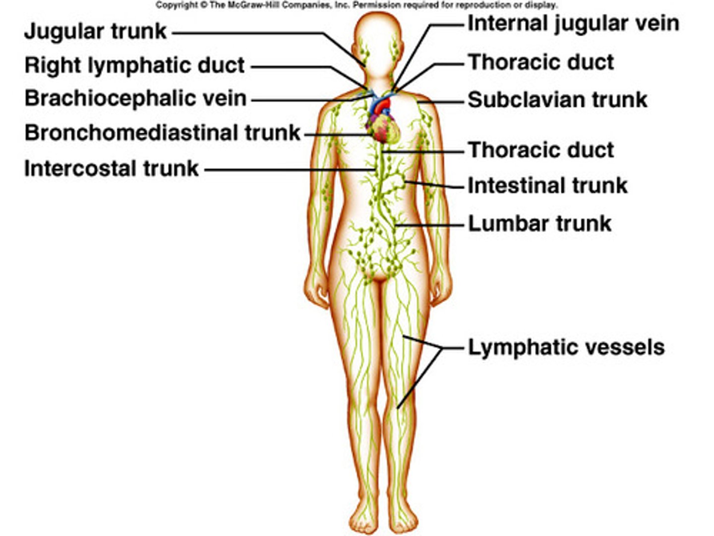
Lymph is eventually delivered to one of two large ducts in the thoracic region. What are their names?
- The right lymphatic duct
- The thoracic duct (much larger)
The right lymphatic duct drains lymph from the...
right upper limb and the right side of the head and thorax.
The much larger thoracic duct receives lymph from...
the rest of the body (left side and lower right side).
Each terminal duct empties its lymph into the venous circulation at the junction of the _______ ______ _____ and _______ ______ on its own side of the body
internal jugular vein, subclavian vein
lymphangitis
Inflammation of lymphatic vessels; visable red streaks on the skin
T/F: . When physical activity or passive movements increase, lymph flows much more rapidly.
True, reason why it is a good idea to immobilize a badly infected body part to hinder flow of inflammatory material from that region.
Lymphedema
swelling due to an abnormal accumulation of lymph fluid within the tissues
Name for functions of the lymphatic system
- Return excess tissue fluid to the blood
- Return leaked proteins to the blood
- Carry pathogens to lymph nodes
- Carry absorbed fat from the intestine to the blood
Lymphocytes are the main warriors of the immune system. What are the two different types?
T lymphocytes (T cells) and B lymphocytes (B cells) (protect the body against antigens).
Activated T cells
manage the immune response, and some of them directly attack and destroy infected cells.
Activated B cells
protect the body by producing plasma cells, daughter cells that secrete antibodies into the blood (or other body fluids)
Macrophages play a crucial role in body protection and the immune response by...
phagocytizing foreign substances and by helping to activate T cells
What do spiny-looking dendritic cells do?
capture antigens and bring them back to the lymph nodes.
Reticular cells
Produce the reticular fiber stroma, which is the network that supports the other cell types in lymphoid organs and tissues.
Lymphoid tissue, largely composed of loose connective tissue called...
reticular connective tissue (dominates all the lymphoid organs except the thymus).
How does our body ensure that lymphocytes reach infected or damaged sites quickly?
The cycling of lymphocytes between the circulatory vessels, lymphoid tissues, and loose connective tissues
Types of lymphoid tissue
- Diffuse lymphoid tissue
- Lymphoid follicles (nodules)
Diffuse lymphoid tissue
- A loose arrangement of lymphoid cells and some reticular fibers—is found in virtually every body organ.
- Largely in the lamina propria and the mucous membranes of the digestive tract
Lymphoid follicles
solid, spherical bodies consisting of tightly packed lymphoid cells and reticular fibers.
Follicles often have lighter-staining germinal centers where proliferating (rapidly dividing) ___________ predominate.
B cells
isolated aggregations of lymphoid follicles occur where?
- Peyer's patches
- Appendix
The lymphoid organs are grouped into two functional categories.
- The primary lymphoid organs: where B and T cells mature.
- The secondary lymphoid organs: where mature lymphocytes first encounter their antigens and are activated.
What are the primary lymphoid organs?
1. Red bon marrow (where B cells mature)
2. The thymus (where T cells mature)
What are the secondary lymphoid organs?
Lymph nodes, spleen, MALT (tonsils, Peyer's patches, appendix).
The most important of the secondary lymphoid organs in the body are the...
lymph nodes, which cluster along the lymphatic vessels of the body.
Large clusters of lymph nodes occur near the body surface in the _______, ___________, _________ places where the collecting lymphatic vessels converge to form trunks
inguinal, axillary, and cervical regions,
Lymph nodes have two basic protective functions
1. Cleansing the lymph
2. Immune system activation
Trabeculae
supporting bundles of bony fibers in cancellous bone
A lymph node has two histologically distinct regions what are they?
- Cortex
- Medulla
Cortex
- The superficial part contains densely packed follicles, many with germinal centers.
- The deeper part primarily houses T cells in transit.
- Dendritic cells are abundant.
Medulla
Consists of medullary cords, thin inward extensions from the cortical lymphoid tissue. (contains both B and T cells)
Lymph sinuses
Large lymphatic capillaries spanned by crisscrossing reticular fibers.
T/F: There are fewer efferent vessels draining the node than afferent vessels feeding it
True: this allows time for the lymphocytes and macrophages to carry out their protective functions.
lymphadenopathy
- Swollen "glands"
- The bubonic plague
The soft, blood-rich spleen is about the size of a fist and is the...
largest lymphoid organ.
Besides extracting aged and defective blood cells (RBC graveyard) and platelets from the blood the spleens macrophages remove....
debris and foreign matter
Three additional functions of the spleen?
- Recycles the breakdown products of red blood cells for later reuse.
- Stores blood platelets and monocytes for release into the blood when needed.
- May be a site of erythrocyte production in the fetus.
The spleen consists of two components?
white pulp and red pulp.
white pulp of spleen
Where immune functions take place, so it is composed mostly of lymphocytes suspended on reticular fibers.
red pulp of spleen
Where worn-out red blood cells and bloodborne pathogens are destroyed, so it contains huge numbers of erythrocytes and the macrophages that engulf them.
MALT helps protect us from
pathogens that seek to enter our bodies.
Tonsils
Form a ring of lymphoid tissue around the entrance to the pharynx (throat), where they appear as swellings of the mucosa.
These are the largest tonsils and the ones most often infected.
The paired palatine tonsils
Name the different types of tonsils
- The paired palatine tonsils
- The lingual tonsil
- The pharyngeal tonsil
- The tiny tubal tonsils
Where are peyer's patches located?
in the wall of the distal portion of the small intestine.
What are two supposed functions of the appendix?
(1) to prevent bacteria from breaching the intestinal wall
(2) to generate many "memory" lymphocytes for long-term immunity.
Thymic corpuscles
involved in the development of regulatory T cells (a class of T lymphocytes that are important for preventing autoimmune responses).
The thymus is a primary lymphoid organ and differs from secondary lymphoid organs in three important ways:
- Has no follicles because it lacks B cells.
- Does not directly fight antigens.
- The stroma of the thymus consists of epithelial cells rather than reticular fibers.