Lower Limb/Back/Suboccipital Triangle Muscles

0.0(0)
Studied by 1 person
call kaiCall Kai
Locked
learnLearn
examPractice Test
spaced repetitionSpaced Repetition
heart puzzleMatch
flashcardsFlashcards
GameKnowt Play
Card Sorting

1/76

encourage image

There's no tags or description

Looks like no tags are added yet.

Last updated 3:13 PM on 7/25/26
Name
Mastery
Learn
Test
Matching
Spaced
Call with Kai
Chat

No analytics yet

Send a link to your students to track their progress

77 Terms

1
New cards

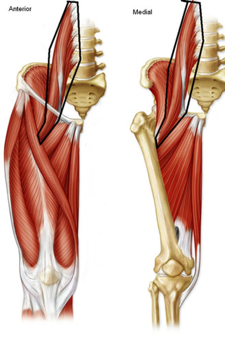
Psoas Major

Origin: sides of T12-L5 vertebrae and the discs between them; transverse processes of all lumbar vertebrae

Insertion: lesser trochanter of femur

Innervation: anterior rami of lumbar nerve (L1, L2, L3)

Action: acts conjointly with the iliacus to flex and stabalize the hip joint; postural muscle that helps control deviation of the trunk and is active during standing
<p>Origin: sides of T12-L5 vertebrae and the discs between them; transverse processes of all lumbar vertebrae</p><p>Insertion: lesser trochanter of femur</p><p>Innervation: anterior rami of lumbar nerve (<strong>L1</strong>, <strong>L2</strong>, L3)</p><p>Action: acts conjointly with the iliacus to flex and stabalize the hip joint; postural muscle that helps control deviation of the trunk and is active during standing</p>
2
New cards

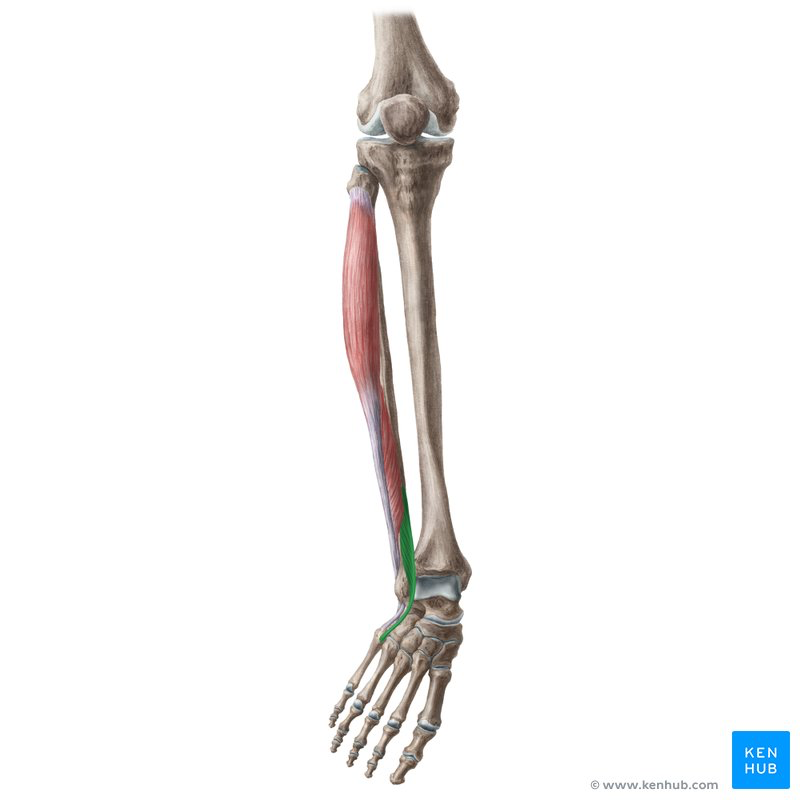
Fibularis Tertius

Origin: inferior third of anterior surface of fibula and interosseous membrane

Insertion: dorsom of base of 5th metatarsal

Innervation: deep fibular nerve (L5, S1)

Action: dorsiflexes ankle; aids in eversion of foot
<p>Origin: inferior third of anterior surface of fibula and interosseous membrane</p><p>Insertion: dorsom of base of 5th metatarsal</p><p>Innervation: deep fibular nerve (L5, S1)</p><p>Action: dorsiflexes ankle; aids in eversion of foot</p>
3
New cards

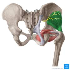
Gluteus Minimus

Origin: external surface of ilium between anterior and inferior gluteal lines

Insertion: anterior surface of greater trochanter of femur

Innervation: superior gluteal nerve (L4, L5, S1)

Action: abduct and anterior portions medially rotate hip joint; keep pelvis level when opposite limb is elevated
<p>Origin: external surface of ilium between anterior and inferior gluteal lines</p><p>Insertion: anterior surface of greater trochanter of femur</p><p>Innervation: superior gluteal nerve (L4, L5, S1)</p><p>Action: abduct and anterior portions medially rotate hip joint; keep pelvis level when opposite limb is elevated</p>
4
New cards

Obturator externus

Origin: margins of obturator foramen and obturator membrane

Insertion: trochanteric fossa of femur

Innervation: obturator nerve (L3, L4)

Action: laterally rotates hip joint; pulls head of femur into acetabulum holding pelvis steady
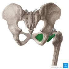
<p>Origin: margins of obturator foramen and obturator membrane</p><p>Insertion: trochanteric fossa of femur</p><p>Innervation: obturator nerve (L3, <strong>L4</strong>)</p><p>Action: laterally rotates hip joint; pulls head of femur into acetabulum holding pelvis steady</p>
5
New cards

Adductor Brevis

Origin: body and inferior ramus of pubis

Insertion: pectineal line and proximal part of linea aspera of femur

Innervation: obturator line (L2, L3, L4)

Action: adducts hip joint and to some extent flexes it
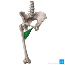
<p>Origin: body and inferior ramus of pubis</p><p>Insertion: pectineal line and proximal part of linea aspera of femur</p><p>Innervation: obturator line (L2, <strong>L3</strong>, L4)</p><p>Action: adducts hip joint and to some extent flexes it</p>
6
New cards

Iliacus

Origin: iliac crest, iliac fossa, ala of sacrum, and anterior sacro-iliac ligaments

Insertion: tendon of psoas major, lesser trochanter of femur and distal to it

Innervation: femoral nerve (L2, L3)

Action: acts conjointly with psoas major to flex and stabilize the hip joint
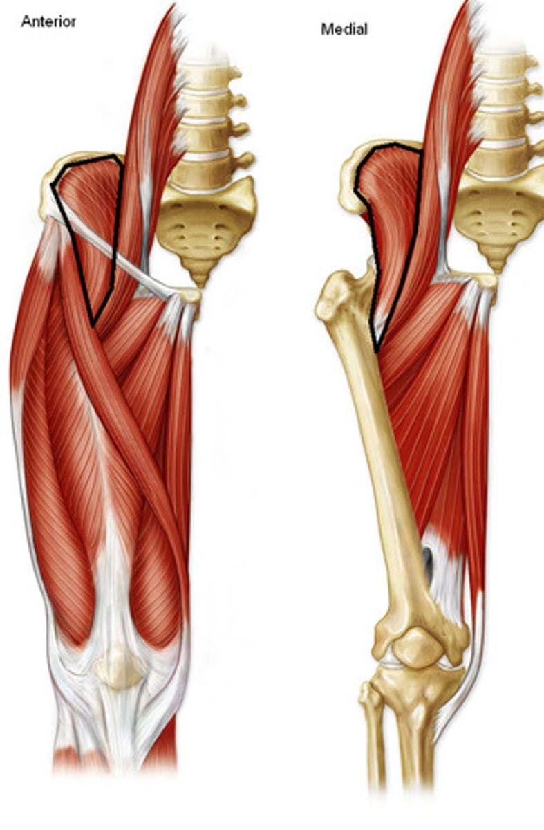
<p>Origin: iliac crest, iliac fossa, ala of sacrum, and anterior sacro-iliac ligaments</p><p>Insertion: tendon of psoas major, lesser trochanter of femur and distal to it</p><p>Innervation: femoral nerve (<strong>L2, </strong>L3)</p><p>Action: acts conjointly with psoas major to flex and stabilize the hip joint</p>
7
New cards

Iliopsoas

psoas major and iliacus muscles combine to form this muscle as it enters the thigh region
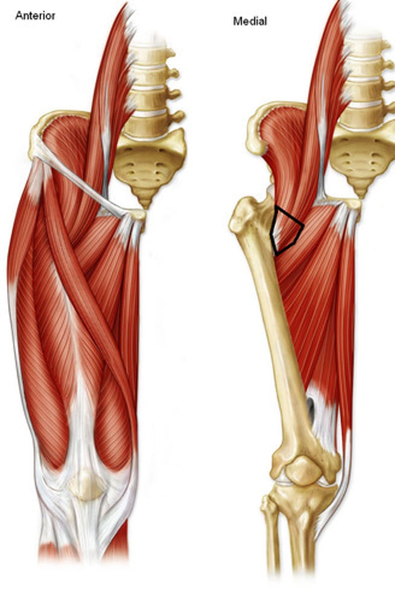
<p>psoas major and iliacus muscles combine to form this muscle as it enters the thigh region</p>
8
New cards

Gluteus Maximus

Origin: ilium posterior to posterior gluteal line, dorsal surface of sacrum and coccyx; and sacrotuberous ligament

Insertion: most fibers end in iliotibial tract, which inserts into lateral condyle of tibia; some fibers insert on gluteal tuberosity of femur

Innervation: inferior gluteal nerve (L5, S1, S2)

Action: extends hip joint between flexed and standing positions and assists in its lateral rotation; steadies thigh and assists in rising from sitting position
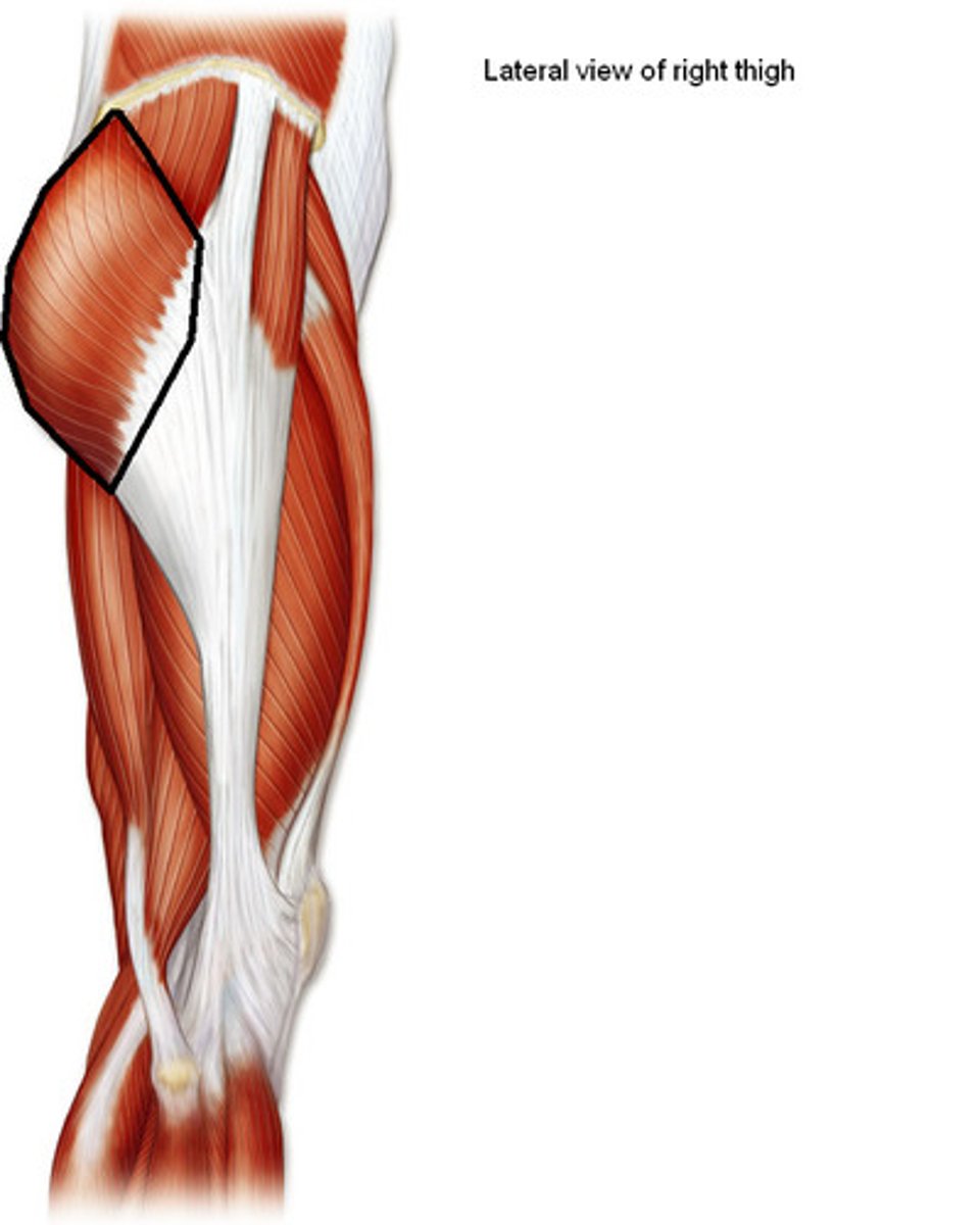
<p>Origin: ilium posterior to posterior gluteal line, dorsal surface of sacrum and coccyx; and sacrotuberous ligament</p><p>Insertion: most fibers end in iliotibial tract, which inserts into lateral condyle of tibia; some fibers insert on gluteal tuberosity of femur</p><p>Innervation: inferior gluteal nerve (L5, <strong>S1</strong>, <strong>S2</strong>)</p><p>Action: extends hip joint between flexed and standing positions and assists in its lateral rotation; steadies thigh and assists in rising from sitting position</p>
9
New cards

Gluteus Medius

Origin: external surface of ilium between anterior and posterior gluteal lines

Insertion: lateral surface of greater trochanter of femur

Innervation: superior gluteal nerve (L4, L5, S1)

Action: abducts and anterior portions medially rotate hip joint; keeps pelvis level when opposite limb is elevated
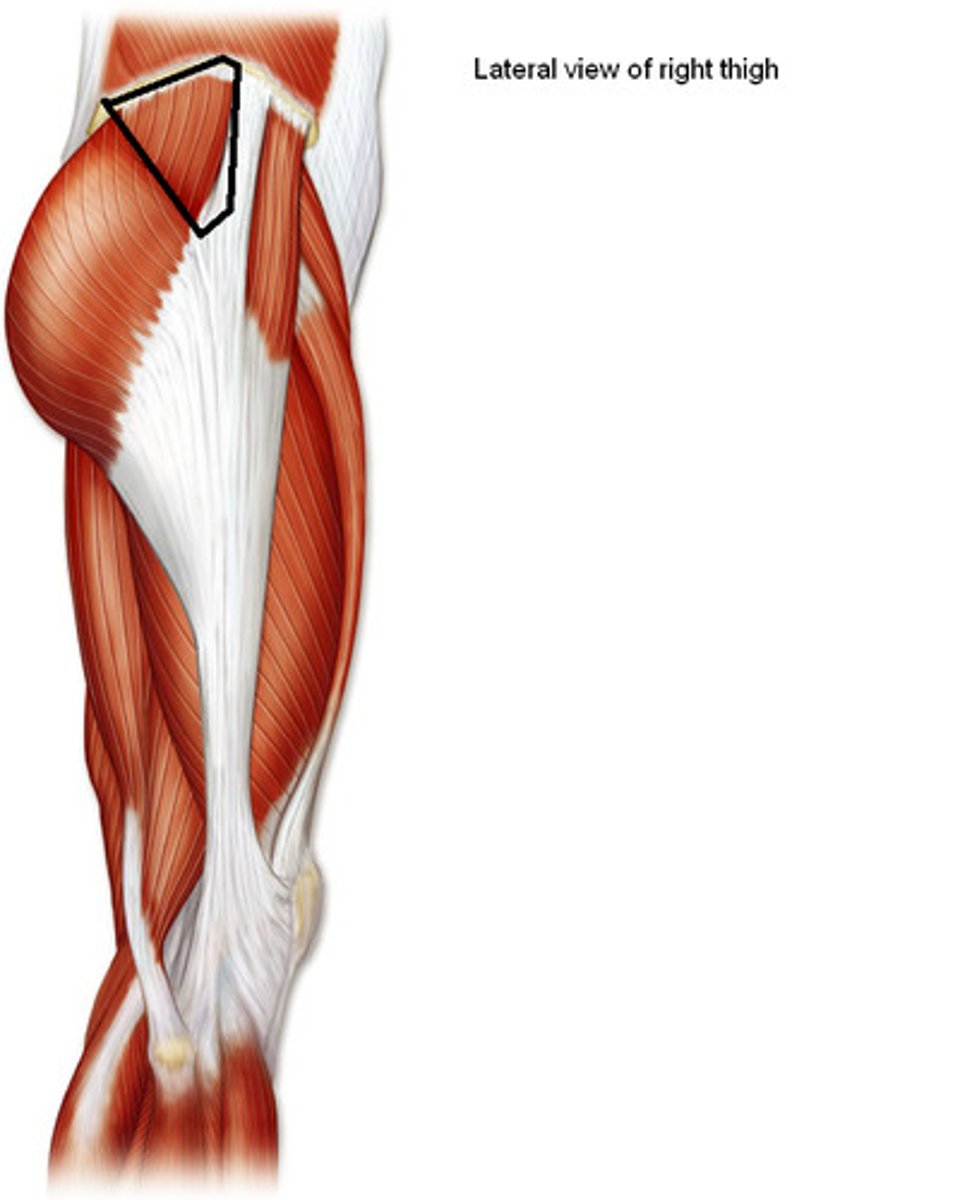
<p>Origin: external surface of ilium between anterior and posterior gluteal lines</p><p>Insertion: lateral surface of greater trochanter of femur</p><p>Innervation: superior gluteal nerve (L4, L5, S1)</p><p>Action: abducts and anterior portions medially rotate hip joint; keeps pelvis level when opposite limb is elevated</p>
10
New cards

Tensor Fascia Latae

Origin: anterior superior iliac spine; anterior part of iliac crest

Insertion: iliotibial tract, which attaches to lateral condyle of tibia (gerdy tubercle)

Innervation: superior gluteal nerve (L4, L5, S1)

Action: flexes hip joint; acts with gluteus maximus to stabilize the extended knee joint; pulls IT band
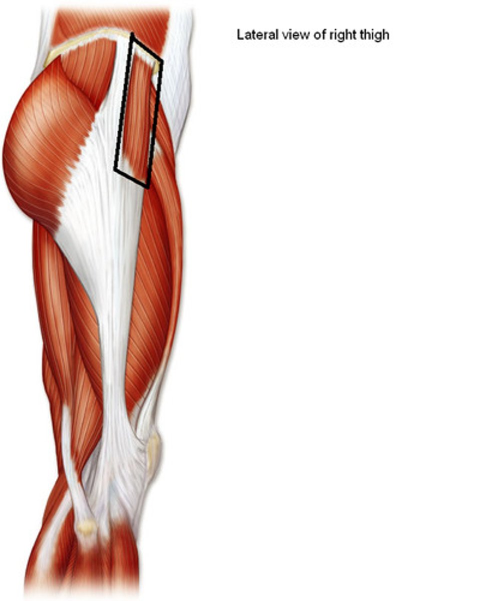
<p>Origin: anterior superior iliac spine; anterior part of iliac crest</p><p>Insertion: iliotibial tract, which attaches to lateral condyle of tibia (gerdy tubercle)</p><p>Innervation: superior gluteal nerve (L4, L5, S1)</p><p>Action: flexes hip joint; acts with gluteus maximus to stabilize the extended knee joint; pulls IT band</p>
11
New cards

Fascia Lata

Layer of connective tissue overlying thigh muscles
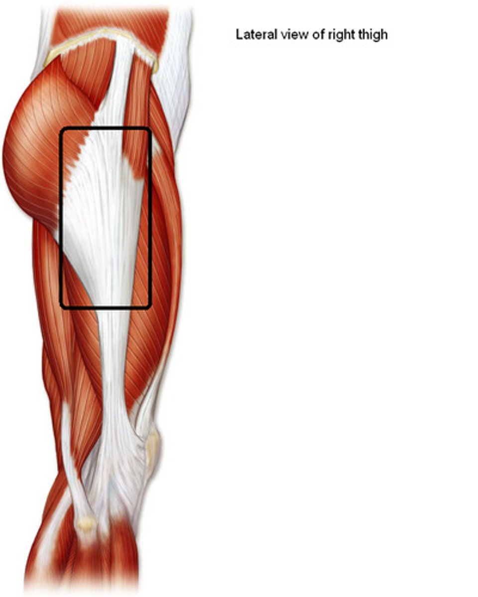
<p>Layer of connective tissue overlying thigh muscles</p>
12
New cards

Iliotibial tract

Band of longitudinal fibers on lateral side of fascia lata, connects distally to the lateral condyle of tibia
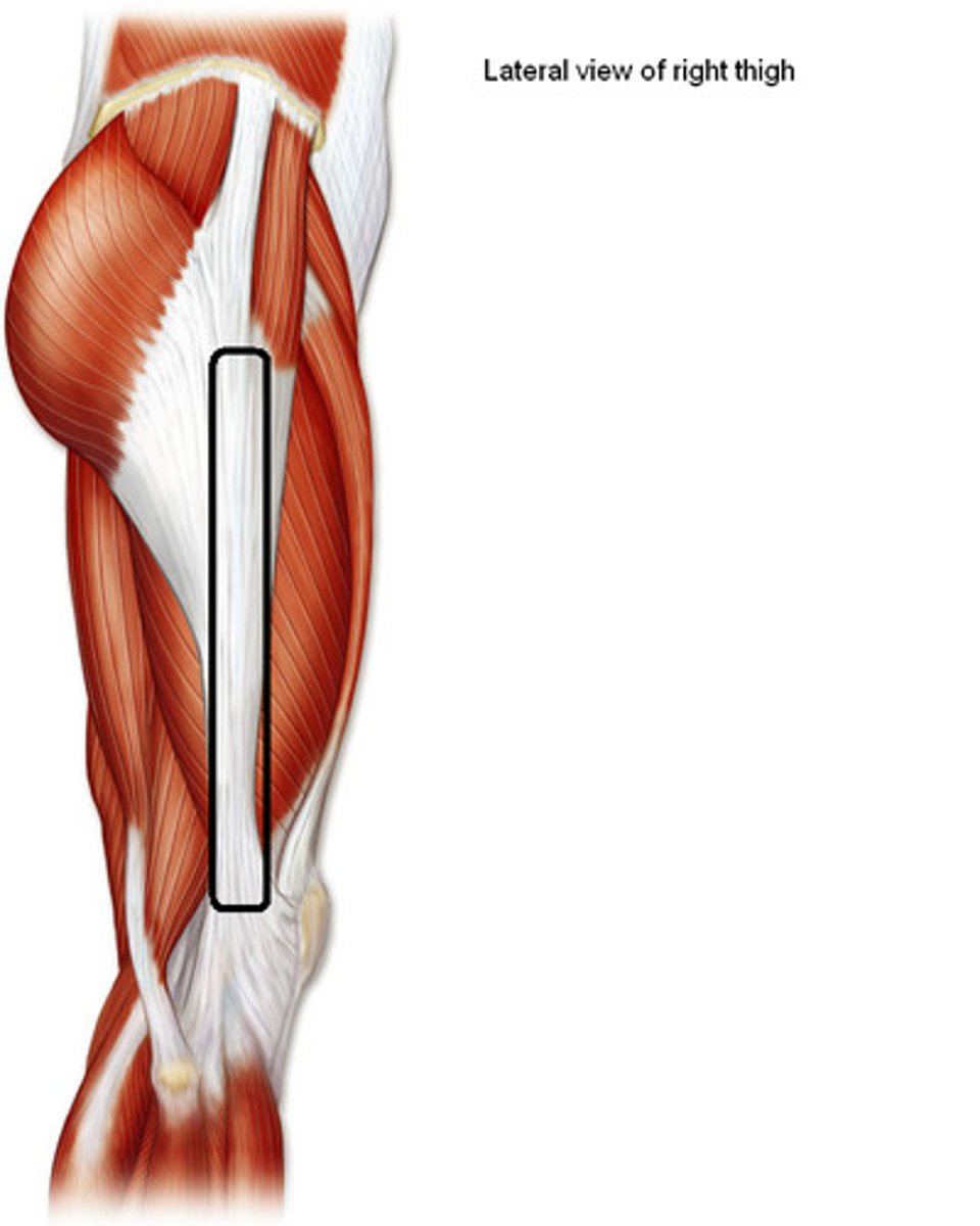
<p>Band of longitudinal fibers on lateral side of fascia lata, connects distally to the lateral condyle of tibia</p>
13
New cards

Piriformis

Origin: anterior surface of 2nd - 4th sacral segments; superior margin of greater sciatic notch and sacrotuberous ligament

Insertion: superior border of greater trochanter of femur

Innervation: branches of anterior rami of S1, S2

Action: laterally rotate extended hip joint; abduct flexed hip joint; steady femoral head in acetabulum (stabilizes hip joint)
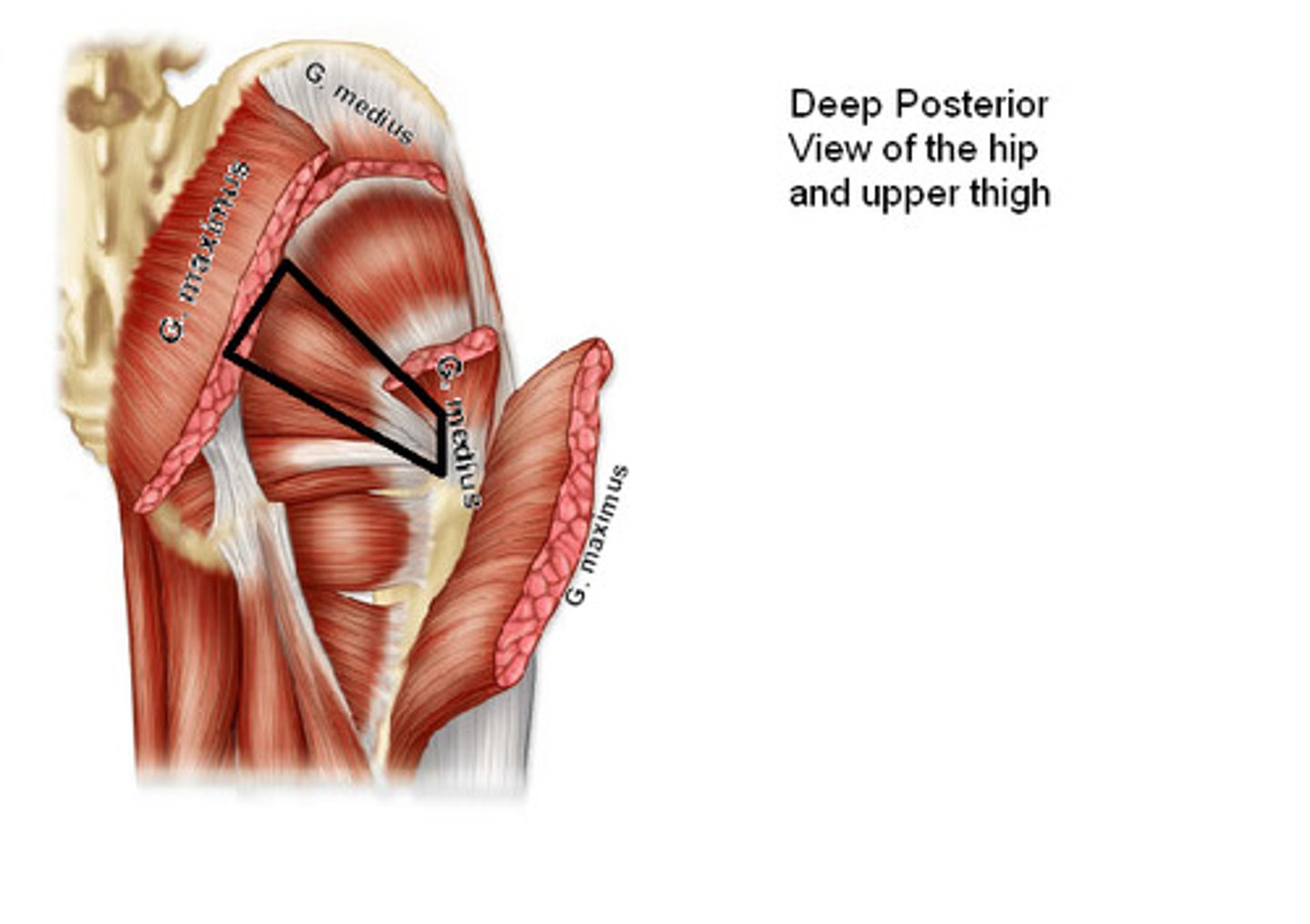
<p>Origin: anterior surface of 2nd - 4th sacral segments; superior margin of greater sciatic notch and sacrotuberous ligament</p><p>Insertion: superior border of greater trochanter of femur</p><p>Innervation: branches of anterior rami of <strong>S1</strong>, S2</p><p>Action: laterally rotate extended hip joint; abduct flexed hip joint; steady femoral head in acetabulum (stabilizes hip joint)</p>
14
New cards

Gemellus Superior

Origin: ischial spine

Insertion: medial surface of greater trochanter (trochanteric fossa) of femur

Innervation: nerve to obturator internus (L5, S1)

Action: laterally rotate extended hip joint; abduct flexed hip joint; steady femoral head in acetabulum (stabilizes hip joint)
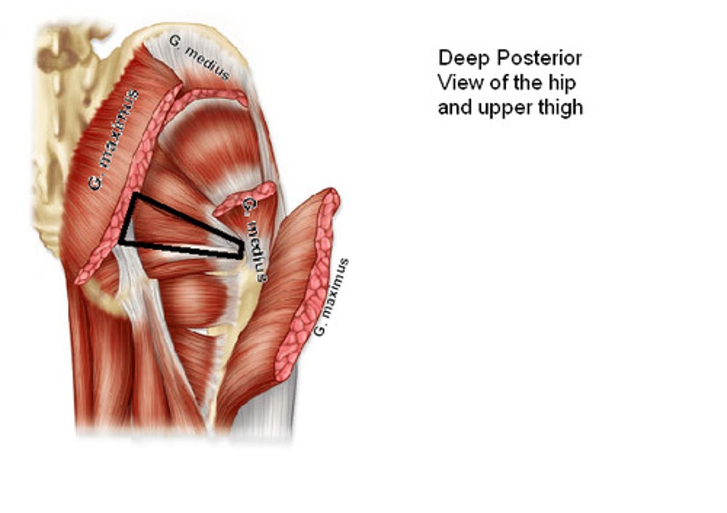
<p>Origin: ischial spine</p><p>Insertion: medial surface of greater trochanter (trochanteric fossa) of femur</p><p>Innervation: nerve to obturator internus (L5, <strong>S1</strong>)</p><p>Action: laterally rotate extended hip joint; abduct flexed hip joint; steady femoral head in acetabulum (stabilizes hip joint)</p>
15
New cards

Obturator Internus

Origin: pelvic surface of ilium and ischium; obturator membrane

Insertion: medial surface of greater trochanter (trochanteric fossa) of femur

Innervation: nerve of obturator internus (L5, S1)

Action: laterally rotate extended hip joint; abduct flexed hip joint; steady femur head into acetabulum (stabilizes hip joint)
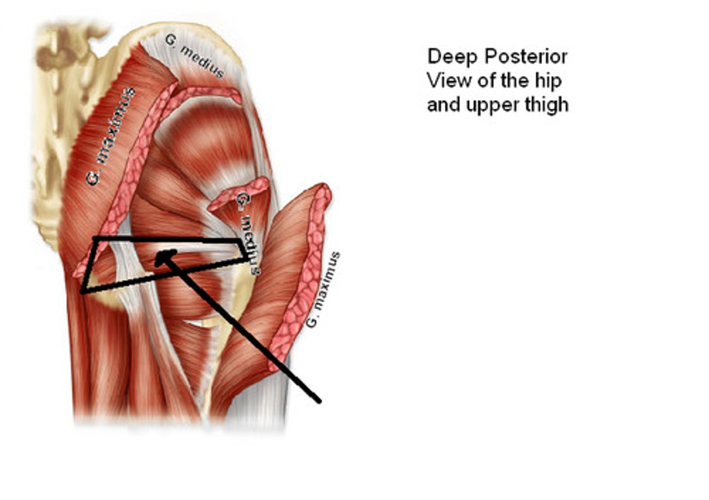
<p>Origin: pelvic surface of ilium and ischium; obturator membrane</p><p>Insertion: medial surface of greater trochanter (trochanteric fossa) of femur</p><p>Innervation: nerve of obturator internus (L5, <strong>S1</strong>)</p><p>Action: laterally rotate extended hip joint; abduct flexed hip joint; steady femur head into acetabulum (stabilizes hip joint)</p>
16
New cards

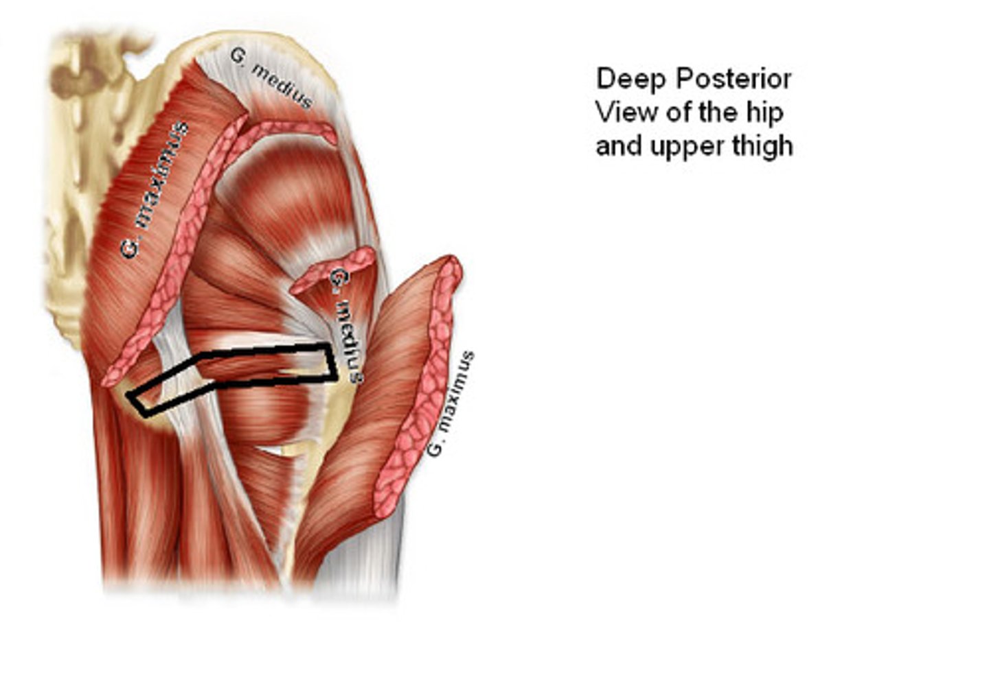
Gemellus Inferior

Origin: ischial tuberosity

Insertion: medial surface of greater trochanter (trochanteric fossa) of femur

Innervation: nerve to quadratus femoris (L5, S1)

Action: laterally rotate extended hip joint; abduct flexed hip joint; steady femoral head in acetabulum (stabilizes hip joint)
<p>Origin: ischial tuberosity</p><p>Insertion: medial surface of greater trochanter (trochanteric fossa) of femur</p><p>Innervation: nerve to quadratus femoris (L5, S1)</p><p>Action: laterally rotate extended hip joint; abduct flexed hip joint; steady femoral head in acetabulum (stabilizes hip joint)</p>
17
New cards

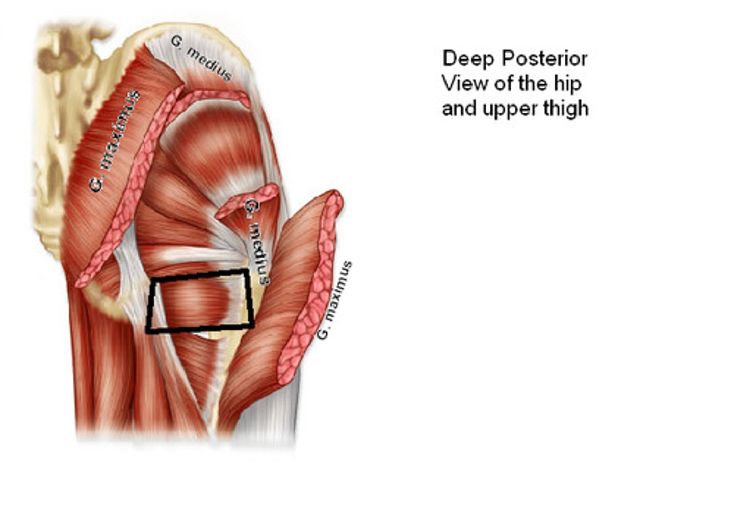
Quadratus Femoris

Origin: lateral border of ischial tuberosity

Insertion: quadrate tubercle on intertrochanteric crest of femur

Innervation: nerve to quadratus femoris (L5, S1)

Action: laterally rotates hip joint; also pulls femoral head into acetabulum to stabilize hip joint
<p>Origin: lateral border of ischial tuberosity</p><p>Insertion: quadrate tubercle on intertrochanteric crest of femur</p><p>Innervation: nerve to quadratus femoris (L5, S1)</p><p>Action: laterally rotates hip joint; also pulls femoral head into acetabulum to stabilize hip joint</p>
18
New cards

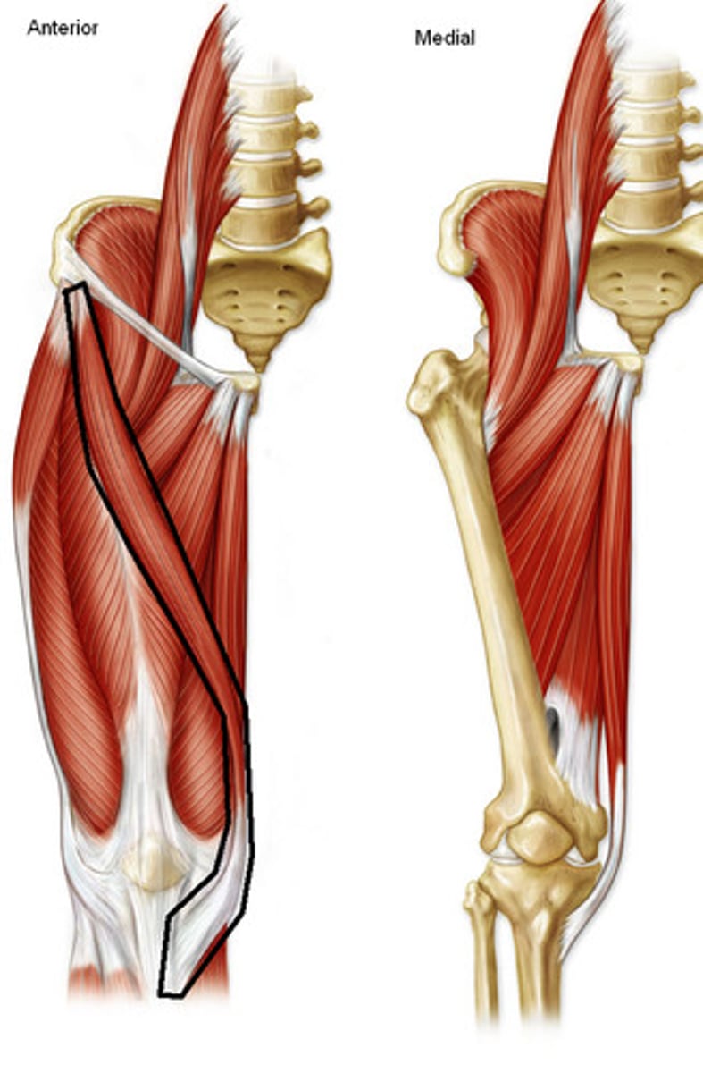
Sartorius

Origin: anterior superior iliac spine and superior part of notch inferior to it

Insertion: superior part of medial surface of tibia

Innervation: femoral nerve (L2, L3)

Action: flexes, abducts and laterally rotates hip joint; flexes knee joint
<p>Origin: anterior superior iliac spine and superior part of notch inferior to it</p><p>Insertion: superior part of medial surface of tibia</p><p>Innervation: femoral nerve (L2, L3)</p><p>Action: flexes, abducts and laterally rotates hip joint; flexes knee joint</p>
19
New cards

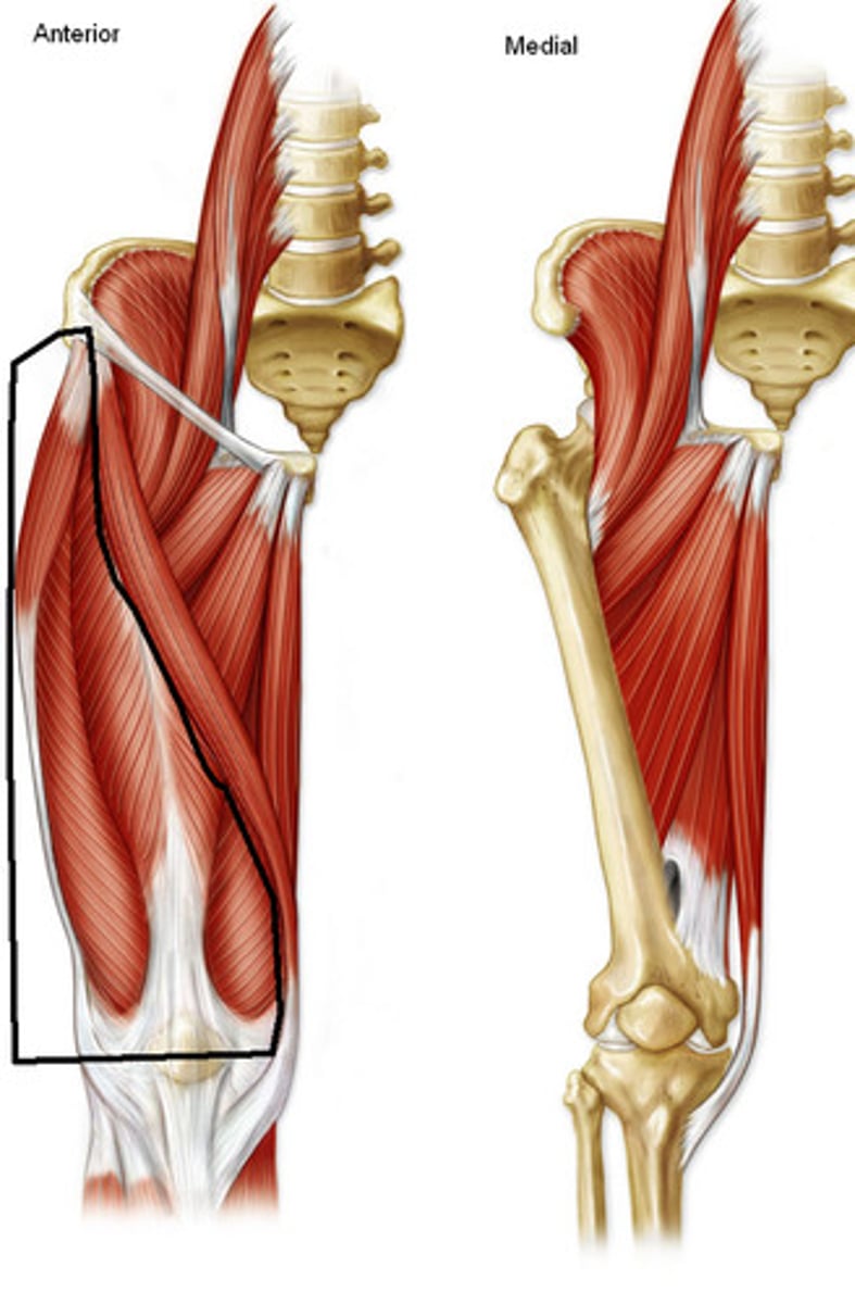
Quadriceps Femoris

Muscle group associated with movement of the knee (and hip)
<p>Muscle group associated with movement of the knee (and hip)</p>
20
New cards

Rectus Femoris

Origin: anterior inferior iliac spine and ilium superior to acetabulum

Insertion: via common tendinous (quadriceps tendon) and independent attachments to base of patella; indirectly via patellar ligament to tibial tuberosity

Innervation: femoral nerve (L2, L3, L4)

Action: extends knee joint; stabilizes hip joint and helps iliopsoas flex hip joint
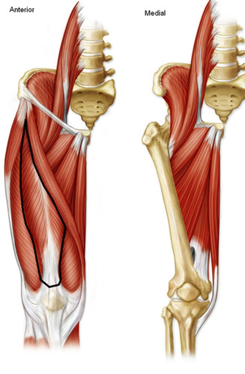
<p>Origin: anterior inferior iliac spine and ilium superior to acetabulum</p><p>Insertion: via common tendinous (quadriceps tendon) and independent attachments to base of patella; indirectly via patellar ligament to tibial tuberosity</p><p>Innervation: femoral nerve (L2, <strong>L3, L4</strong>)</p><p>Action: extends knee joint; stabilizes hip joint and helps iliopsoas flex hip joint</p>
21
New cards

Vastus Intermedius

Origin: anterior and lateral surfaces of shaft of femur

Insertion: via common tendinous (quadriceps tendon) and independent attachments to base of patella; indirectly via patellar ligament to tibial tuberosity

Innervation: femoral nerve (L2, L3, L4)

Action: extends knee joint
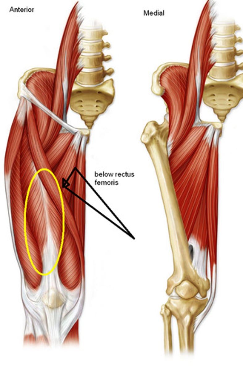
<p>Origin: anterior and lateral surfaces of shaft of femur</p><p>Insertion: via common tendinous (quadriceps tendon) and independent attachments to base of patella; indirectly via patellar ligament to tibial tuberosity</p><p>Innervation: femoral nerve (L2, <strong>L3, L4)</strong></p><p>Action: extends knee joint</p>
22
New cards

Vastus Lateralis

Origin: greater trochanter of femur and lateral lip of linea aspera

Insertion: via common tendinous (quadriceps tendon) and independent attachments to base of patella; indirectly via patellar ligament to tibial tuberosity; tibia and patella via aponeorses (lateral patella retinacula)

Innervation: femoral nerve (L2, L3, L4)

Action: extends knee joint
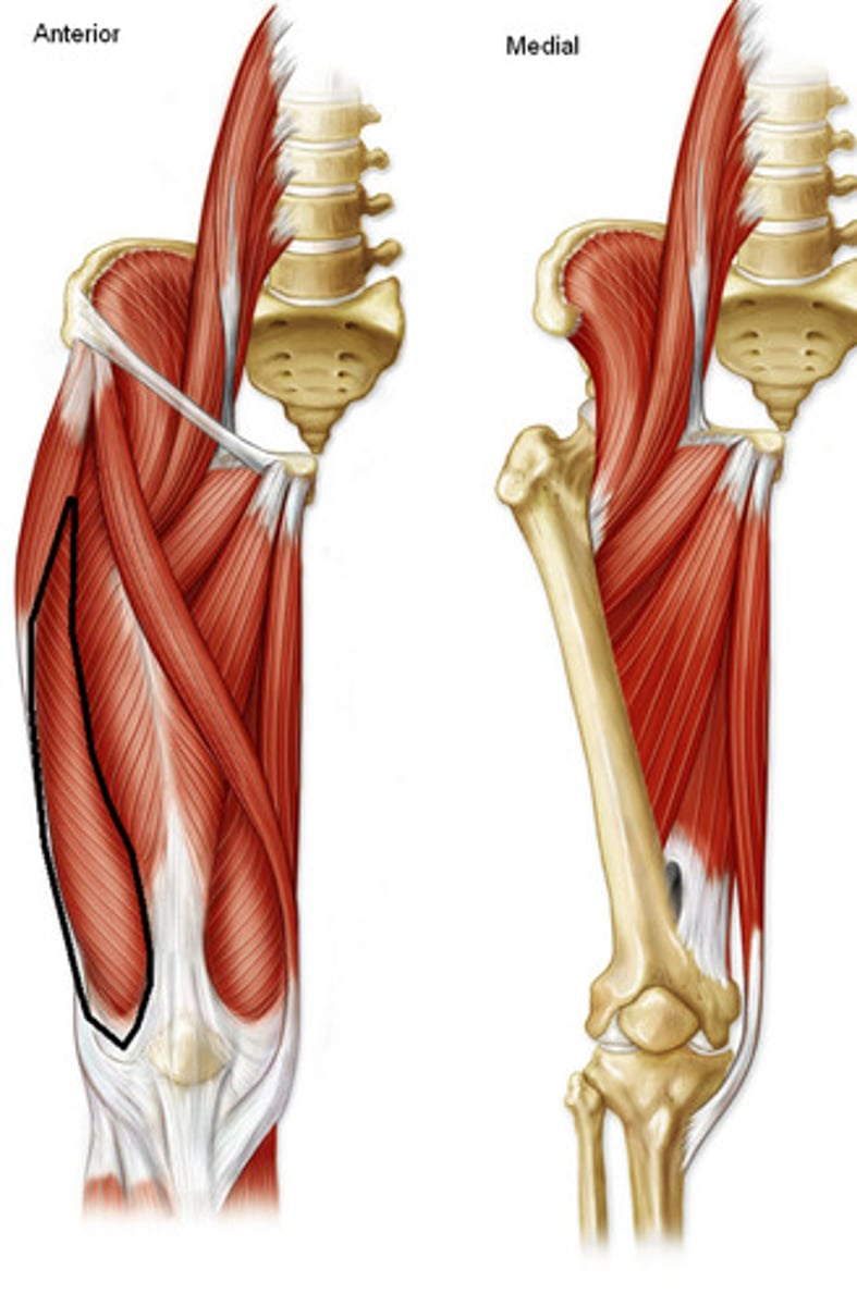
<p>Origin: greater trochanter of femur and lateral lip of linea aspera</p><p>Insertion: via common tendinous (quadriceps tendon) and independent attachments to base of patella; indirectly via patellar ligament to tibial tuberosity; tibia and patella via aponeorses (lateral patella retinacula)</p><p>Innervation: femoral nerve (L2, <strong>L3, L4)</strong></p><p>Action: extends knee joint</p>
23
New cards

Vastus Medialis

Origin: intertrochanteric line and medial lip of linea aspera

Insertion: via common tendinous (quadriceps tendon) and independent attachments to base of patella; indirectly via patellar ligament to tibial tuberosity; tibia and patella via aponeorses (medial patella retinacula)

Innervation: femoral nerve (L2, L3, L4)

Action: extends knee joint
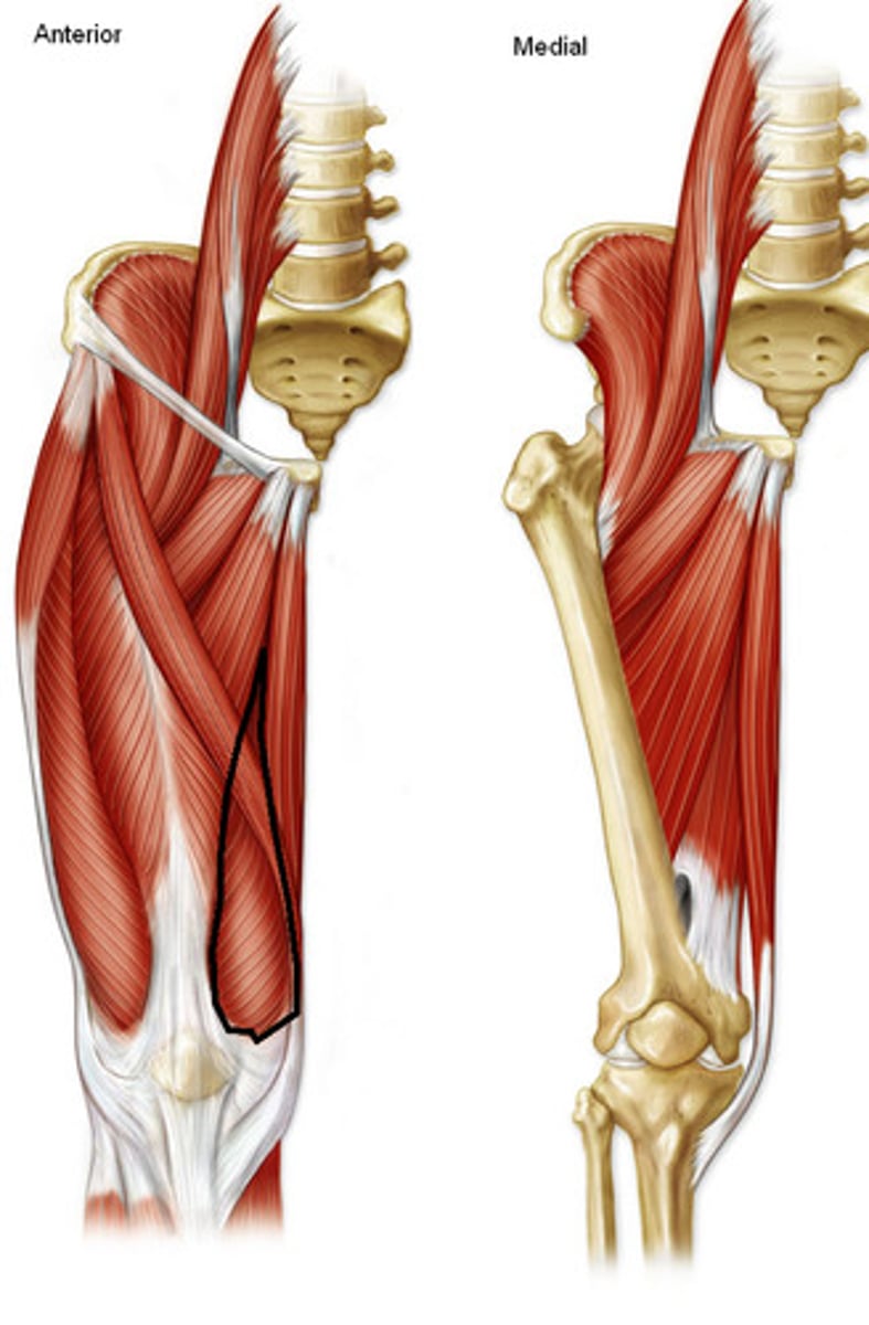
<p>Origin: intertrochanteric line and medial lip of linea aspera</p><p>Insertion: via common tendinous (quadriceps tendon) and independent attachments to base of patella; indirectly via patellar ligament to tibial tuberosity; tibia and patella via aponeorses (medial patella retinacula)</p><p>Innervation: femoral nerve (L2, <strong>L3, L4)</strong></p><p>Action: extends knee joint</p>
24
New cards

Quadriceps Tendon

common tendon for quadriceps group, attaches to patella
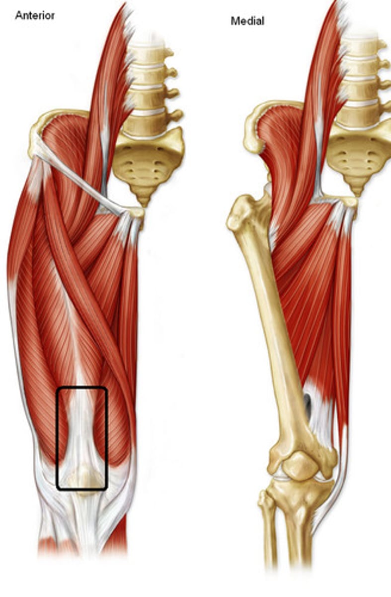
<p>common tendon for quadriceps group, attaches to patella</p>
25
New cards

Patellar Ligament

transfers action of quadriceps muscles from patella to tibia
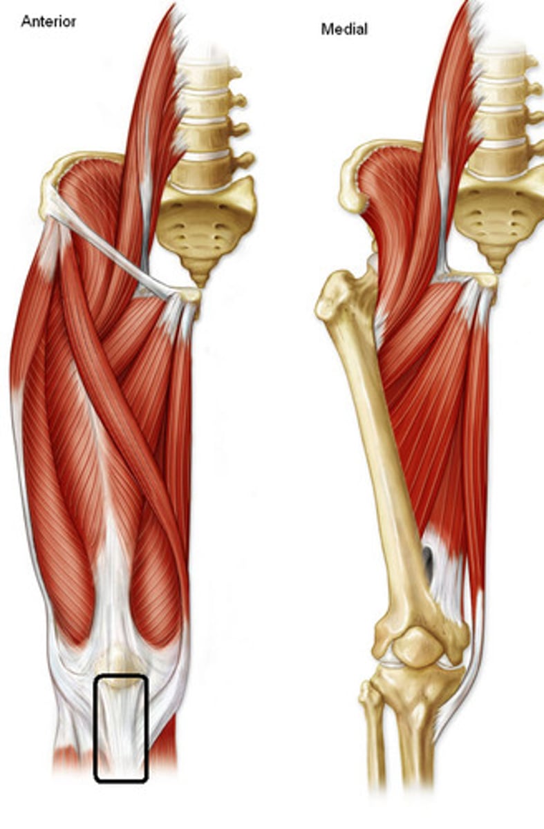
<p>transfers action of quadriceps muscles from patella to tibia</p>
26
New cards

Pectineus

Origin: superior ramus of pubis

Insertion: pectineal line of femur, just inferior to lesser trochanter

Innervation: femoral nerve (L2, L3)

Action: adducts and flexes hip joint; assists with medial rotation of hip joint
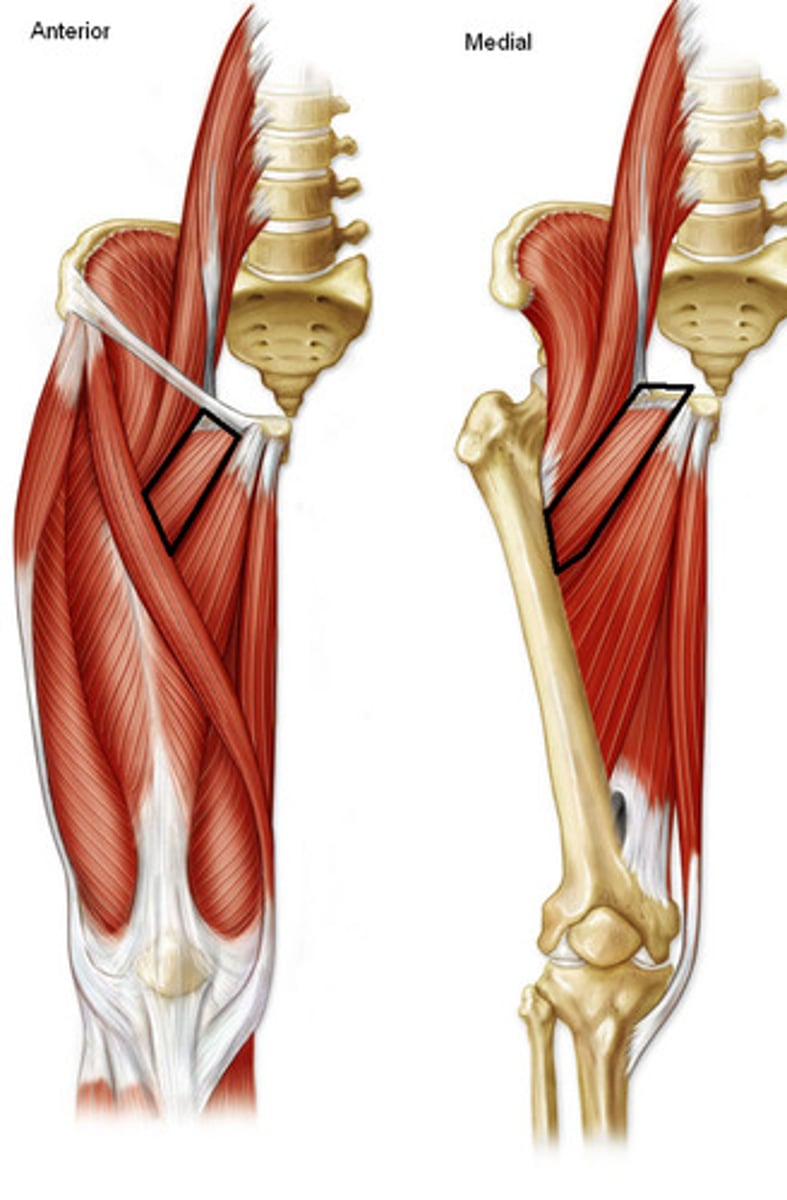
<p>Origin: superior ramus of pubis</p><p>Insertion: pectineal line of femur, just inferior to lesser trochanter</p><p>Innervation: femoral nerve (<strong>L2</strong>, L3)</p><p>Action: adducts and flexes hip joint; assists with medial rotation of hip joint</p>
27
New cards

Adductor Longus

Origin: body of pubis inferior to pubic crest

Insertion: middle third of linea aspera of femur

Innervation: obturator nerve (L2, L3, L4)

Action: adducts hip joint
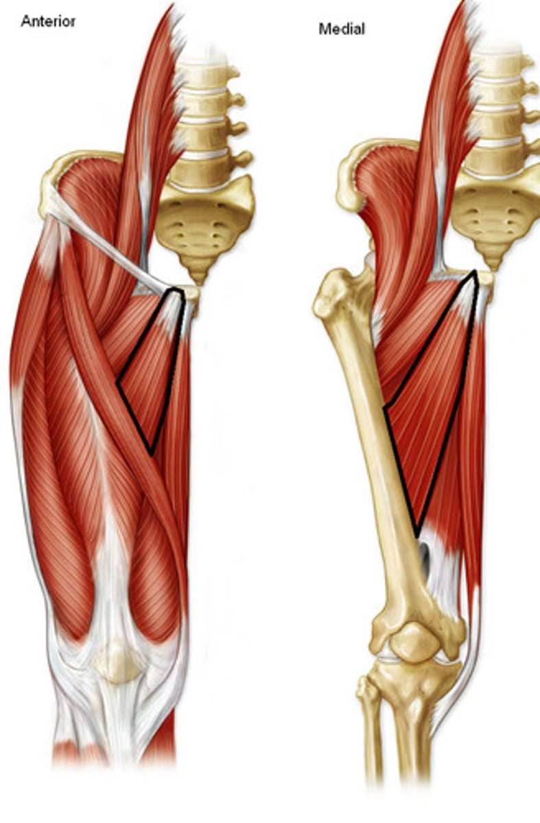
<p>Origin: body of pubis inferior to pubic crest</p><p>Insertion: middle third of linea aspera of femur</p><p>Innervation: obturator nerve (L2, <strong>L3</strong>, L4)</p><p>Action: adducts hip joint</p>
28
New cards

Adductor Magnus

Origin: inferior ramus of pubis and ramus of ischium (adductor part), ischial tuberosity (hamstring part)

Insertion: gluteal tuberosity, linea aspera, and medial supracondylar line(adductor part), adductor tubercle of femur (hamstring part)

Innervation: obturator nerve(L2, L3, L4) (adductor part) tibial part of sciatic nerve (L4) (hamstring part)

Action: adducts hip joint; adductor part flexes hip joint, hamstring part extends hip joint
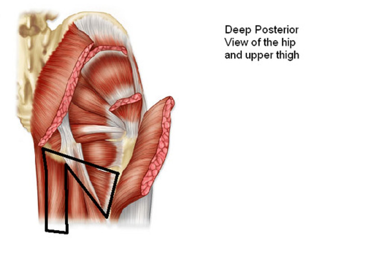
<p>Origin: inferior ramus of pubis and ramus of ischium (adductor part), ischial tuberosity (hamstring part)</p><p>Insertion: gluteal tuberosity, linea aspera, and medial supracondylar line(adductor part), adductor tubercle of femur (hamstring part)</p><p>Innervation: obturator nerve(L2, <strong>L3</strong>, <strong>L4</strong>) (adductor part) tibial part of sciatic nerve (<strong>L4</strong>) (hamstring part)</p><p>Action: adducts hip joint; adductor part flexes hip joint, hamstring part extends hip joint</p>
29
New cards

Gracilis

Origin: body and inferior ramus of pubis

Insertion: superior part of medial surface of tibia

Innervation: obturator nerve (L2, L3)

Action: adducts hip joint, flexes knee joint and helps rotate it medially
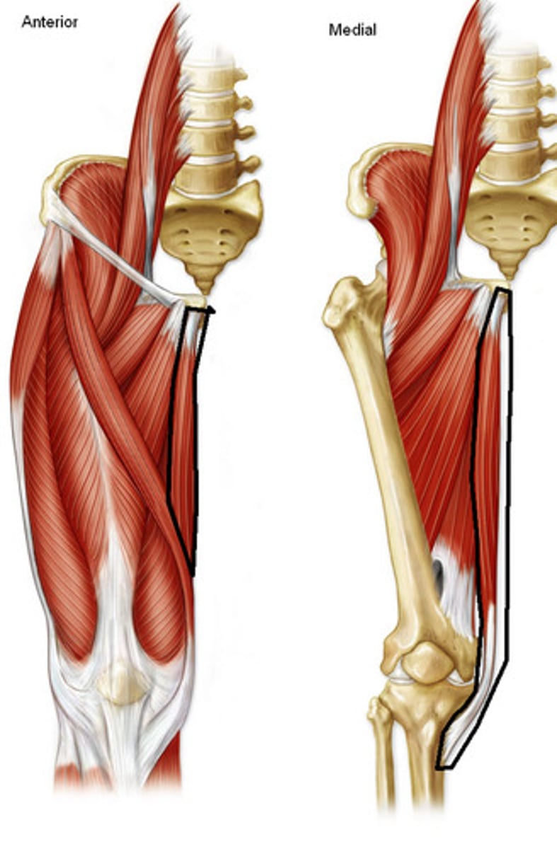
<p>Origin: body and inferior ramus of pubis</p><p>Insertion: superior part of medial surface of tibia</p><p>Innervation: obturator nerve (<strong>L2</strong>, L3)</p><p>Action: adducts hip joint, flexes knee joint and helps rotate it medially</p>
30
New cards

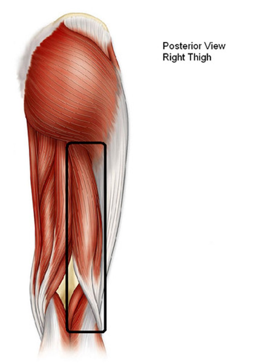
Biceps Femoris

knowt flashcard image
31
New cards

Biceps Femoris (Long Head)

Origin: ischial tuberosity

Insertion: lateral side of head of fibula; tendon is split at this site by fibular collateral ligament of knee

Innervation: tibial division of sciatic nerve (L5, S1, S2)

Action: flexes knee joint and rotates it laterally when knee is flexed; extends hip joint
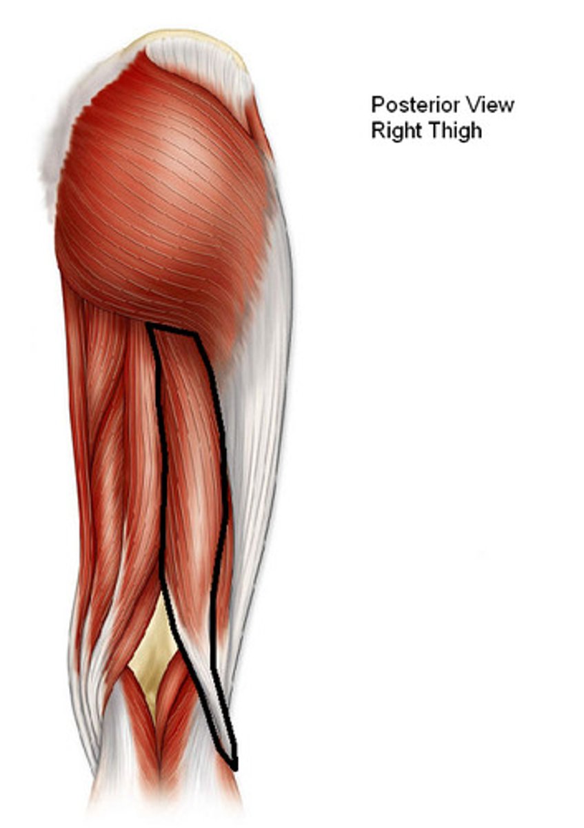
<p>Origin: ischial tuberosity</p><p>Insertion: lateral side of head of fibula; tendon is split at this site by fibular collateral ligament of knee</p><p>Innervation: tibial division of sciatic nerve (L5, <strong>S1</strong>, S2)</p><p>Action: flexes knee joint and rotates it laterally when knee is flexed; extends hip joint</p>
32
New cards

Biceps Femoris (Short Head)

Origin: linea aspera and lateral supracondylar line of femur

Insertion: lateral side of head of fibula; tendon is split at this site by fibular collateral ligament of knee

Innervation: common fibular division of sciatic nerve (L5, S1, S2)

Action: flexes knee joint and rotates it laterally when knee is flexed; extends hip joint
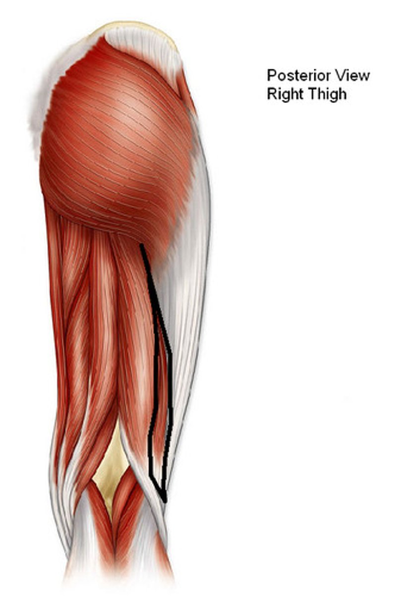
<p>Origin: linea aspera and lateral supracondylar line of femur</p><p>Insertion: lateral side of head of fibula; tendon is split at this site by fibular collateral ligament of knee</p><p>Innervation: common fibular division of sciatic nerve (L5, <strong>S1</strong>, S2)</p><p>Action: flexes knee joint and rotates it laterally when knee is flexed; extends hip joint</p>
33
New cards

Semimembranosis

Origin: ischial tuberosity

Insertion: posterior part of medial condyle of tibia; reflected attachment forms oblique popliteal ligament (to lateral femoral condyle)

Innervation: tibial division of sciatic nerve (L5, S1, S2)

Action: extends hip joint; flex knee joint and rotate the leg medially when knee is flexed; when hip and knee are flexed, can extend trunk
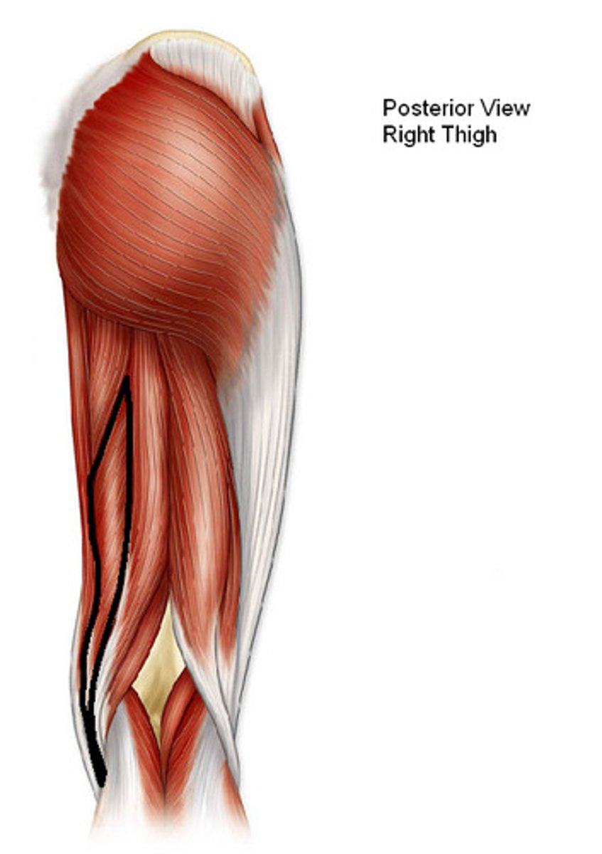
<p>Origin: ischial tuberosity</p><p>Insertion: posterior part of medial condyle of tibia; reflected attachment forms oblique popliteal ligament (to lateral femoral condyle)</p><p>Innervation: tibial division of sciatic nerve (<strong>L5</strong>, <strong>S1</strong>, S2)</p><p>Action: extends hip joint; flex knee joint and rotate the leg medially when knee is flexed; when hip and knee are flexed, can extend trunk</p>
34
New cards

Semitendinosus

Origin: ischial tuberosity

Insertion: medial surface of superior part of tibia

Innervation: tibial division of sciatic nerve (L5, S1, S2)

Action: extend hip joint; flex knee joint and rotate the leg medially when knee is flexed; when hip and knee are flexed, can extend trunk
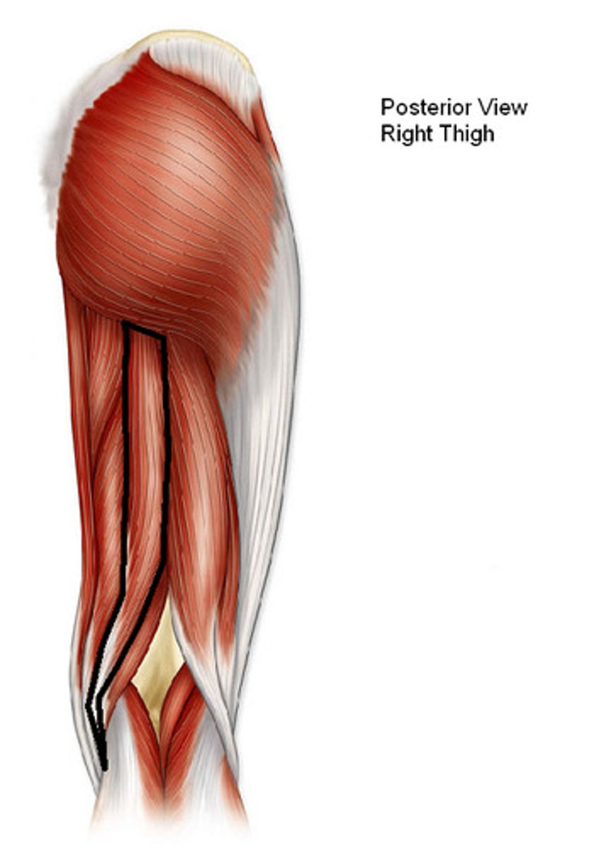
<p>Origin: ischial tuberosity</p><p>Insertion: medial surface of superior part of tibia</p><p>Innervation: tibial division of sciatic nerve (<strong>L5</strong>, <strong>S1</strong>, S2)</p><p>Action: extend hip joint; flex knee joint and rotate the leg medially when knee is flexed; when hip and knee are flexed, can extend trunk</p>
35
New cards

Tibialis Anterior (TA)

Origin: lateral condyle and superior 2/3 of lateral tibal surface, interroseous membrane

Insertion: medial and inferior suface of medial cuneiform and base of 1st metatarsal

Innervation: deep fibular nerve (L4, L5)

Action: primary dorsiflexor of ankle; primary inverter of foot; supports medial longitudinal ankle
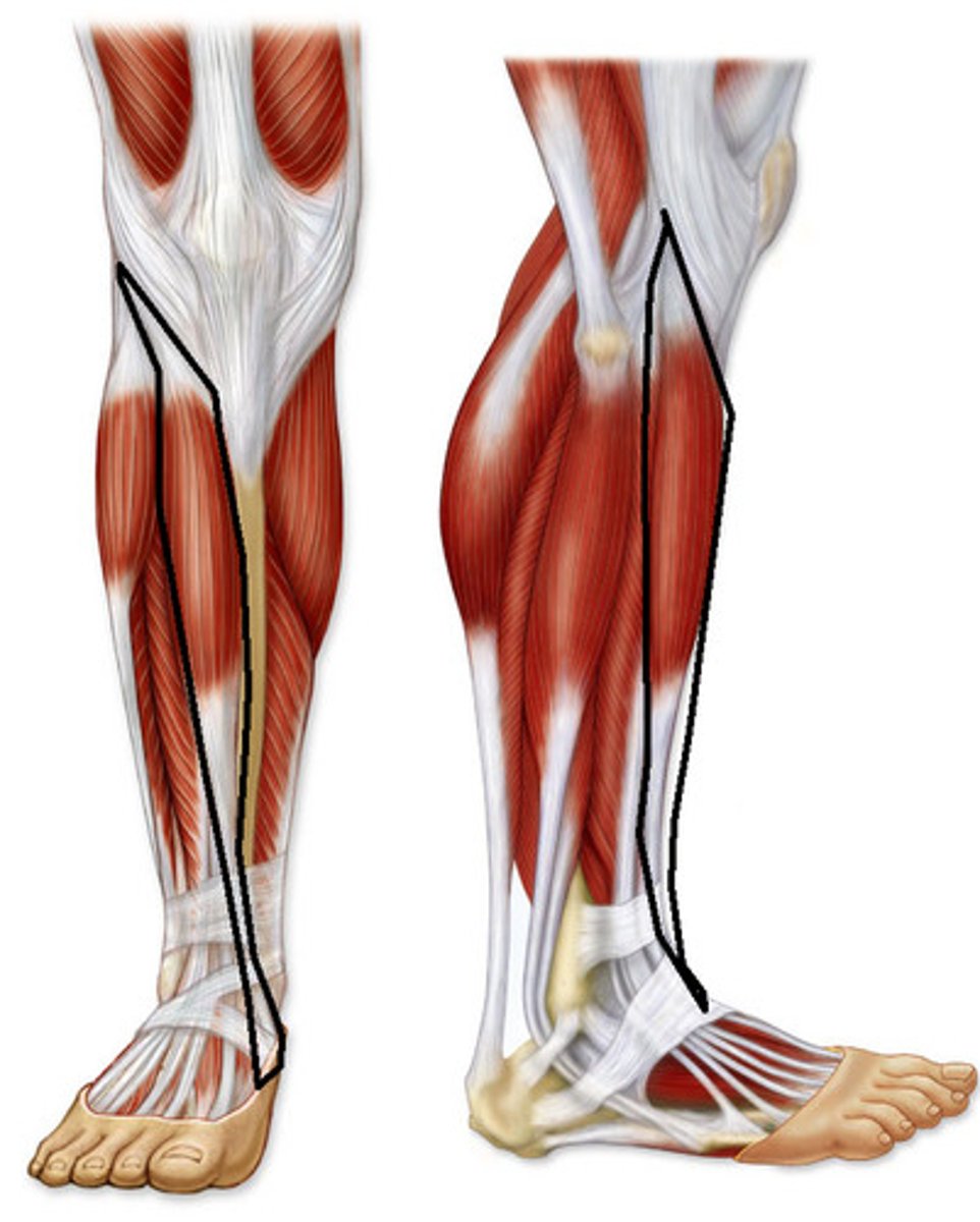
<p>Origin: lateral condyle and superior 2/3 of lateral tibal surface, interroseous membrane</p><p>Insertion: medial and inferior suface of medial cuneiform and base of 1st metatarsal</p><p>Innervation: deep fibular nerve (<strong>L4</strong>, L5)</p><p>Action: primary dorsiflexor of ankle; primary inverter of foot; supports medial longitudinal ankle</p>
36
New cards

Extensor Digitorum Longus (EDL)

Origin: lateral condyle of tibia and superior 2/3 of anterior suface of fibula and interosseous membrane

Insertion: middle and distal phalanges of lateral four digits (2-5)

Innervation: deep fibular nerve (L5, S1)

Action: extends lateral four digits; secondary dorsiflexor of ankle
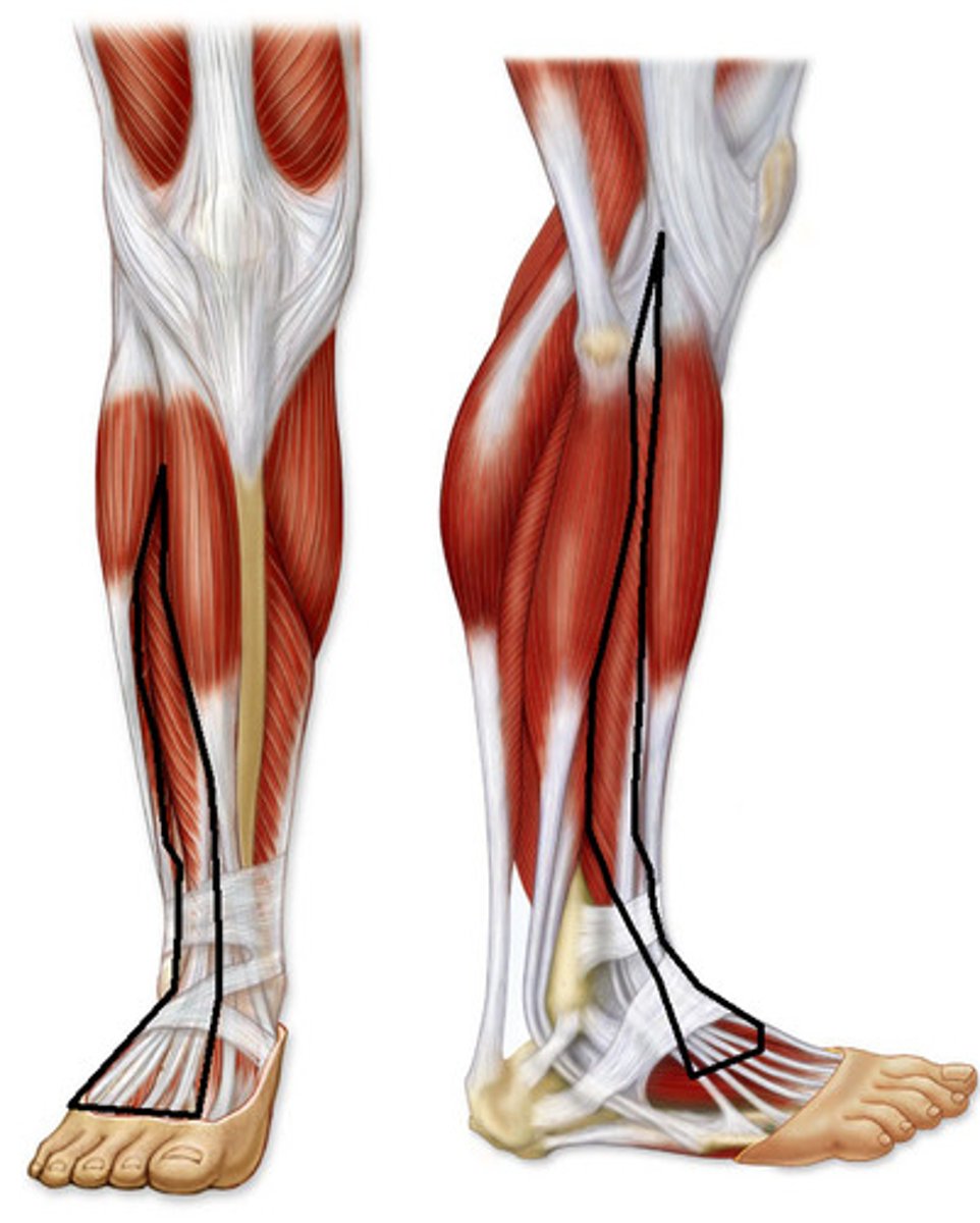
<p>Origin: lateral condyle of tibia and superior 2/3 of anterior suface of fibula and interosseous membrane</p><p>Insertion: middle and distal phalanges of lateral four digits (2-5)</p><p>Innervation: deep fibular nerve (L5, S1)</p><p>Action: extends lateral four digits; secondary dorsiflexor of ankle</p>
37
New cards

Extensor Hallucis Longus (EHL)

Origin: middle part of anterior surface of fibula and interosseous membrane

Insertion: dorsal aspect of base of distal phalanx of great toe (hallux)

Innervation: deep fibular nerve (L5, S1)

Action: extends great toe; secondary dorsiflexor of ankle
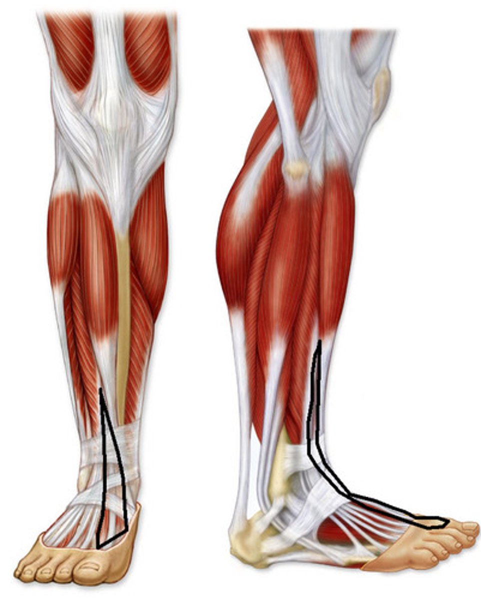
<p>Origin: middle part of anterior surface of fibula and interosseous membrane</p><p>Insertion: dorsal aspect of base of distal phalanx of great toe (hallux)</p><p>Innervation: deep fibular nerve (L5, S1)</p><p>Action: extends great toe; secondary dorsiflexor of ankle</p>
38
New cards

Fibularis Longus (FL)

Origin: head and superior 2/3 of lateral surface of fibula

Insertion: base of 1st metatarsal and medial cuneiform

Innervation: superficial fibular nerve (L5, S1, S2)

Action: evert foot, weakly plantarflexes ankle; supports transverse arch of foot
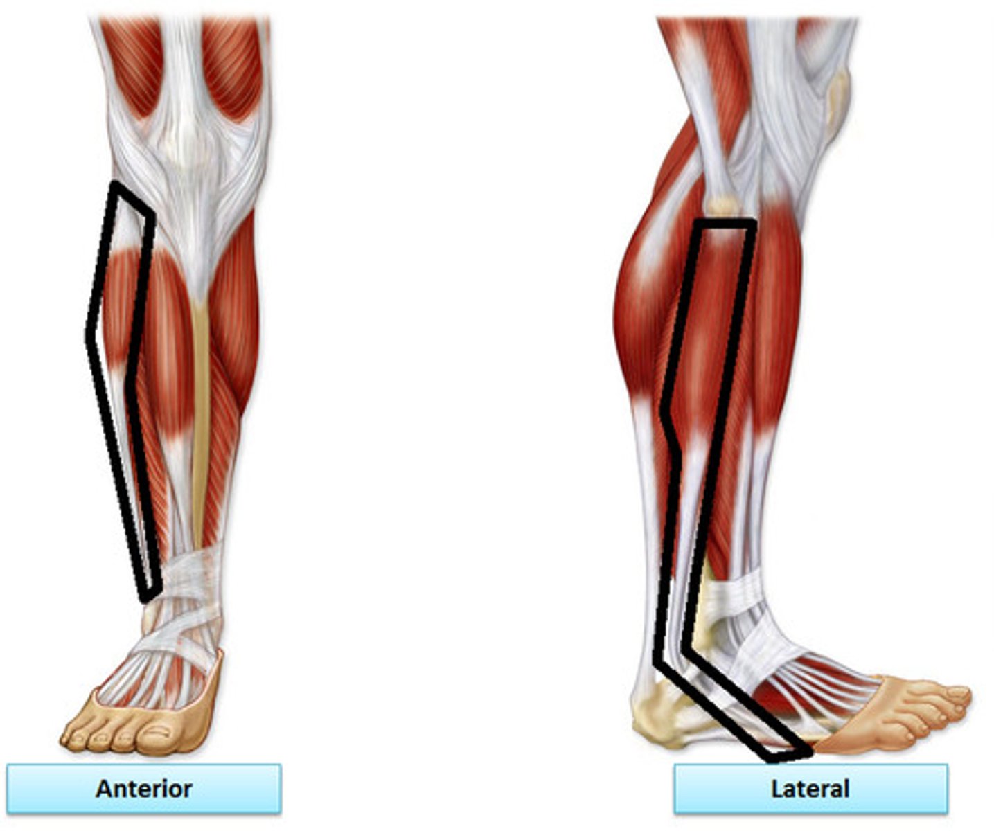
<p>Origin: head and superior 2/3 of lateral surface of fibula</p><p>Insertion: base of 1st metatarsal and medial cuneiform </p><p>Innervation: superficial fibular nerve (<strong>L5</strong>, <strong>S1</strong>, S2)</p><p>Action: evert foot, weakly plantarflexes ankle; supports transverse arch of foot</p>
39
New cards

Fibularis Brevis (FB)

Origin: middle part of lateral surface of fibula

Insertion: dorsal surface of tuberosity of base of 5th metatarsal

Innervation: superficial fibular nerve (L5, S1, S2)

Action: evert foot; weakly plantar flex ankle
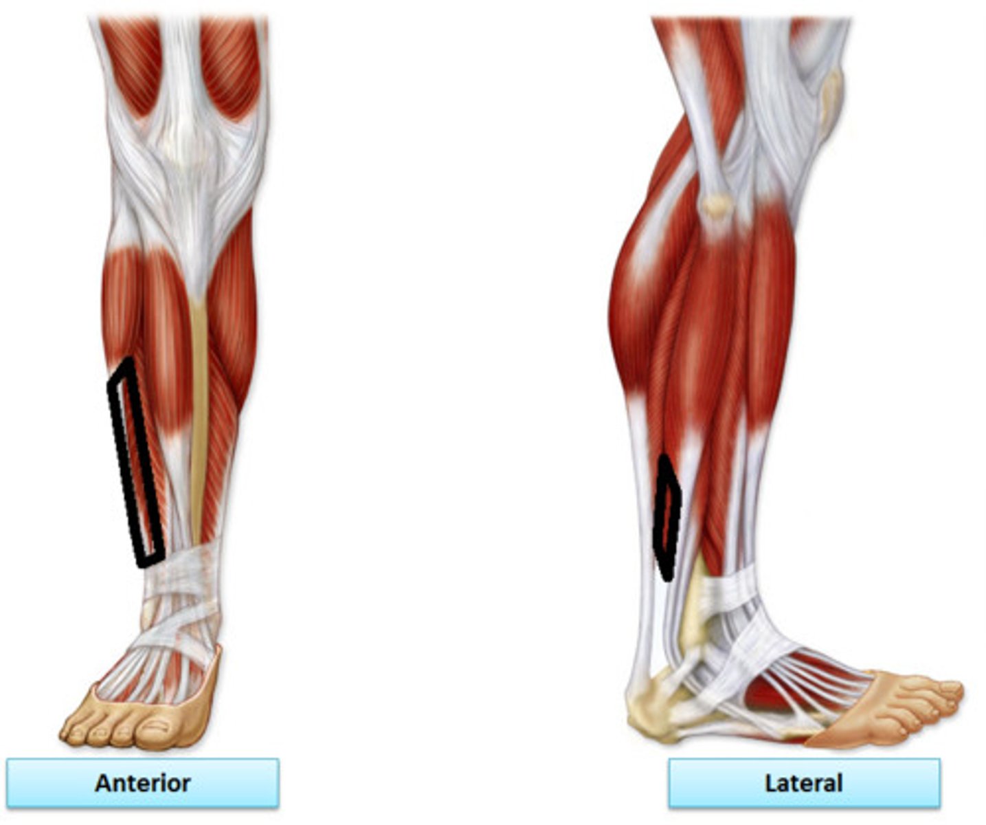
<p>Origin: middle part of lateral surface of fibula</p><p>Insertion: dorsal surface of tuberosity of base of 5th metatarsal</p><p>Innervation: superficial fibular nerve (<strong>L5</strong>, <strong>S1</strong>, S2)</p><p>Action: evert foot; weakly plantar flex ankle</p>
40
New cards

Gastrocnemius

Origin: lateral aspect of lateral condyle of femur (lateral head), popliteal surface of femur, superior to medial condyle (medial head)

Insertion: posterior surface of calcaneus via calcaneal tendon

Innervation: tibial nerve (S1, S2)

Action: plantarflexes ankle when knee is extended; raises heel during walking; and flexes knee joint
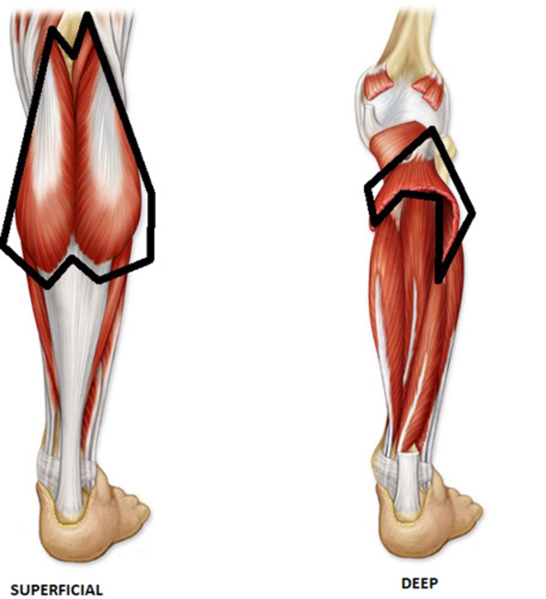
<p>Origin: lateral aspect of lateral condyle of femur (lateral head), popliteal surface of femur, superior to medial condyle (medial head)</p><p>Insertion: posterior surface of calcaneus via calcaneal tendon</p><p>Innervation: tibial nerve (S1, S2)</p><p>Action: plantarflexes ankle when knee is extended; raises heel during walking; and flexes knee joint</p>
41
New cards

Soleus

Origin: posterior aspect of head of fibula, superior quarter of posterior surface of fibula, soleal line, and medial border of tibia

Insertion: posterior surface of calcaneus via calcaneal tendon

Innervation: tibial nerve (S1, S2)

Action: plantar flexes ankle; steadies leg on foot
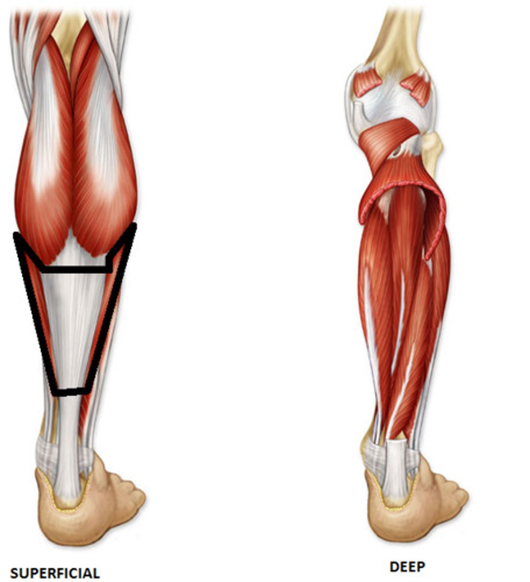
<p>Origin: posterior aspect of head of fibula, superior quarter of posterior surface of fibula, soleal line, and medial border of tibia</p><p>Insertion: posterior surface of calcaneus via calcaneal tendon</p><p>Innervation: tibial nerve (S1, S2)</p><p>Action: plantar flexes ankle; steadies leg on foot</p>
42
New cards

Plantaris

Origin: inferior end of lateral supracondylar line of femur and oblique popliteal ligament

Insertion: posterior surface of calcaneus via calcaneal tendon

Innervation: tibial nerve (S1, S2)

Action: weakly assists gastocnemius in plantarflexing ankle; function is probably mainly proprioceptive
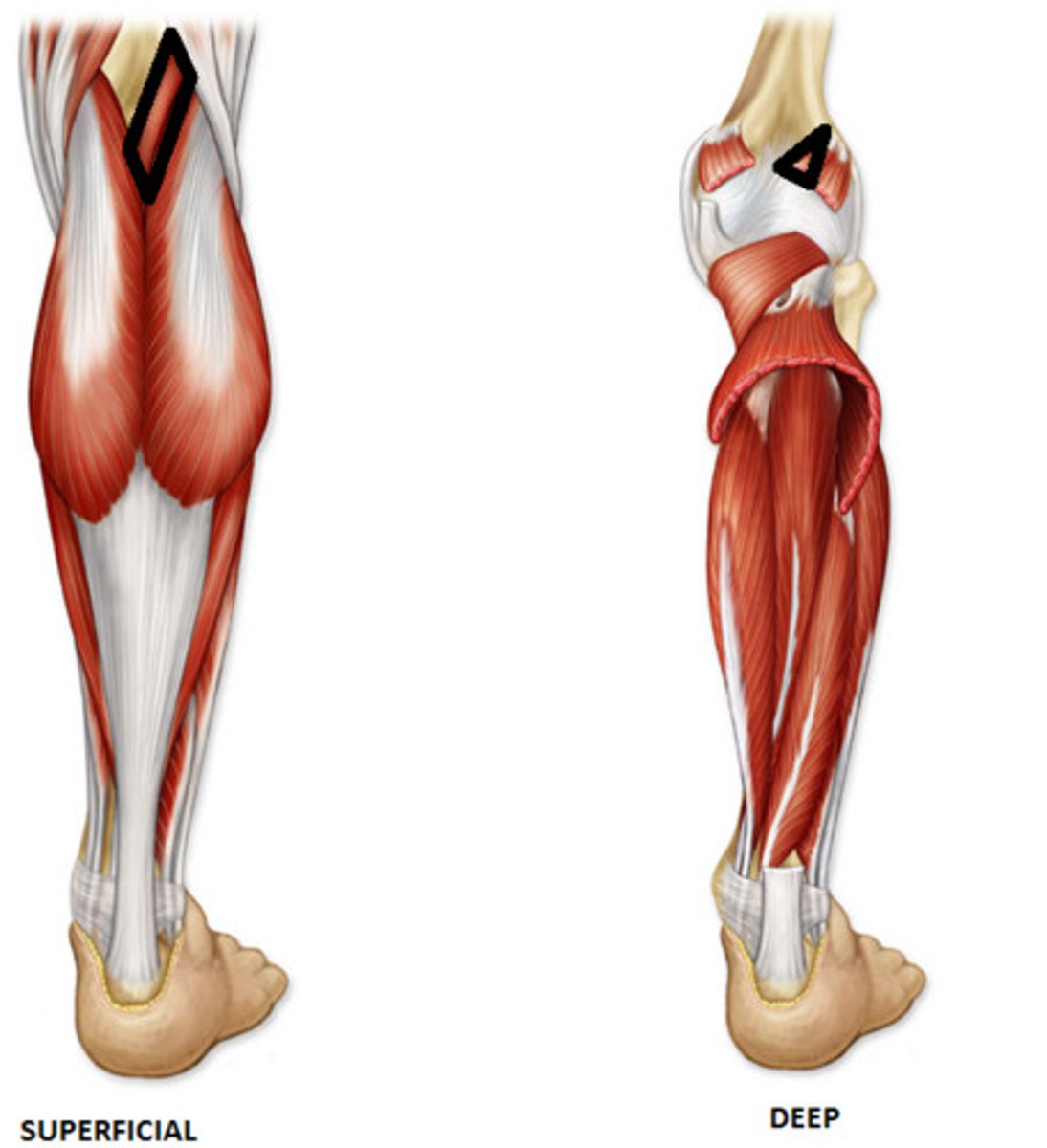
<p>Origin: inferior end of lateral supracondylar line of femur and oblique popliteal ligament</p><p>Insertion: posterior surface of calcaneus via calcaneal tendon</p><p>Innervation: tibial nerve (S1, S2)</p><p>Action: weakly assists gastocnemius in plantarflexing ankle; function is probably mainly proprioceptive</p>
43
New cards

Calcaneal Tendon

common tendon for the superficial posterior leg muscles
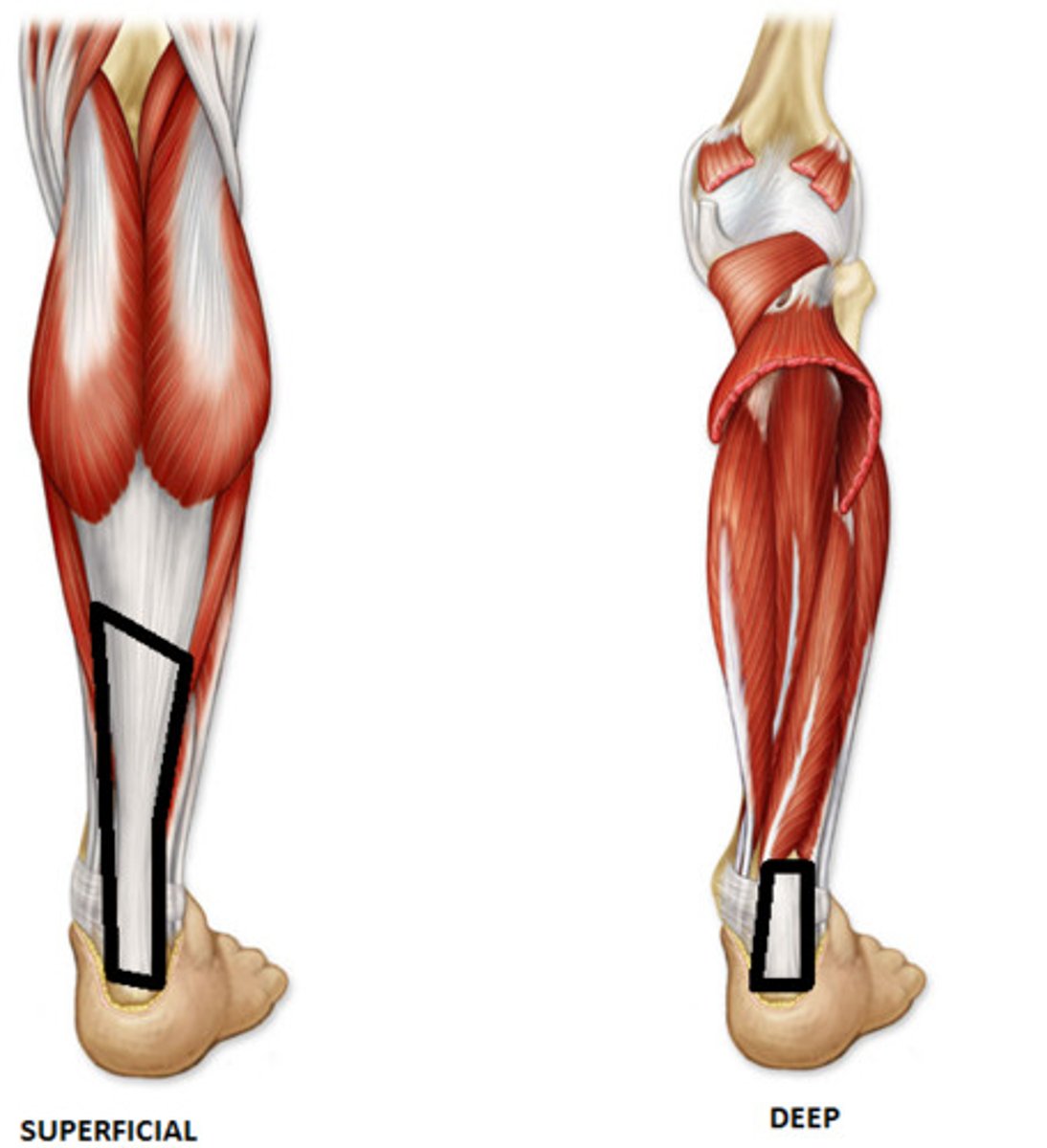
<p>common tendon for the superficial posterior leg muscles</p>
44
New cards

Popliteus

Origin: lateral surface of lateral condyle of femur and lateral meniscus (intra-articular; within cavity of knee joint)

Insertion: posterior surface of tibia, superior to soleal line

Innervation: tibial nerve (L4, L4, S1)

Action: weakly flexes knee and unlocks it by laterally rotating femur on fixed tibia, may also medially rotate tibia of unplanted limb (“key to the knee”)
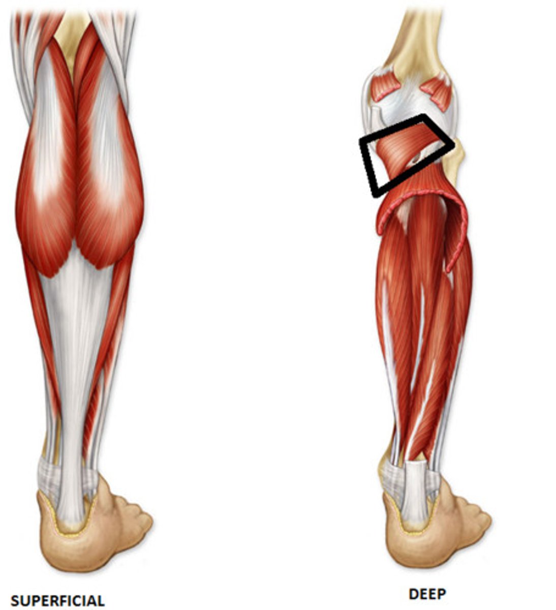
<p>Origin: lateral surface of lateral condyle of femur and lateral meniscus (intra-articular; within cavity of knee joint)</p><p>Insertion: posterior surface of tibia, superior to soleal line</p><p>Innervation: tibial nerve (L4, L4, S1)</p><p>Action: weakly flexes knee and unlocks it by laterally rotating femur on fixed tibia, may also medially rotate tibia of unplanted limb (“key to the knee”)</p>
45
New cards

Tibialis Posterior (TP)

Origin: interosseous membrane, posterior surface of tibia inferior to soleal line, and posterior surface of fibula

Insertion: primarily to tuberosity of navicular; also to cuneiforms, cuboid, and bases of 2nd - 4th metatarsals

Innervation: tibial nerve (L4, L5)

Action: plantarflexes ankle; primary inverter of foot; supports medial longitudinal arch of foot
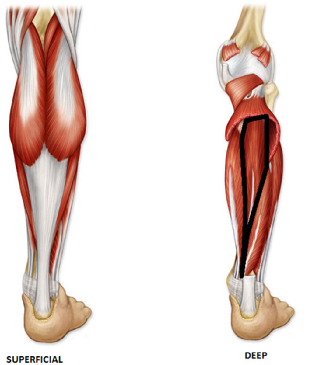
<p>Origin: interosseous membrane, posterior surface of tibia inferior to soleal line, and posterior surface of fibula</p><p>Insertion: primarily to tuberosity of navicular; also to cuneiforms, cuboid, and bases of 2nd - 4th metatarsals</p><p>Innervation: tibial nerve (L4, L5)</p><p>Action: plantarflexes ankle; primary inverter of foot; supports medial longitudinal arch of foot</p>
46
New cards

Flexor Digitorum Longus

Origin: medial part of posterior surface of tibia inferior to soleal line and by a broad tendon to fibula

Insertion: bases of distal phalanges of lateral four digits

Innervation: tibial nerve (S2, S3)

Action: flexes lateral four digits; plantar flexes ankle; supports longitudinal arches of foot
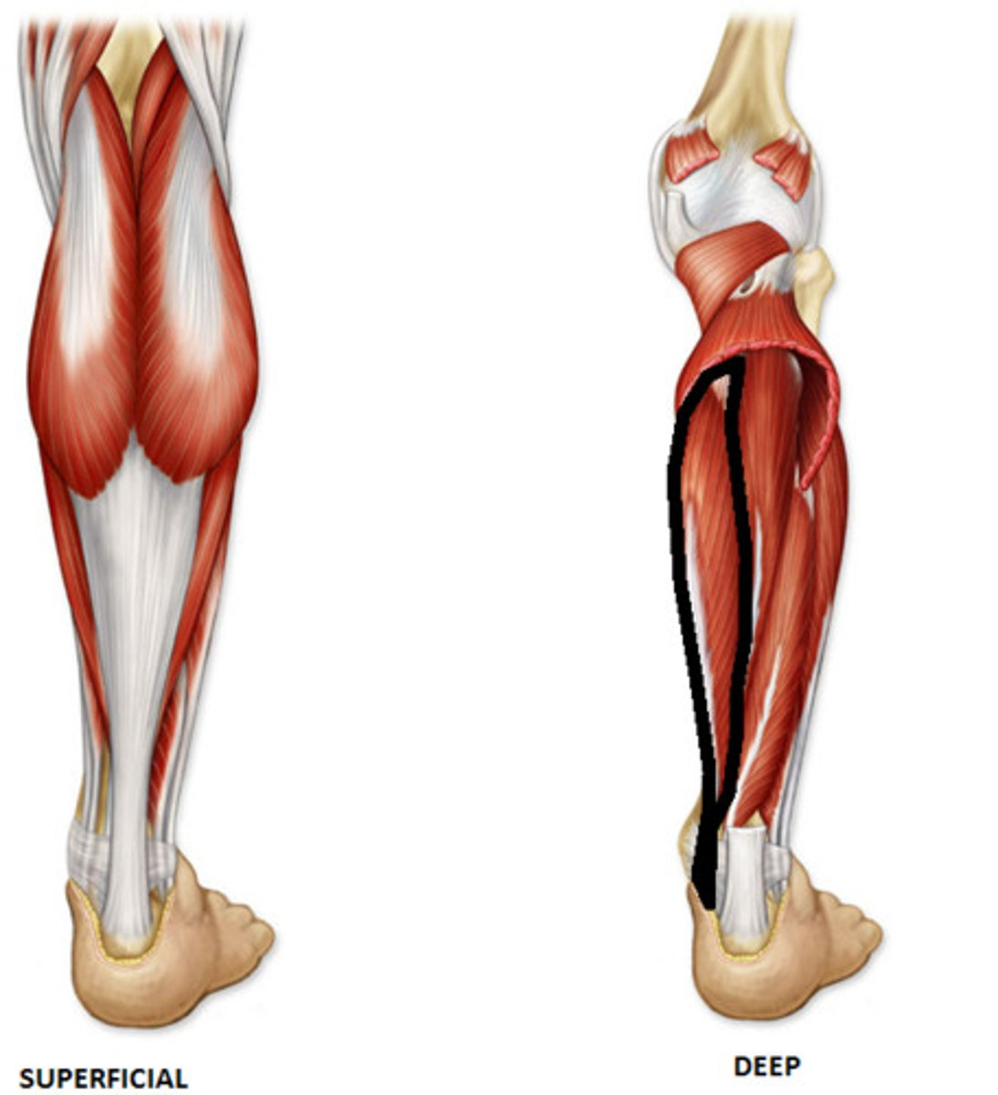
<p>Origin: medial part of posterior surface of tibia inferior to soleal line and by a broad tendon to fibula</p><p>Insertion: bases of distal phalanges of lateral four digits</p><p>Innervation: tibial nerve (<strong>S2</strong>, S3)</p><p>Action: flexes lateral four digits; plantar flexes ankle; supports longitudinal arches of foot</p>
47
New cards

Flexor Hallucis Longus

Origin: inferior 2/3 of posterior surface of fibula and inferior part of interosseous membrane

Insertion: base of distal phalanx of great toe (hallux)

Innervation: tibial nerve (S2, S3)

Action: flexes great toe at all joints; weakly plantarflexes ankle; supports medial longitudinal arch of foot
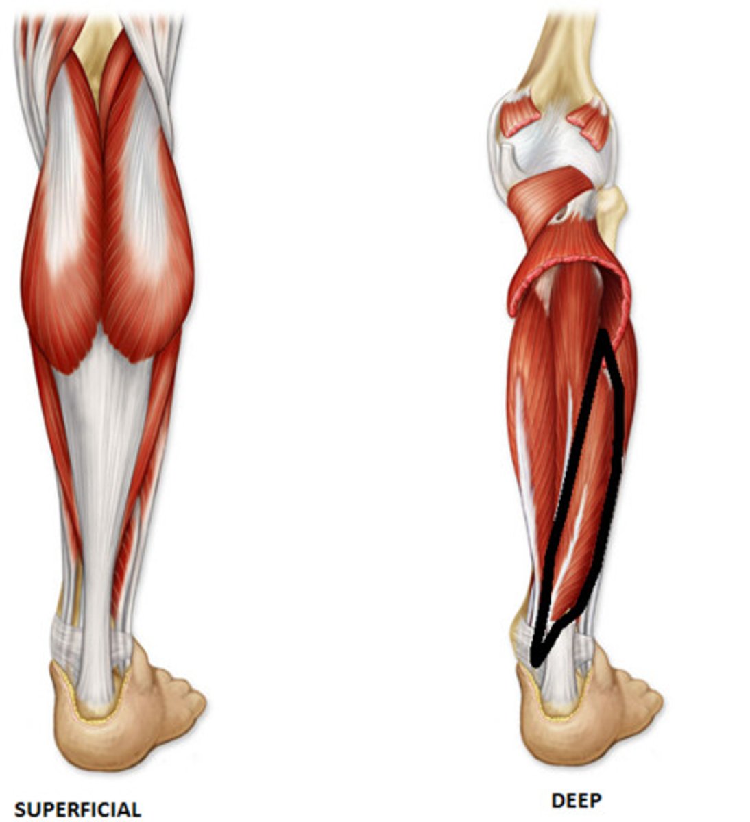
<p>Origin: inferior 2/3 of posterior surface of fibula and inferior part of interosseous membrane</p><p>Insertion: base of distal phalanx of great toe (hallux)</p><p>Innervation: tibial nerve (<strong>S2</strong>, S3)</p><p>Action: flexes great toe at all joints; weakly plantarflexes ankle; supports medial longitudinal arch of foot</p>
48
New cards

Extensor Digitorum Brevis

Origin: dorsal surface of calcaneus

Insertion: 2nd to 4th toes (at dorsal aponeurosis and bases of the middle phalanges)

Innervation: deep fibular nerve (L5, S1)

Action: extends the metatarsophalangeal (MTP) and proximal interphangeal (PIP) joints of the 2nd to 4th toes
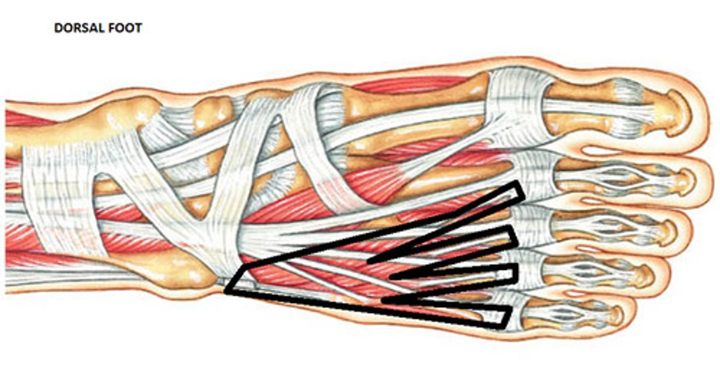
<p>Origin: dorsal surface of calcaneus</p><p>Insertion: 2nd to 4th toes (at dorsal aponeurosis and bases of the middle phalanges)</p><p>Innervation: deep fibular nerve (L5, S1)</p><p>Action: extends the metatarsophalangeal (MTP) and proximal interphangeal (PIP) joints of the 2nd to 4th toes</p>
49
New cards

Extensor Hallucis Brevis

Origin: dorsal surface of calcaneus

Insertion: 1st toe (at dorsal aponeurosis and proximal phalanx)

Innervation: deep fibular nerve (L5, S1)

Action: extends metotarsophalangeal (MTP) joints of 1st toe
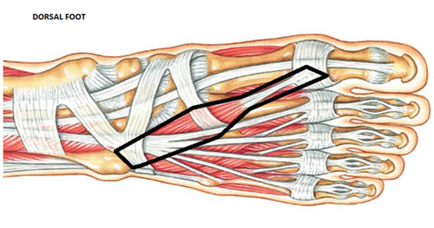
<p>Origin: dorsal surface of calcaneus</p><p>Insertion: 1st toe (at dorsal aponeurosis and proximal phalanx)</p><p>Innervation: deep fibular nerve (L5, S1)</p><p>Action: extends metotarsophalangeal (MTP) joints of 1st toe</p>
50
New cards

Abductor Hallucis

Origin: medial tubercle of tuberosity of calcaneus, flexor retinaculum, and plantar aponeurosis

Insertion: medial side of base of proximal phalanx of 1st digit

Innervation: medial plantar nerve (S2, S3)

Action: abducts and flexes hallux
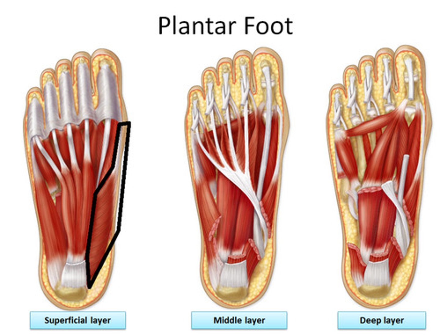
<p>Origin: medial tubercle of tuberosity of calcaneus, flexor retinaculum, and plantar aponeurosis</p><p>Insertion: medial side of base of proximal phalanx of 1st digit</p><p>Innervation: medial plantar nerve (S2, <strong>S3</strong>)</p><p>Action: abducts and flexes hallux</p>
51
New cards

Adductor Hallucis

Origin: oblique head from base of metatarsals 2-4; transverse head from plantar ligaments of 3rd-5th metatarsophalangeal joints

Insertion: tendons of both heads attach to lateral side of base of proximal phalanx of 1st digit

Innervation: deep branch of lateral plantar nerve (S2, S3)

Action: adducts 1st digit; assists in maintaining transverse arch of foot
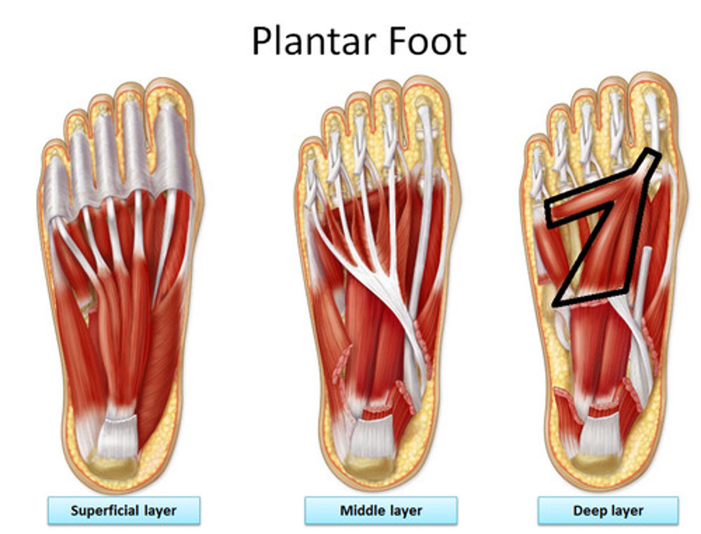
<p>Origin: oblique head from base of metatarsals 2-4; transverse head from plantar ligaments of 3rd-5th metatarsophalangeal joints</p><p>Insertion: tendons of both heads attach to lateral side of base of proximal phalanx of 1st digit</p><p>Innervation: deep branch of lateral plantar nerve (S2, <strong>S3</strong>)</p><p>Action: adducts 1st digit; assists in maintaining transverse arch of foot</p>
52
New cards

Flexor Hallucis Brevis

Origin: plantar surfaces of cuboid and lateral cuneiform

Insertion: both sides of base of proximal phalanx of 1st digit

Innervation: medial plantar nerve (S2, S3)

Action: flexes proximal phalanx of 1st digit
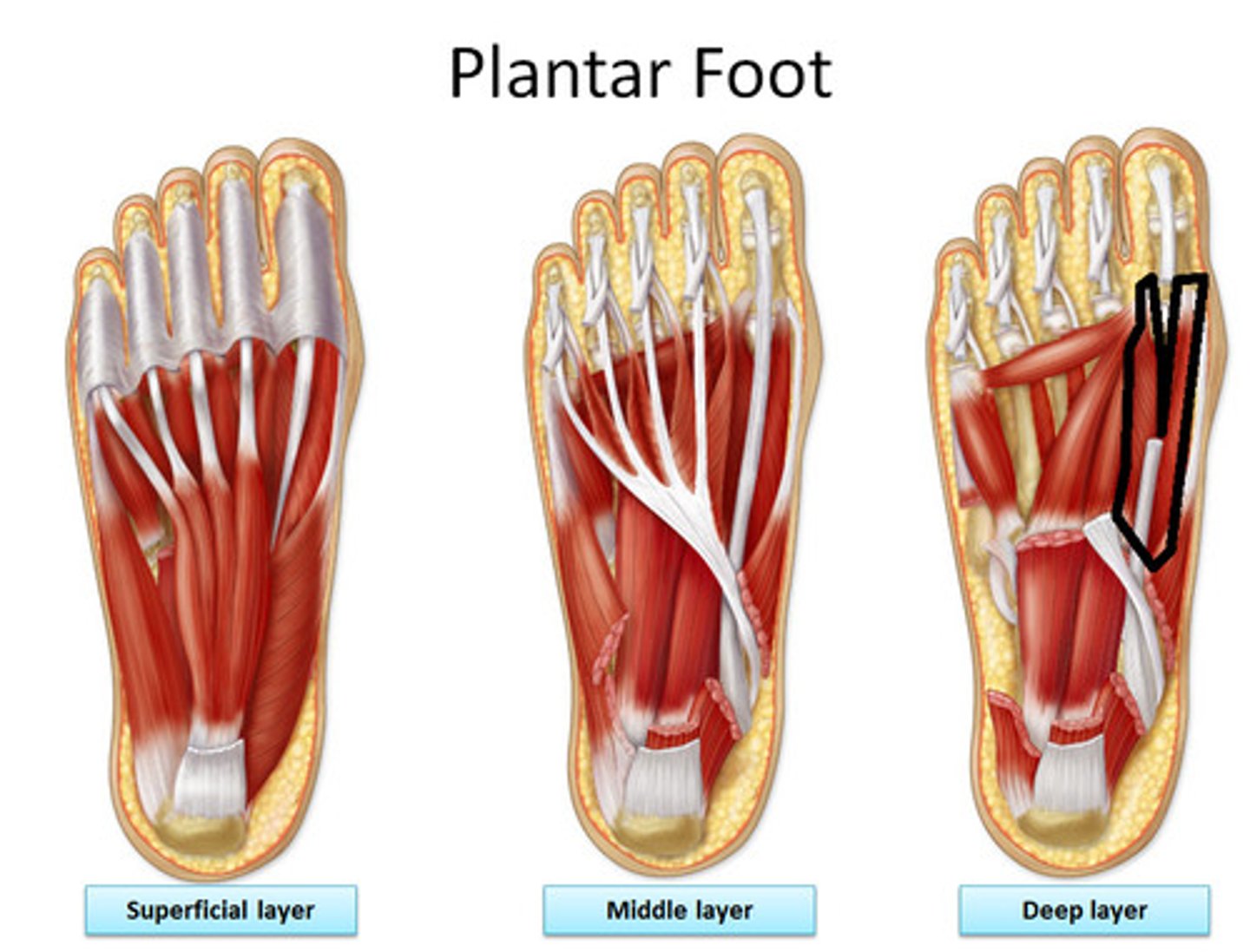
<p>Origin: plantar surfaces of cuboid and lateral cuneiform</p><p>Insertion: both sides of base of proximal phalanx of 1st digit</p><p>Innervation: medial plantar nerve (S2, <strong>S3</strong>)</p><p>Action: flexes proximal phalanx of 1st digit</p>
53
New cards

Abductor Digiti Minimi

Origin: medial and lateral tubercles of tuberosity of calcaneus, plantar aponeurosis, and intermuscular septa

Insertion: lateral side of base of proximal phalanx of 5th digit

Action: abducts and flexes 5th digit
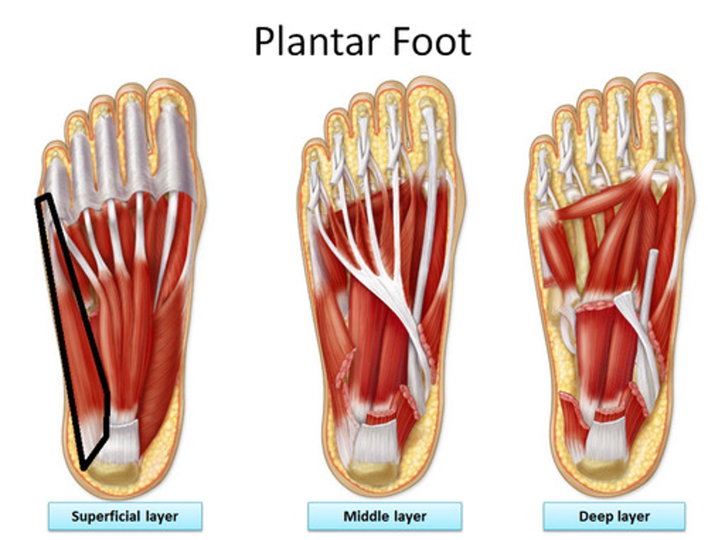
<p>Origin: medial and lateral tubercles of tuberosity of calcaneus, plantar aponeurosis, and intermuscular septa</p><p>Insertion: lateral side of base of proximal phalanx of 5th digit</p><p>Action: abducts and flexes 5th digit</p>
54
New cards

Flexor Digiti Minimi Brevis

Origin: base of 5th metatarsal

Insertion: base of proximal phalanx of 5th digit

Innervation: superficial branch of lateral plantar nerve (S2, S3)

Action: flexes proximal phalanx of 5th digit, thereby assisting with its flexion
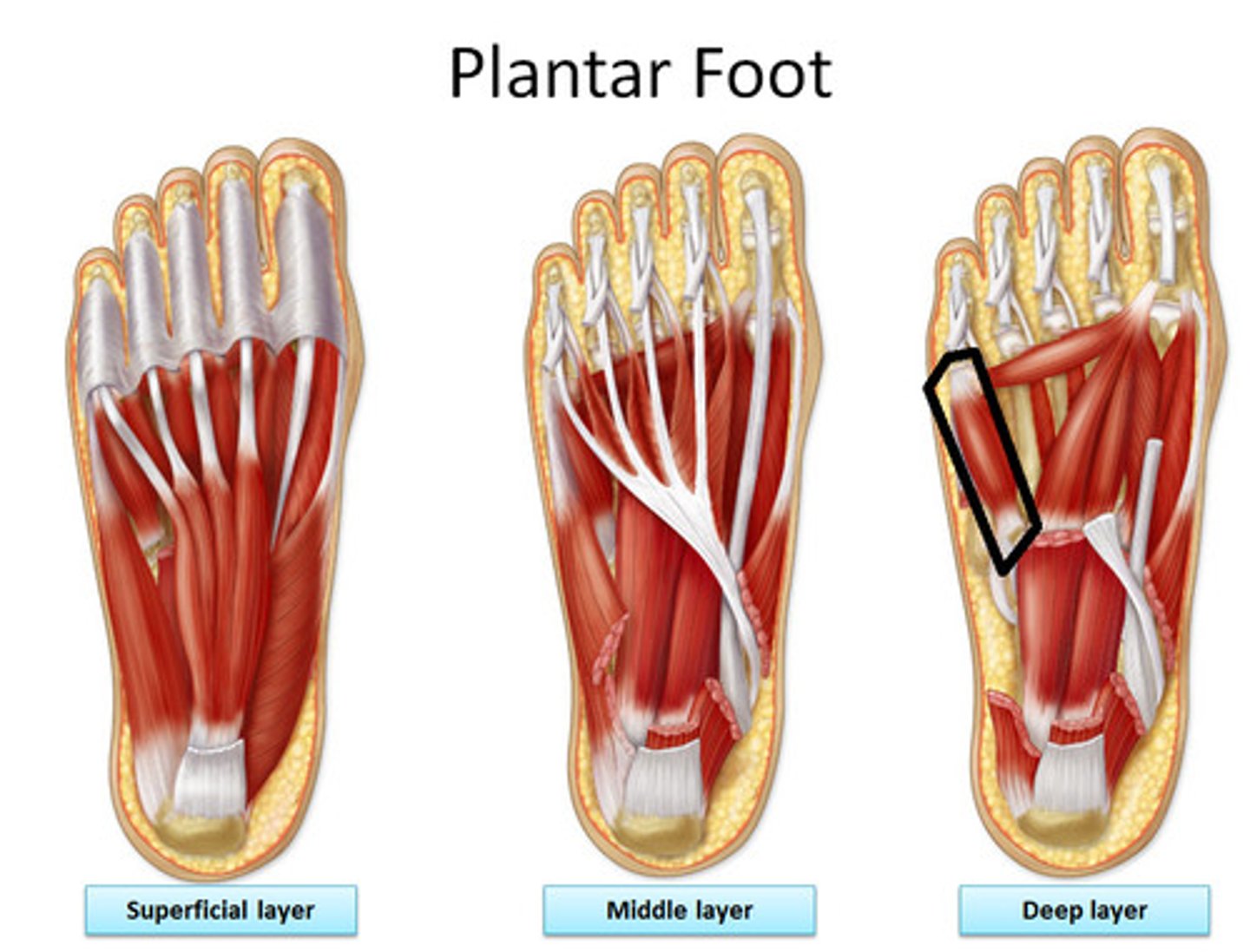
<p>Origin: base of 5th metatarsal</p><p>Insertion: base of proximal phalanx of 5th digit</p><p>Innervation: superficial branch of lateral plantar nerve (S2, <strong>S3</strong>)</p><p>Action: flexes proximal phalanx of 5th digit, thereby assisting with its flexion</p>
55
New cards

Plantar Aponeurosis

broad CT sheet coverin plantar surface of foot
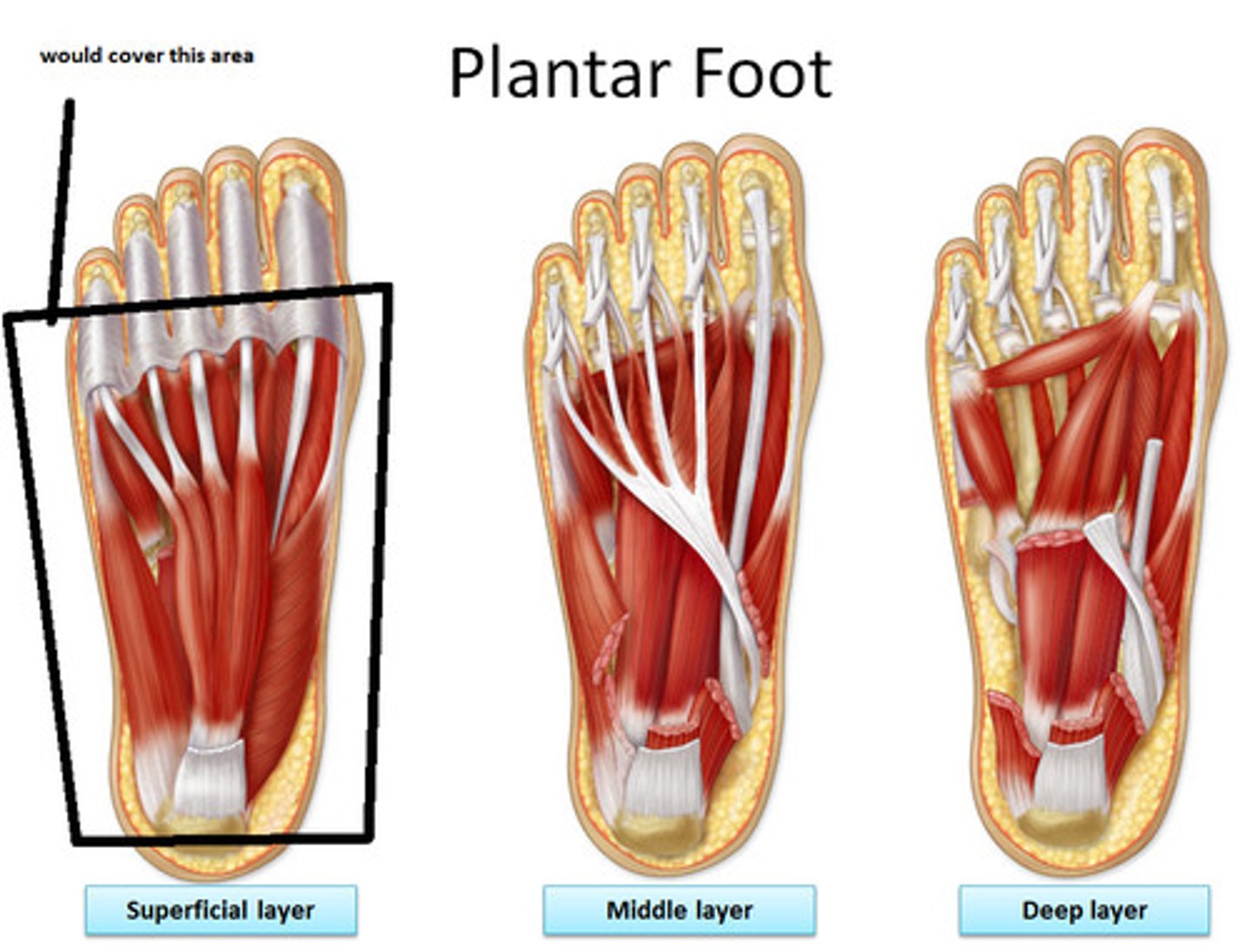
<p>broad CT sheet coverin plantar surface of foot</p>
56
New cards

Flexor Digitorum Brevis

Origin: medial tubercle of tubersoty of calcaneus, plantar aponeurosis, and intermuscular septa

Insertion: both sides of middle phalanges of lateral four digits

Innervation: medial plantar nerve (S2, S3)

Action: flexes digits II-V (lateral four digits)
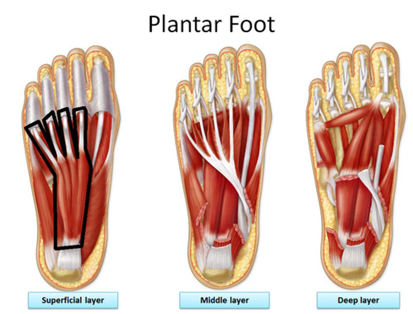
<p>Origin: medial tubercle of tubersoty of calcaneus, plantar aponeurosis, and intermuscular septa</p><p>Insertion: both sides of middle phalanges of lateral four digits</p><p>Innervation: medial plantar nerve (S2, <strong>S3</strong>)</p><p>Action: flexes digits II-V (lateral four digits)</p>
57
New cards

Quadratus Plantae

Origin: medial surface and lateral margin of plantar surface of calcaneus

Insertion: posterolateral margin of tendon of flexor digitorum longus

Innervation: lateral plantar nerve (S2, S3)

Action: assits flexor digitorum longus in flexing lateral four digits
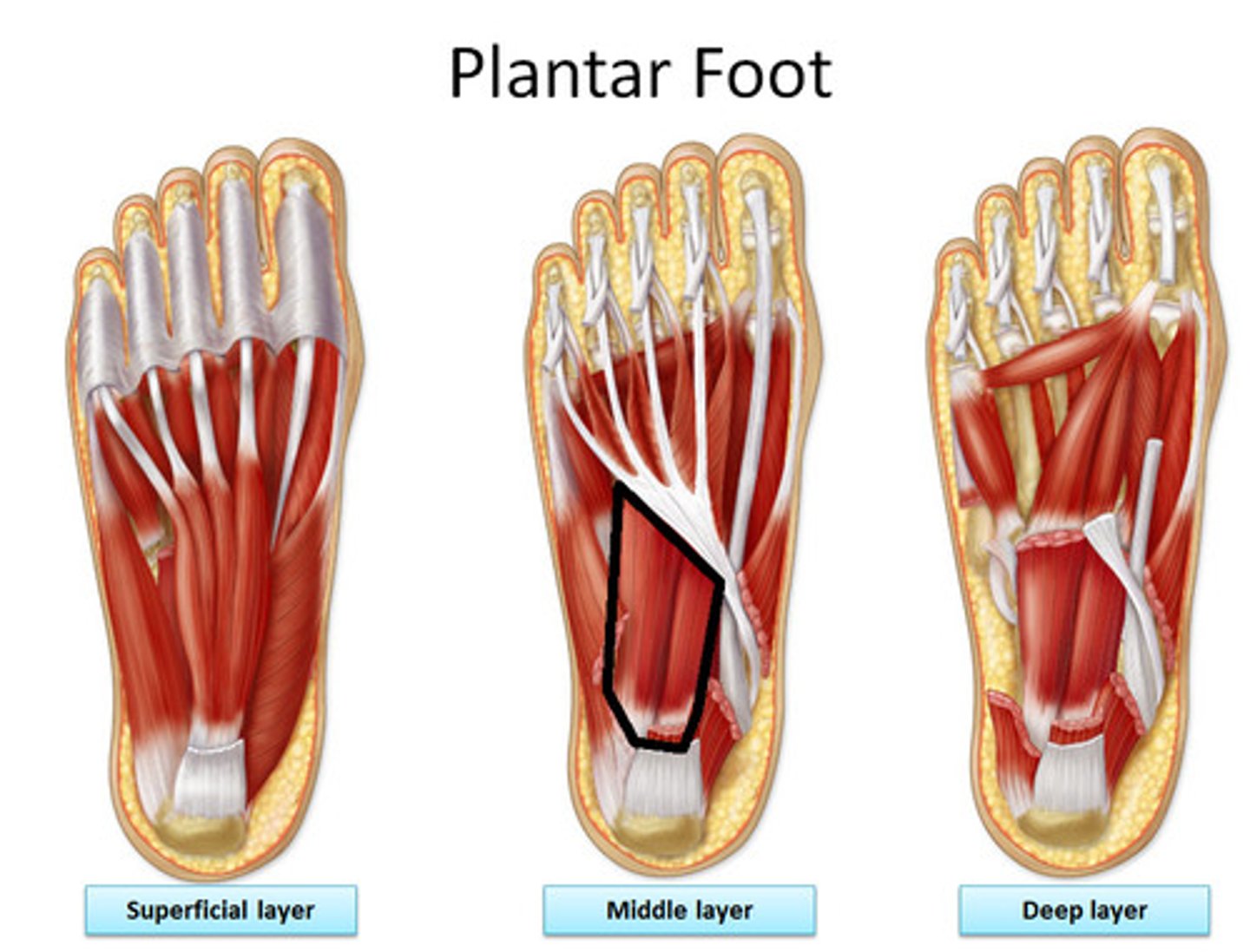
<p>Origin: medial surface and lateral margin of plantar surface of calcaneus</p><p>Insertion: posterolateral margin of tendon of flexor digitorum longus</p><p>Innervation: lateral plantar nerve (S2, <strong>S3</strong>)</p><p>Action: assits flexor digitorum longus in flexing lateral four digits</p>
58
New cards

Lumbricals (4)

Origin: tendons of flexor digitorum longus

Inserion: medial aspect of expansion over lateral four digits

Innervation: medial plantar nerve (S2, S3; medial one) lateral plantar nerve (S2, S3; lateral three)

Action: flex proximal phalanges; extend middle and distal phalanges of lateral four digits
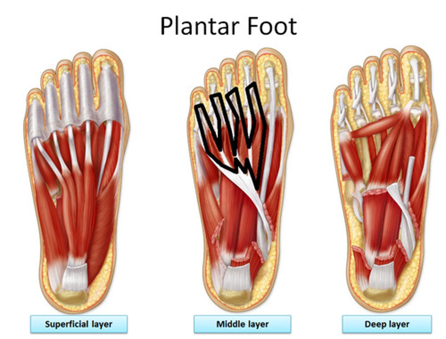
<p>Origin: tendons of flexor digitorum longus</p><p>Inserion: medial aspect of expansion over lateral four digits</p><p>Innervation: medial plantar nerve (S2, <strong>S3</strong>; medial one) lateral plantar nerve (S2, <strong>S3</strong>; lateral three)</p><p>Action: flex proximal phalanges; extend middle and distal phalanges of lateral four digits</p>
59
New cards

Plantar Interossei (three muscles)

Origin: bases and medial sides of metatarsals 3-5

Insertion: medial side of bases of proximal phalanges of 3rd-5th digits

Innervation: lateral plantar nerve (S2, S3)

Action: adduct digits (3-5) and flex metatarsophalangeal joints

60
New cards

Dorsal Interossei (four muscles)

Origin: adjacent sides of metatarsals 1-5

Insertion: medial side of proximal phalanx of 2nd digit (first), lateral sides of 2nd-4th digits (second to fourth)

Innervation: lateral plantar nerve (S2, S3)

Action: abduct digits (2-4) and flex metatarsophalangeal joints

61
New cards

Superficial Layer of Back Muscles

Splenius Capitis and Splenius Cervicis
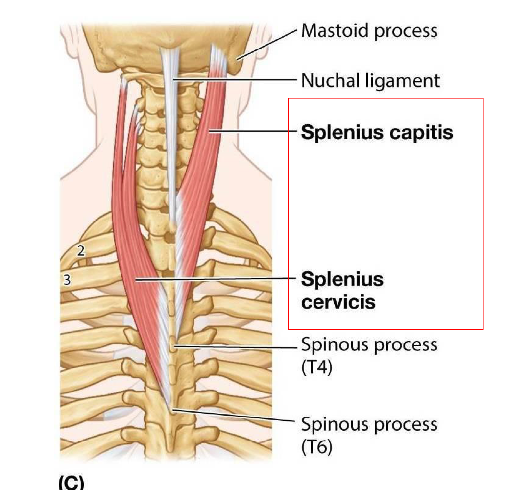
<p>Splenius Capitis and Splenius Cervicis</p>
62
New cards

Splenius (capitis and cervicis)

Origin: arises from nuchal ligament and spinous processes of C7-T6 vertebrae

Insertion: mastoid process of temporal bone and superior nuchal line of occipital bone (capitis), tubercles of transverse processes of C1-C3 or C4 vertebrae (cervicis)

Innervation: posterior rami of spinal nerves

Action: laterally flex, rotate to same side (unilateral), extend head and neck (bilateral)
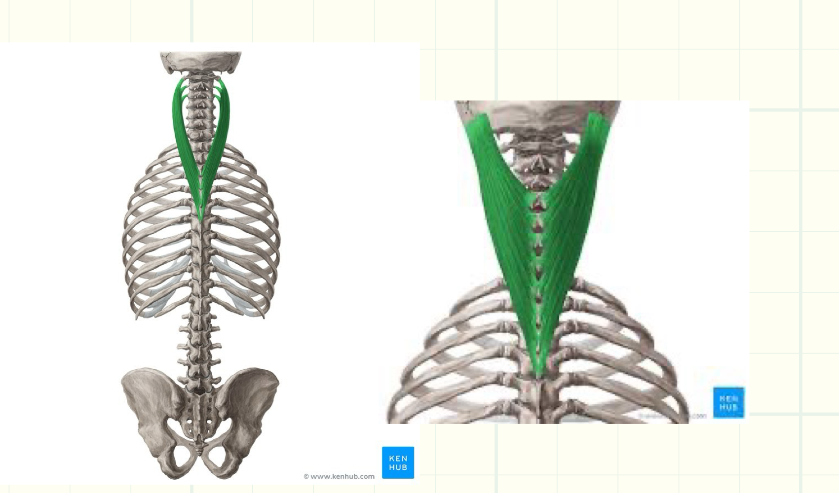
<p>Origin: arises from nuchal ligament and spinous processes of C7-T6 vertebrae</p><p>Insertion: mastoid process of temporal bone and superior nuchal line of occipital bone (capitis), tubercles of transverse processes of C1-C3 or C4 vertebrae (cervicis)</p><p>Innervation: posterior rami of spinal nerves</p><p>Action: laterally flex, rotate to same side (unilateral), extend head and neck (bilateral)</p>
63
New cards

Intermediate Layer of Back Muscles

Erector Spinae: Iliocostalis, Longissimus, Spinalis (lateral to medial)
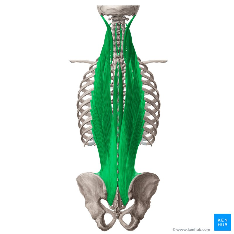
<p>Erector Spinae: Iliocostalis, Longissimus, Spinalis (lateral to medial)</p>
64
New cards

Erector Spinae

Iliocostalis, Longissimus, Spinalis

Origin: posterior part of iliac crest, posterior surface of sacrum, sacro-iliac ligaments, sacral and inferior lumbar spinous processes and supraspinous ligaments

Insertion: lower ribs and cervical transverse processes (iliocostalis; luborum, thoraces, and cervicis), ribs and transverse processes in thoracic and cervical regions and mastoid process of temporal bone (longissimus; thoracis, cervicis, and capitis), spinous processes in upper thoracic region to cranium (spinalis; thoracis, cervicis, and capitis)

Innervation: posterior rami of spinal nerves

Action: laterally flex, rotate to same side (unilateral), extend (bilateral)
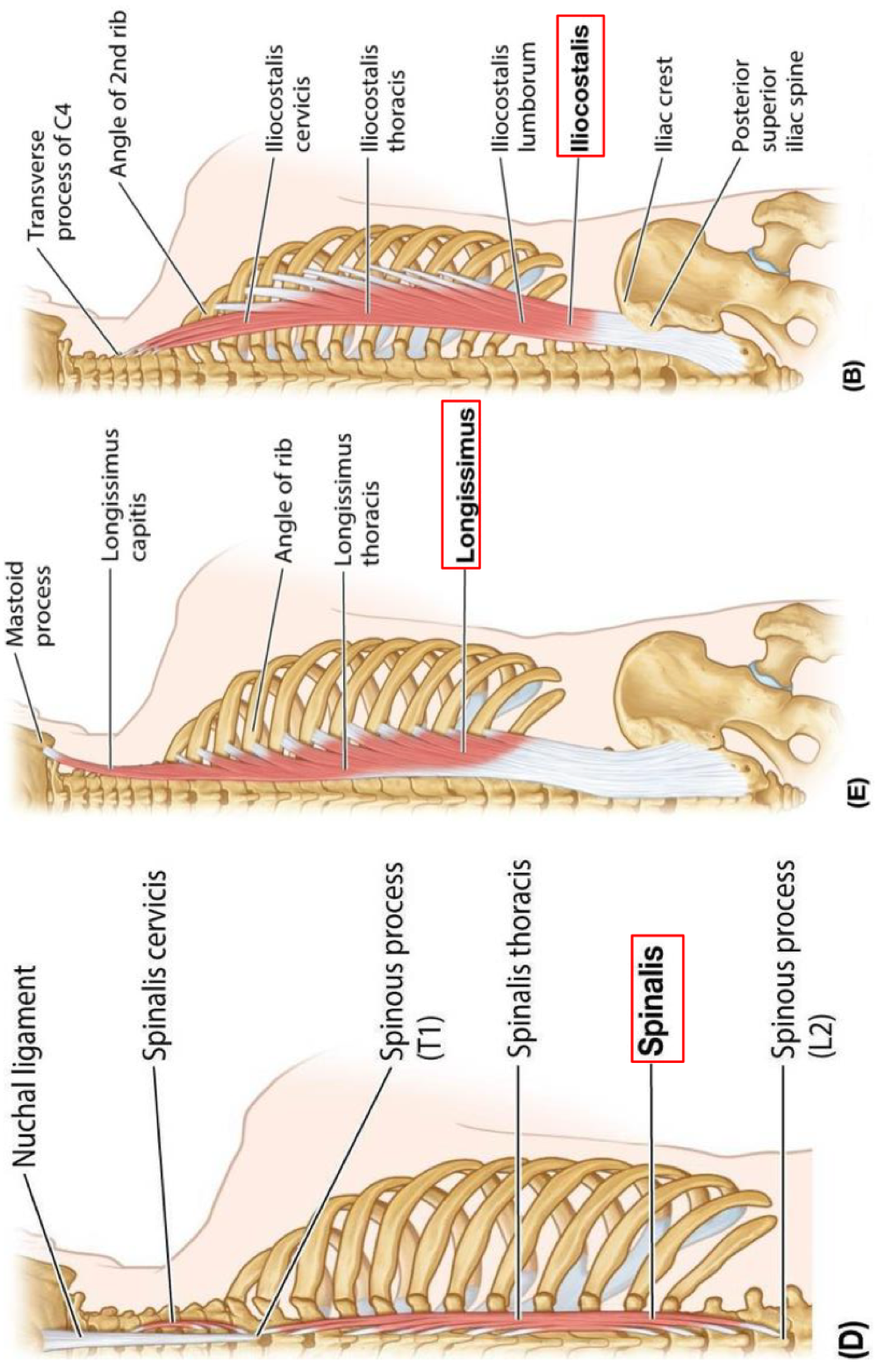
<p>Iliocostalis, Longissimus, Spinalis</p><p>Origin: posterior part of iliac crest, posterior surface of sacrum, sacro-iliac ligaments, sacral and inferior lumbar spinous processes and supraspinous ligaments</p><p>Insertion: lower ribs and cervical transverse processes (iliocostalis; luborum, thoraces, and cervicis), ribs and transverse processes in thoracic and cervical regions and mastoid process of temporal bone (longissimus; thoracis, cervicis, and capitis), spinous processes in upper thoracic region to cranium (spinalis; thoracis, cervicis, and capitis)</p><p>Innervation: posterior rami of spinal nerves</p><p>Action: laterally flex, rotate to same side (unilateral), extend (bilateral)</p>
65
New cards

Deep Layer of Back Muscles

Transversospinales: Semispinalis (thoracis, cervicis, and capitis), Multifidus, Rotores (brevis and longus)

66
New cards

Transversospinales

Semispinalis, Multifidus, Rotatores → stability and posture muscles of back

67
New cards

Semispinalis (thoracis, cervicis, and captitis)

Origin: transverse processes of C4-T10

Insertion: occipital bone, spinous processes in upper thoracic and cervical regions

Innervation: posterior rami of spinal nerves
Action: facilitates contralateral rotation (unilateral), extends head, neck, and thoracic regions (bilateral); stability and posture
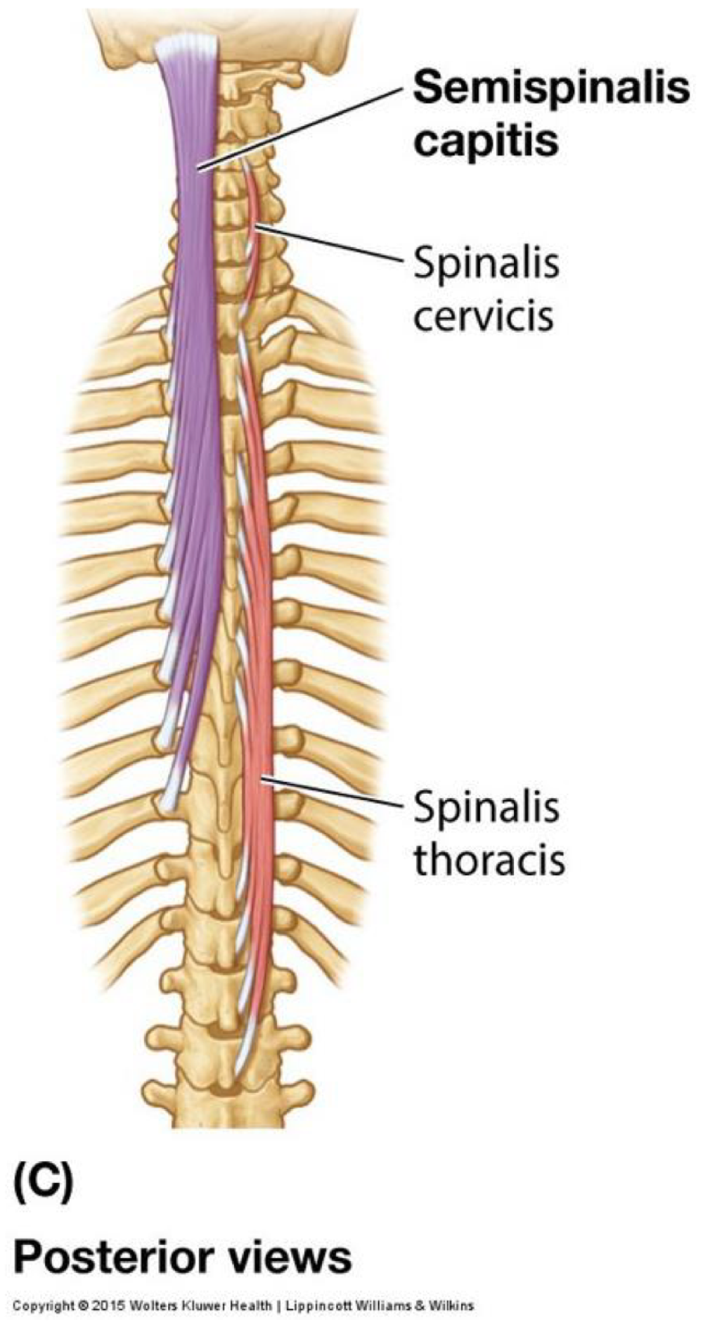
<p>Origin: transverse processes of C4-T10</p><p>Insertion: occipital bone, spinous processes in upper thoracic and cervical regions</p><p>Innervation: posterior rami of spinal nerves <br>Action: facilitates contralateral rotation (unilateral), extends head, neck, and thoracic regions (bilateral); stability and posture</p>
68
New cards

Multifidus

Origin: posterior sacrum, superior iliac spine of ilium, aponeurosis of erector spinae, sacro-iliac ligaments, mammilary processes of lumbar vertebrae, transverse processes of thoracic vertebrae, and articular processes of C4-C7

Insertion: spinous processes of vertebrae located two to four segments superior to origin

Innervation: posterior rami of spinal nerves

Action: laterally flex, rotate to opposite side (unilateral), extend (bilateral); stability and posture
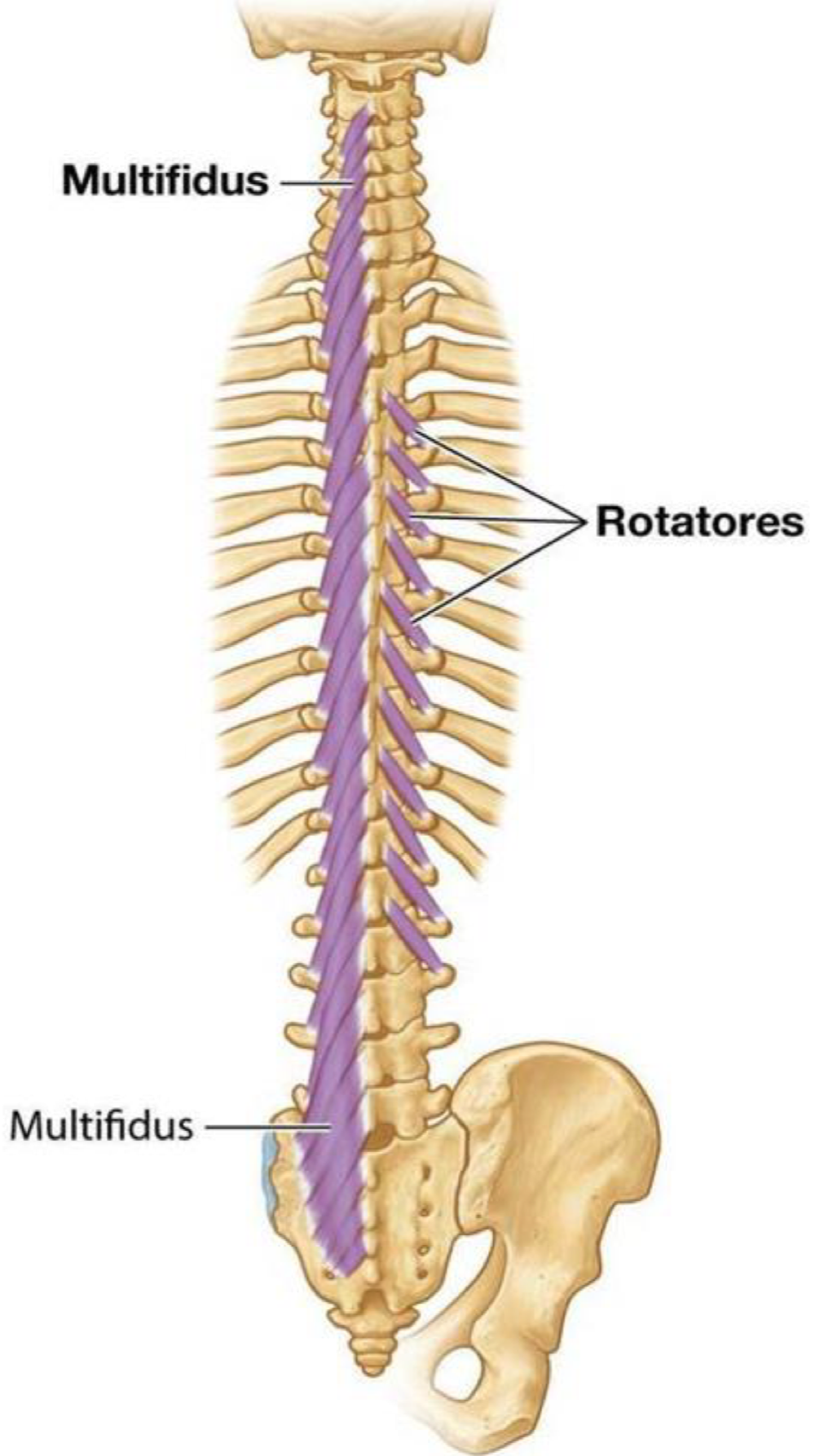
<p>Origin: posterior sacrum, superior iliac spine of ilium, aponeurosis of erector spinae, sacro-iliac ligaments, mammilary processes of lumbar vertebrae, transverse processes of thoracic vertebrae, and articular processes of C4-C7</p><p>Insertion: spinous processes of vertebrae located two to four segments superior to origin</p><p>Innervation: posterior rami of spinal nerves</p><p>Action: laterally flex, rotate to opposite side (unilateral), extend (bilateral); stability and posture</p>
69
New cards

Rotares (brevis and longus)

Origin: transverse processes of (thoracic) vertebrae

Insertion: spinous process of vertebra immediately (brevis) or two segments (longus) superior to vertebra of origin

Innervation: posterior rami of spinal nerves

Action: laterally flex, rotate to opposite side (unilateral), extend (bilateral); stability and posture
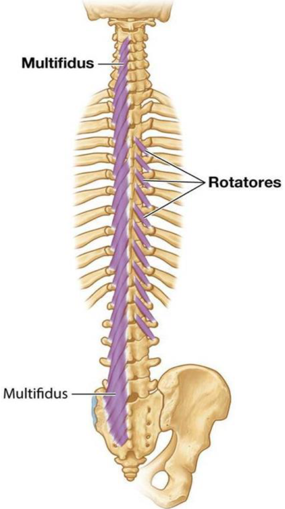
<p>Origin: transverse processes of (thoracic) vertebrae</p><p>Insertion: spinous process of vertebra immediately (brevis) or two segments (longus) superior to vertebra of origin</p><p>Innervation: posterior rami of spinal nerves</p><p>Action: laterally flex, rotate to opposite side (unilateral), extend (bilateral); stability and posture</p>
70
New cards

Minor Deep Layer of Back Muscles

Levator costorum, Lumbar interspinalis, Lumbar intertransversarii

71
New cards

Levator Costorum (action and innervation)

Unilateral: elevate ribs (during inspiration), laterally flex

Bilateral: extension

Innervation: posterior rami of spinal nerves
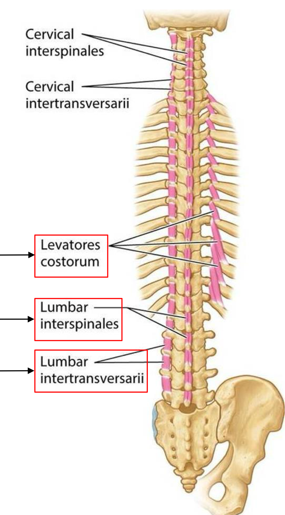
<p>Unilateral: elevate ribs (during inspiration), laterally flex</p><p>Bilateral: extension</p><p>Innervation: posterior rami of spinal nerves</p>
72
New cards

Lumbar Interspinales (action and innervation)

Action: extension

Innervation: posterior rami of spinal nerves
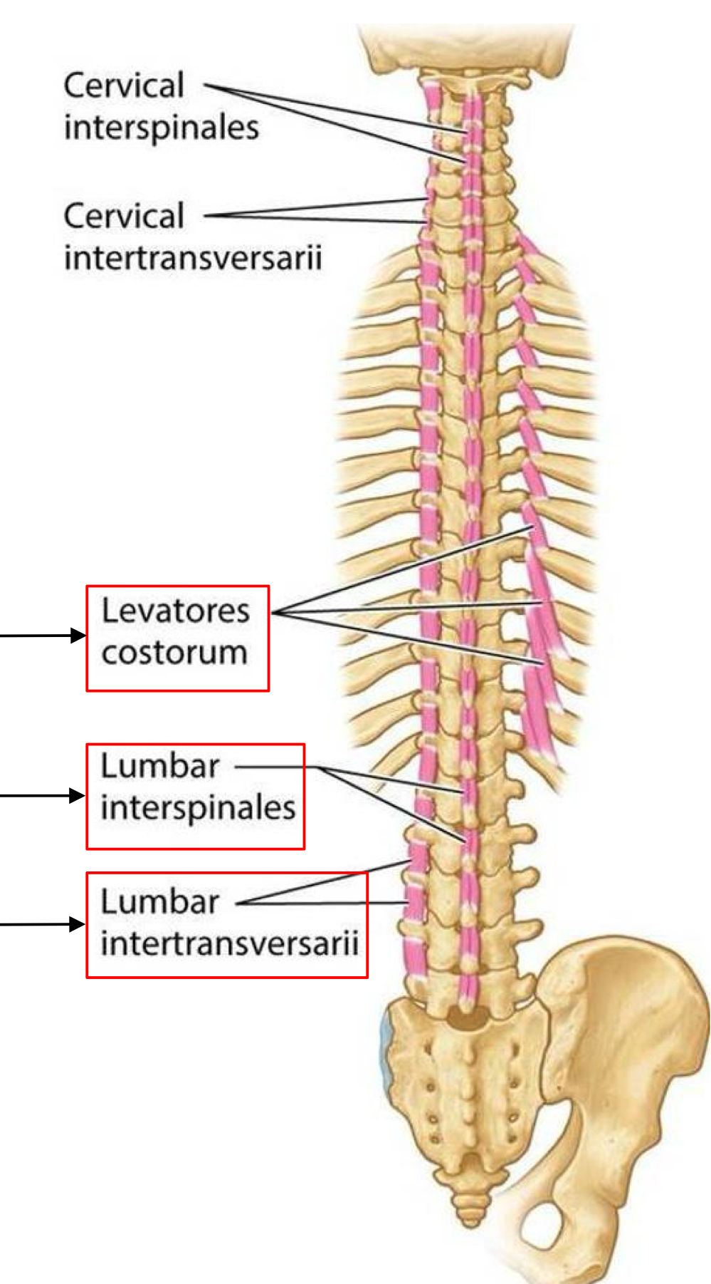
<p>Action: extension</p><p>Innervation: posterior rami of spinal nerves</p>
73
New cards

Lumbar Intertransversarii (action and innervation)

Unilateral: lateral flexion

Bilateral: extension

Innervaiton: posterior and anterior rami of spinal nerves
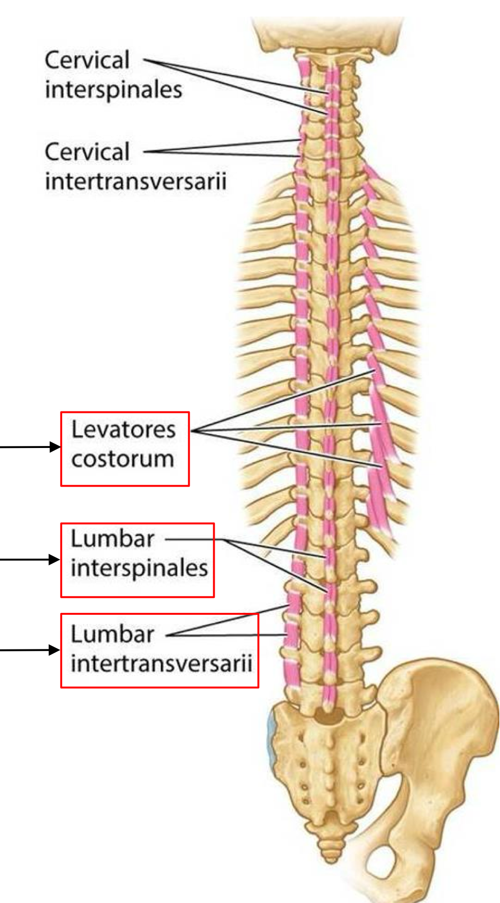
<p>Unilateral: lateral flexion</p><p>Bilateral: extension</p><p>Innervaiton: posterior and anterior rami of spinal nerves</p>
74
New cards

Rectus Capitis Posterior Major (action and innervation)

Action: extends and rotates the head

Innervation: suboccipital nerve (dorsal ramus of C1)
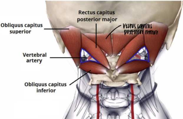
<p>Action: extends and rotates the head</p><p>Innervation: suboccipital nerve (dorsal ramus of C1)</p>
75
New cards

Rectus Capitis Posterior Minor (action and innervation)

Action: extends the head

Innervation: suboccipital nerve (dorsal ramus of C1)
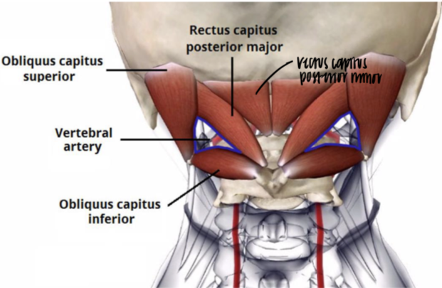
<p>Action: extends the head</p><p>Innervation: suboccipital nerve (dorsal ramus of C1)</p>
76
New cards

Obliquus Capitis Superior (action and innervation)

Action: extends and laterally flexes the head

Innervation: suboccipital nerve (dorsal ramus of C1)
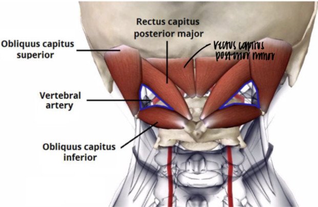
<p>Action: extends and laterally flexes the head</p><p>Innervation: suboccipital nerve (dorsal ramus of C1)</p>
77
New cards

Obliquus Capitis Inferior (action and innervation)

Action: rotates the atlas (C1)

Innervation: suboccipital nerve (dorsal ramus of C1)
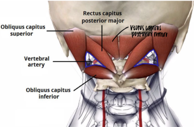
<p>Action: rotates the atlas (C1)</p><p>Innervation: suboccipital nerve (dorsal ramus of C1)</p>