Virus/Host
 Call Kai
 Learn
Learn Practice Test
 Spaced Repetition
 Match
Match Flashcards
 Knowt Play
1/59

There's no tags or description
Looks like no tags are added yet.
Name | Mastery | Learn | Test | Matching | Spaced | Call with Kai | Chat |
|---|
No analytics yet
Send a link to your students to track their progress
60 Terms
What is a virus
Extremely small (20-400 nm)
Nonliving
Acellular
Obligate intracellular pathogens
Compare and contrast viruses and bacteria
Viruses:
Acellular
No “cells”
Nonliving
Hijack their host’s cell system to replicate themselves
DO NOT exhibit metabolism
Made of RNA
Filterable due to size
Has protein capsid coating and nucleic acid (bacteria have cells w/o nuclei or membrane-bound organelles)
Virus can be composed of both DNA and RNA (bacteria w/DNA)
Describe a viral structure
A virion is a single, infectious virus particle (virus in the singular)
Has a capsid (protective exterior protein)
Has genetic material (DNA or RNA)
SOME have outer lipid envelope
Has spikes (protruding from capsid or outer lipid envelope)
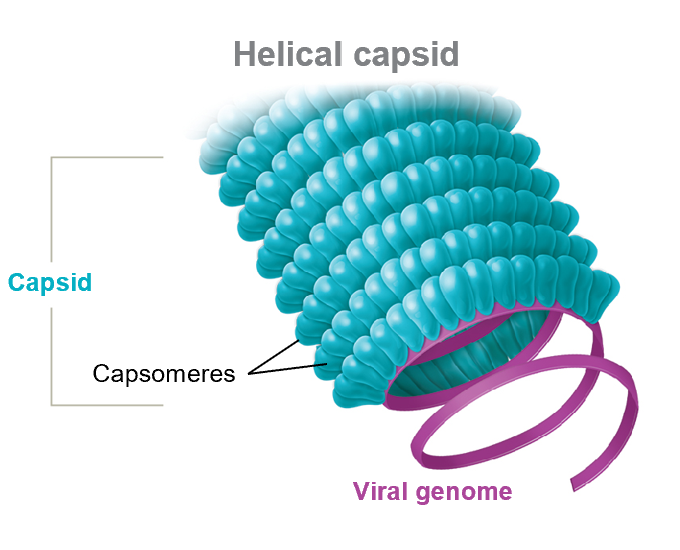
Helical capsid
Looks like a hollow tube
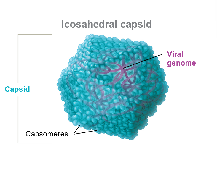
Icosahedral capsid
Looks like a 3-D polygon
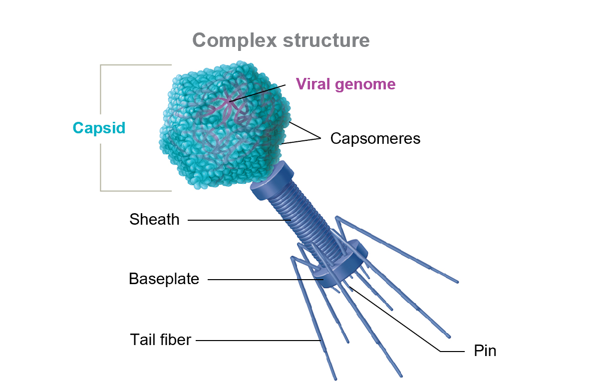
Complex capsid
Deviation from the other 2 possible structures of capsids (helical or icosahedral)
List 4 characteristics or features that are used to name and classify viruses?
Type of nucleic acid present (RNA or DNA)
Capsid symmetry (helical, icosahedral, or complex)
Presence or absence of an envelope (enveloped or naked)
Genome architecture (ssDNA, ssRNA, etc.)
Why are bacteriophages medically important
They do not infect humans, but they can serve as a means for bacteria to evolve.
Bacteriophages facilitate transduction, which enables bacteria to develop new genetic combinations despite their inability to reproduce sexually
Describe the Lytic and Lysogenic cycles of bacteriophages
Lytic pathway: Replication of bacteriophages (bacterial viruses) that kill the host cells
Lysogenic pathway: Replication of temperate phages, a type of bacteriophage that has lambda
Attachment phase (adsorption)
A phage binds to bacteria
Bacteriophage tail fibers help the virus adhere to specific proteins on the bacterial cell wall surface.
Release phase
Phage encodes and releases lysozyme to break host cell walls (causing bacterial lysis) once phages are mature
Assembly (maturation) phase
Phage parts are replicated
Genome is packed into capsid
Phage parts are assembled to complete virions
Replication phase (synthesis)
Phage commandeers host cell factors to transcribe and translate viral genes
DNAases (DNA-degrading enzymes) are formed
Proteins to build new phage particles
Penetration phase
Phage injects its genome into the host
Empty capsid remains outside the cell
Lytic pathway steps
Attachment
Penetration
Replication (synthesis)
Assembly (maturation)
Release
Lysogenic pathway steps
Attachment
Penetration → phophage or lysogenic replication occurs
Replication
Assembly
Release
What happens during lysogenic replication
Phage DNA integrates into the host genome, forming a prophage
As the bacterial cell divides, it copies its own genome and the prophage.
The phage may enter the lytic pathway if the host is stressed, called the “abandon ship” approach.
This allows for bacteriophages to replicate and find a new host in case their current host dies
Describe a prophage?
The combination of bacteriophage genomes with that of their host cell genome.
Medically important because prophages allow bacterial cells to gain new pathogenic properties, a process called phage conversion (e.g., toxins)
What about a provirus?
Prophages that gave bacteria the ability to make toxins
C. diphtheria→ causes diphtheria toxins
C. Botulinum → causes botulinum toxins
Compare bacteriophage and animal viral multiplication
They both have the same 5 steps, but animal viral multiplication has “uncoating” as an additional step
Attachment of animal viral multiplication
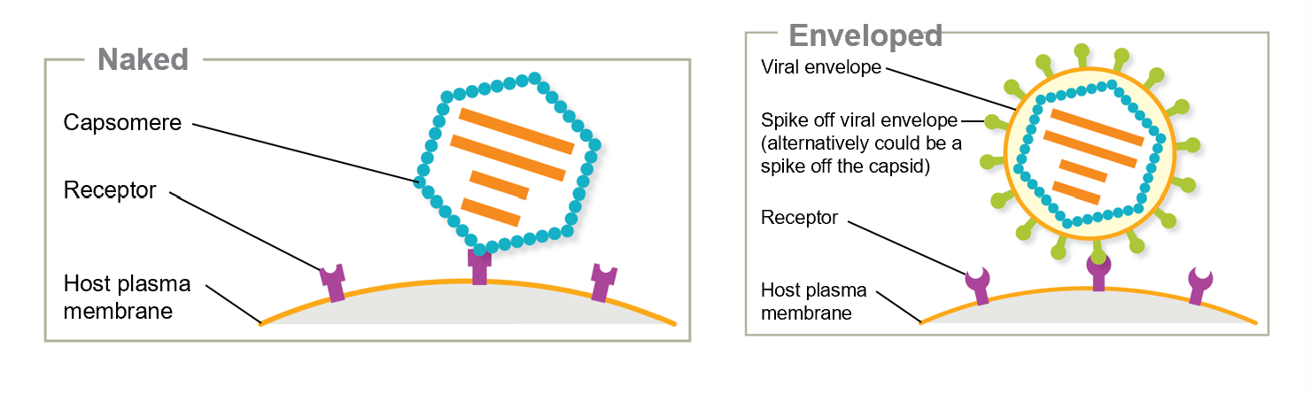
A naked virus attaches to a host cell’s membranes through capsid proteins
Enveloped viruses (and naked viruses) bind via spikes
Knowing this makes the attachment proteins of the virus a target for drug therapy to limit or prevent infection
What drug blocks HIV’s attachment to host cell proteins
Maraviroc (Selzentry)
Penetration of animal viral multiplication
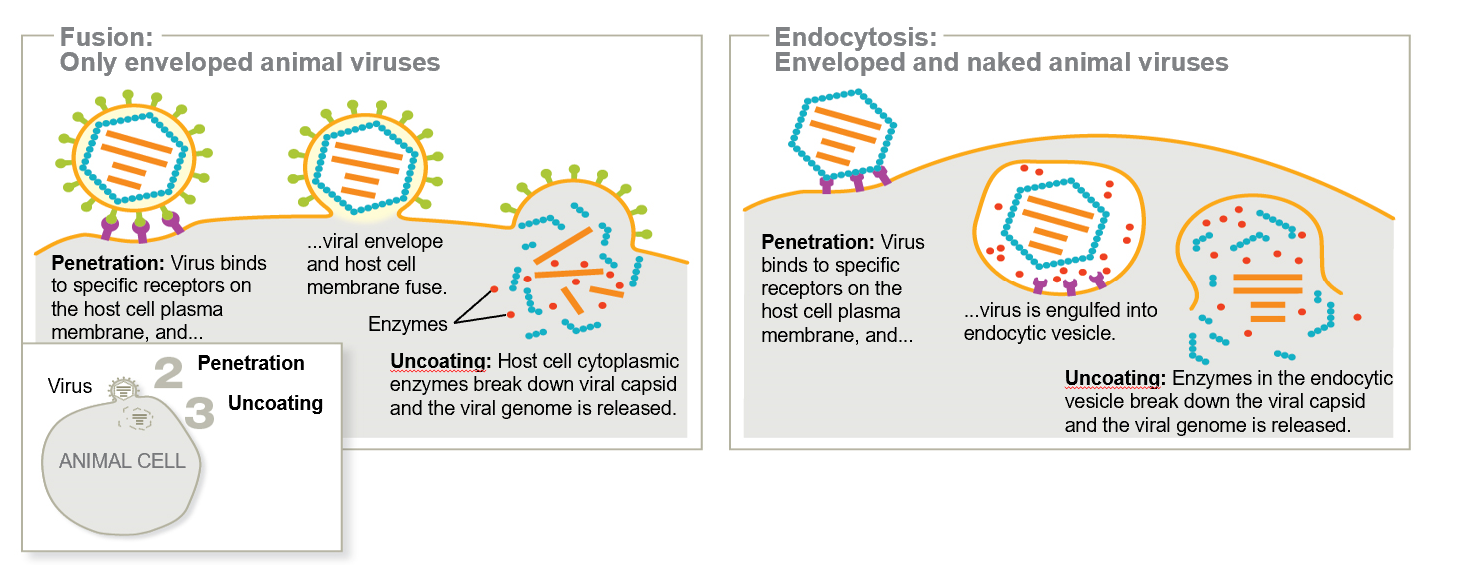
Naked and enveloped viruses enter via Endocytosis or membrane fusion.
Membrane fusion→ Viral envelope blend w/cell’s plasma membrane
Endocytosis →Virus binds to host cell surface receptors, triggering uptake of virus into vesicles
Uncoating of animal viral multiplication
Animal virus capsids DO enter the host cells and are then entirely or partially broken down, releasing the genome.
A virus that entered via membrane fusion may have its capsid degraded in the cell cytoplasm,
A virus entering via endocytosis may have its capsid digested away by enzymes within the vesicle
Poliovirus is an exception and spits out its genome via a pore on the capsid and endocytic vesicle
Viruses’ w/DNA may not let go of their genome until they have reached the nucleus
Replication (synthesis) of animal viral multiplication
The genome is replicated, and viral proteins are made
Viruses’ w/DNA do replication and transcribe in the nucleus
RNA replication and protein synthesis occur in the cytoplasm
What viruses are an exception and replicate their RNA within the nucleus during animal viral multiplication
Orthomyxoviruses (which cause influenza)
Retroviruses such as HIV
Assembly of animal viral multiplication
New virions are formed
Capsids usually build around the genome
Sometimes capsids even have the genome inserted before they are even finished or sealed
Release of animal viral multiplication
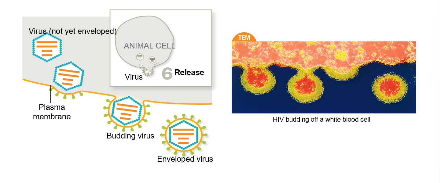
Enveloped virus: Before it is enveloped, viral proteins are embedded in the host cell plasma membrane, and virions usually take a portion of the cell’s plasma membrane, enriched with the viral surface proteins, making it enveloped.
Naked virus: Naked viruses rupture the host cell during release, usually killing the cell
Acute infections
Viruses infect cells and reproduce immediately. Usually cleared by host immune system
E.g. common cold and influenza
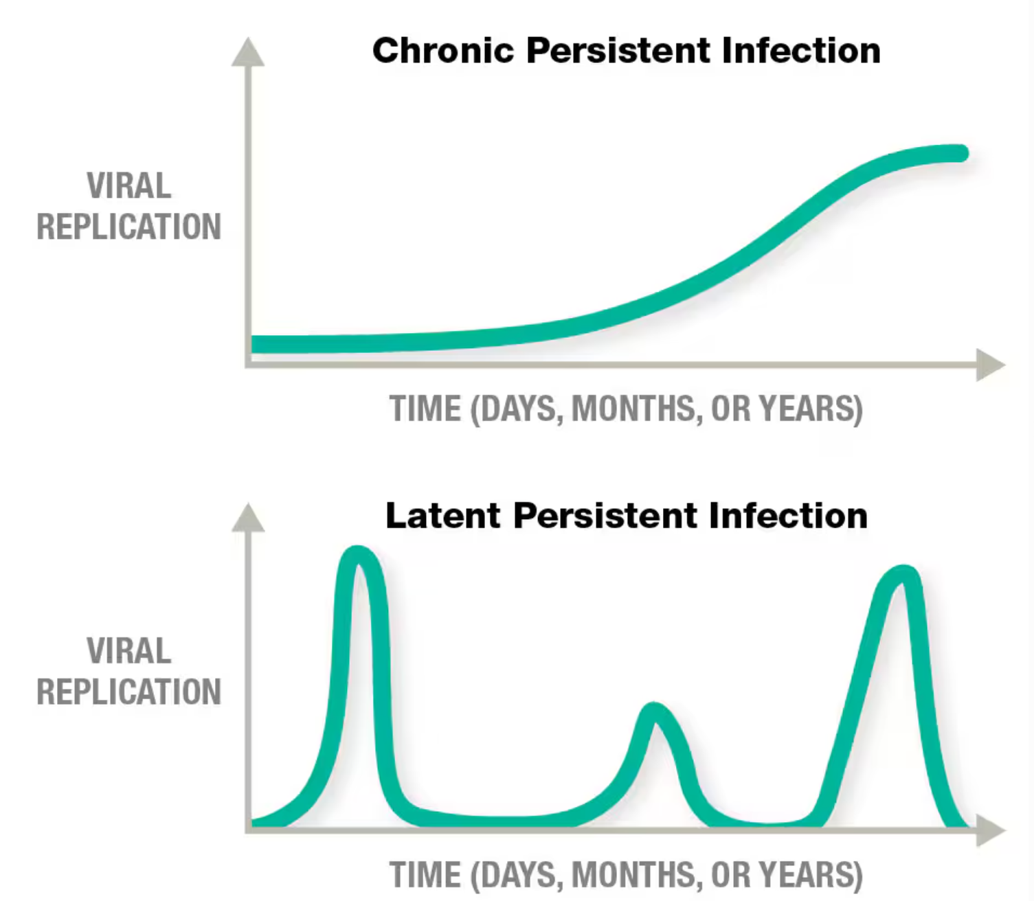
Persistent infections
Viruses have replication strategies that allow them to avoid immune system clearance
Chronic (e.g. HIV)
Continuous release of virions over time and slow progression of the disease
Latent (E.g. Herpes and Cancer)
Flare-ups w/intermittent periods of dormancy
During flare-ups, virions are shed, and the person experiences symptoms
Flare-ups can be triggered by stress (fever, sunburn, hormones)
Human papilloma viruses (HPVs)
Integrates w/host genome
Has a DNA viral genome
Uncontrolled cell division
Cervical, oropharyngeal, anal, and vaginal/penile cancer
Human herpes virus-8
DOESNT integrate w/host genome
Has DNA viral genome
Uncontrolled cell division
Kaposis sarcoma
Epstein-Barr virus
DOESNT integrate w/host genome
Has DNA viral genome
Uncontrolled cell division
B cell and T cell lymphomas and Hodgkin’s disease
Human T lymphotropic viruses (HTLVs)
Integrates w/host genome
Has RNA viral genome
Uncontrolled cell division
Adult T-cell leukemia
Hepatitis B virus
DOESNT integrate w/host genome
Has a DNA viral genome
Chronic inflammation triggers host cell DNA damage and mutations
Liver cancer
Hepadnaviridae family
Hepatitis C virus
DOESNT integrate w/host genome
Has RNA viral genome
Chronic inflammation triggers host cell DNA damage and mutations
Liver cancer
Flaviviridae family
How does oncogenic viruses cause cancer
They stimulate uncontrolled host cell division and decrease host cell responsiveness to death signals
HPVs and HLTVs are some examples
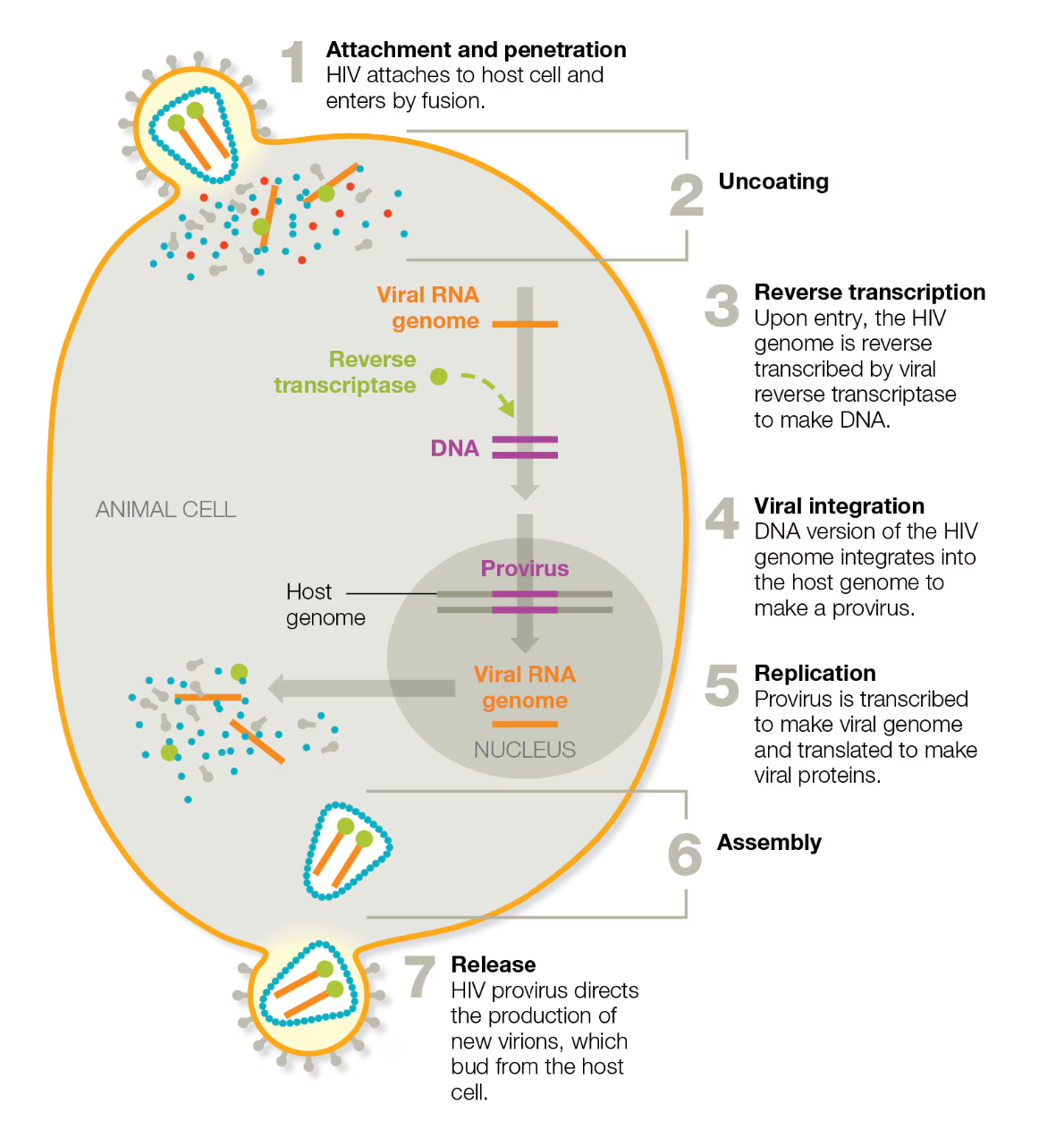
Briefly describe the replication of HIV virus
Attachment → HIV attaches to a host cell and enters by fusion
Uncoating
Reverse transcription → HIV genome is reverse transcribed by viral reverse transcriptase to make DNA
Viral integration → DNA version of the virus integrates into the host cell’s genome to make a provirus
Replication → Provirus is transcribed to make viral genome and translated to make viral proteins
Assembly
Release → HIV provirus directs production of new virions, which bud from the host cell
Herpesviridae family
HSV-1 → cold sores
HSV-2 → genital herpes
HHV-3 → Chickenpox and shingles
What is reverse transcriptase?
An enzyme used in HIV replication to transcribe the RNA genome of HIV into DNA to be read by the nucleus of the host cell and translated into viral genome again for multiplication of the virus
How are viruses cultured?
Bacteriophages use the plaque assay technique
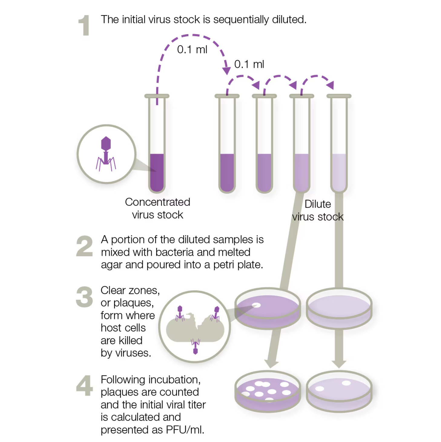
Plaque assay
initial virus stock is diluted sequentially
A portion of the diluted samples is mixed w/bacteria and melted agar and poured into a Petri dish
Clear zones or plaques form where viruses kill host cells
After incubation, plaques are counted, and the initial viral titer is calculated and presented as PFU/ml
Viral titer→ Quantity of virus present in a given volume sample. Determines how much of a given virus is present
PFU →Quantity of bacteriophages in an initial volume of sample that will form a plaque
HeLa cells – what is the historical importance of these cells?
Cancer cells derived from a patient named Henrietta Lacks are among the most common commercially available human cells used in research today.
They may not always support the growth of a virus bc they get used to being outside of the animal they come from and lose the ability to make specific surface factors that the virus requires for attachment
Primary cell lined like white blood cells can instead be used along w/embryonated eggs
Can you use embryonated eggs to grow viruses
Embryonated eggs are useful for growing certain viruses.
Once the egg is injected w/viruses, the egg is incubated for viral replication. The injection site depends on which virus is grown.
Once the virus is grown, they are purified from the eggs
A minuscule amount of egg protein may remain and is the reason why patients are asked about egg allergies
Differentiate between Bacteriophages and animal viruses in how they are cultured.
Animal viruses are grown using tissue culture techniques
Live hosts may be required to support growth of some viruses
Embryonated eggs (fertilized eggs) are also useful for growing animal viruses
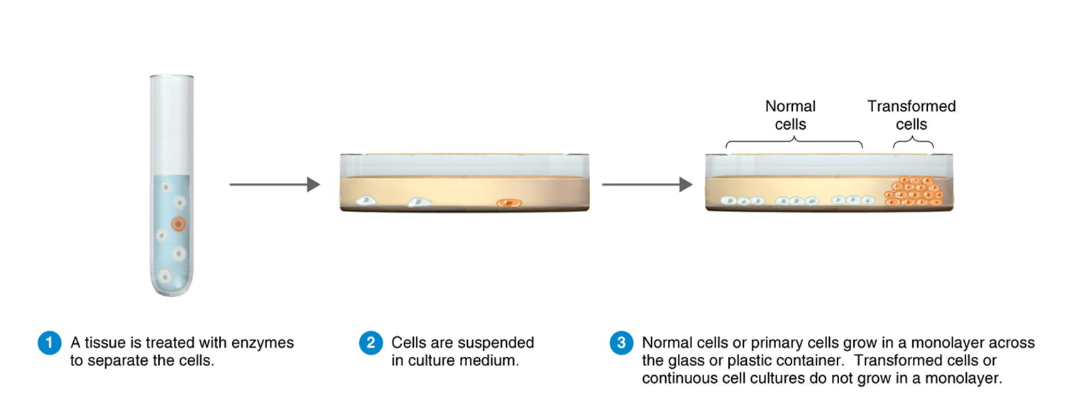
Cell tissue culture
A tissue is treated w/enzymes to separate the cells
Cells are suspended in culture medium
Normal cells grown in a monolayer across the glass/plastic container. Transformed cells or continuous cell cultures do not grow in a monolayer
What is a plaque forming unit?
The quantity of bacteriophages (viruses) in an initial volume of sample that eventually form a plaque
List and explain 3 diagnostic tests used to identify viruses
Agglutination tests
ELISAs
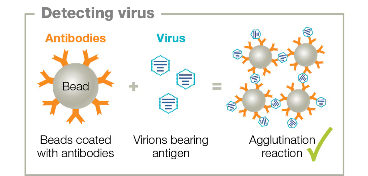
Latex agglutination test
What are agglutination tests
Purified antibodies linked to tiny latex beads (coated w/viral proteins)
Specific antibodies are used to coat latex beads
These beads are exposed to a sample containing antigens
If beads clump (agglutinate), then it indicates that the antigens bound to the antibodies
Used to screen bodily fluids like urine, cerebrospinal fluid, blood for viruses, etc
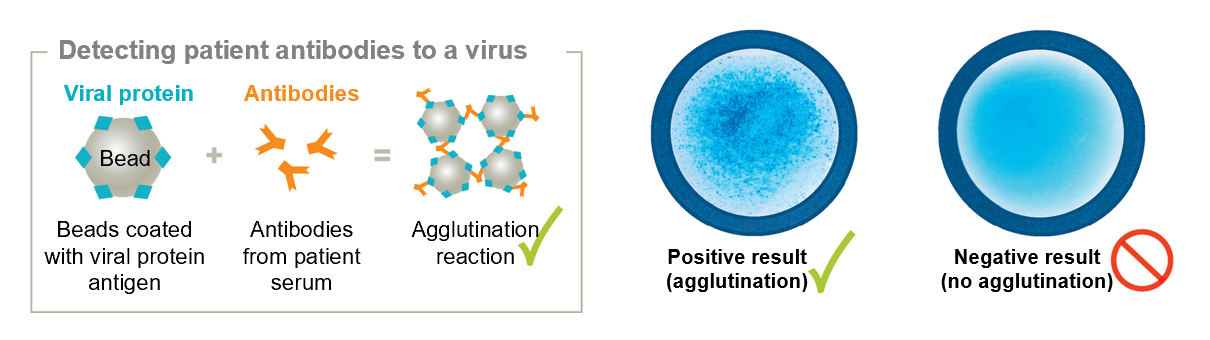
Latex agglutination test
Viral antigens/proteins are linked to tiny latex beads
Mixed with the sample that has antibodies
If the patient has antibodies that bind to the viral antigen, then the beads will agglutinate when exposed to the patient’s serum
ELISA
Can detect either antigens or antibodies in a sample
Instead of clumping beads, ELISAs adhere the antigen or antibody to A SURFACE and usually change color if there is binding
Use of polyclonal antibodies (primary and secondary)
Antibodies bind to antigen
Enzyme-linked antibody interacts w/substrate
Change of color indicates binding
Explain the difference between specificity and sensitivity with regards to diagnostic tests.
Specificity → Identifies only the virus of interest (no false positives)
Sensitivity → Test detects the lowest levels of the target (no false negatives)
Sometimes antibodies bind to viral antigens as well
List 4 DNA oncogenic viruses and 1 RNA oncogenic viruses
HPVs, Human herpes virus-8, epstein-barr virus, and hepatitis B have DNA
HTLVs + Hep C have RNA
Limitations to agglutination tests and ELISA
The sample must be a liquid, and the antigens being detected must be fairly well characterized to ensure detection specificity
IF there is an antigenic shift, it may no longer be detectable
Tests are futile during early infections because it takes a couple of weeks for antibodies to develop, a time period called seroconversion window
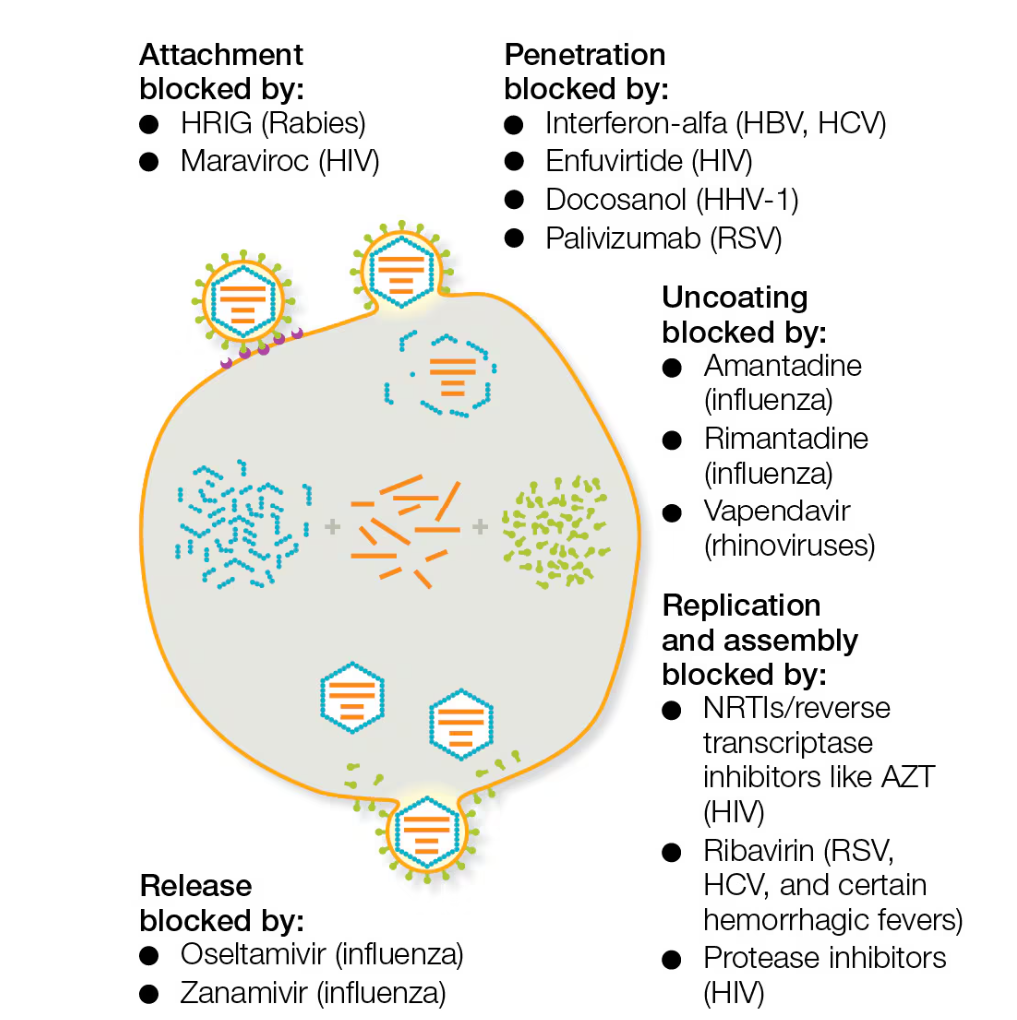
Mechanism of action of selected antiviral drugs
Blocked attachment
Blocked penetration
Blocked Uncoating
Blocked replication
Blocked release
Several antiviral drugs prevent viral entry into cells by blocking the above
Antisense antivirals
Short sequences of nucleotides complementary to the viral DNA
Bind to viral RNA, inhibiting translation
Targeted RNA is destroyed by cellular enzymes
Vitravene
What is a Prion?
Infectious proteins w/no genetic materials
DO NOT replicate
Cause transmissible spongiform encephalopathies (TSEs)
How does a Prion cause a disease?
Spongiform encephalopathy can be inherited or acquired by
Gerstmann-Sträussler-Scheinker syndrome
Fatal familial insomnia
Both inherited
Creuzfeldt-Jakob disease (CJD)
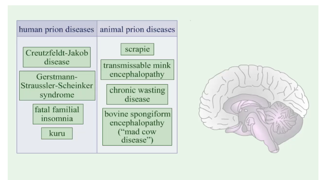
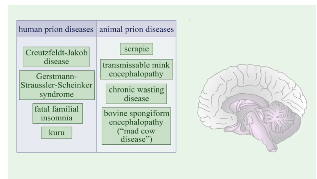
List human and animal diseases associated with prions.
Human:
CJD
Gestmann-Strassler-Schenker syndrome
Fatal familial insomnia
Kuru
Animal:
Scrapie
Transmissible mink encephalopathy
Chronic wasting disease
Bovine spongiform encephalopathy