Anatomy 6511 Back and Spine 낱말 카드
 Call Kai
 Learn
Learn Practice Test
 Spaced Repetition
 Match
Match Flashcards
 Knowt Play
1/124

There's no tags or description
Looks like no tags are added yet.
Name | Mastery | Learn | Test | Matching | Spaced | Call with Kai | Chat |
|---|
No analytics yet
Send a link to your students to track their progress
125 Terms
Paraxial mesoderm
Which germ layer gives rise to the muscles of the back?
Brain (foramen magnum) to L2 vertebra
The spinal cord runs from where to where?
Conus Medullaris
Tapered end of the spinal cord

Filum Terminale
Extension of pia mater that supports the end of the spinal cord

Cauda Equina
Bundle of nerves below the end of the spinal cord

Dorsal root ganglion
What structure is labelled?

Sensory
What type of information do dorsal root ganglion carry?
Below their corresponding vertebrae
Where do spinal nerves, besides cervical, exit in the vertebral column (above or below)?
Above their corresponding vertebrae
Where do cervical spinal nerves exit the vertebral column?
Ventral rami
Do dorsal or ventral rami form nerve plexuses?
Cervical, lumbar, sacral
In which spinal regions are plexuses found?
Thoracic
In which spinal region are intercostal nerves found?
Costal groove of ribs
Where do intercostal nerves run through?
C1-C4 (first four)
Which spinal nerves make up the cervical nerve plexus?
C5-T1 (starts right after cervical nerve plexus/5 levels)
Which spinal nerves make up the brachial nerve plexus?
L1-S3 (8 levels)
Which spinal nerves make up the lumbosacral nerve plexus?
Dorsal
Do sensory neurons enter the spinal cord at the dorsal or ventral side?
Ventral
Do motor neurons exit the spinal cord at the dorsal or ventral side?
Interneurons
What type of neurons are responsible for our reflexes?
Cervical (Most white matter bc it has the most areas to innervate)
Which spinal region of the spinal cord is pictured?

Thoracic
Which spinal region of the spinal cord is pictured?

Lumbar
Which spinal region of the spinal cord is pictured?

Sacral (least amount of white matter)
Which spinal region of the spinal cord is pictured?

Cervical and Lumbar-Sacral (located at levels of nerve plexuses)
Which 2 areas of the spinal cord are dilated in size?
Pia mater (appears shiny)
What is the innermost meningeal layer of the spinal cord?
Arachnoid mater
What is the middle meningeal layer of the spinal cord?
Dura mater
What is the thickest meningeal layer in the spinal cord?
Dura mater
What is the outermost meningeal layer of the spinal cord?
Denticulate ligaments (septa)
What is the term for extensions of pia mater that secure the spinal cord to the dura mater?

Central canal and subarachnoid space
In what 2 locations would you find CSF around the spinal cord?
Aorta
Segmental spinal arteries branch off which structure to supply blood to the spinal cord?
Venous plexus system
What does the spinal cord contain for draining blood/waste?
Attachment and movement of upper limb
What is the function of the superficial back muscles?
Aid in breathing
What is the function of the intermediate back muscles?
Posture
What is the function of the deep back muscles?
Trapezius
Which back muscle is shown?

Levator scapulae
Which back muscle is shown?
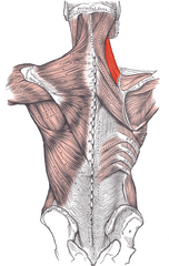
Rhomboid minor
Which back muscle is shown?

Rhomboid major
Which back muscle is shown?

Latissimus dorsi
Which back muscle is shown?

Inferior
Is rhomboid major superior or inferior to Rhomboid minor?
Skull and spine to scapula and clavicle
What does the trapezius connect?
Elevates, retracts, and rotates the scapula
What is the action of the trapezius?
Spinal Accessory nerve (CN XI)
What is the motor innervation of the trapezius?
C3-C4
What is the proprioception innervation of the trapezius?
Transverse cervical artery
What artery supplies blood to the trapezius?
Thyrocervical trunk
Where does the transverse cervical artery branch off from?
Jugular foramen
Which cranial foramen does the spinal accessory nerve travel through?
Spine, ribs, and hips to humerus
What does the latissimus dorsi connect?
Extends, adducts and medially rotates humerus
What is the action of the latissimus dorsi?
Thoracodorsal
What nerve innervates the latissimus dorsi?
C6-C8 and posterior cord
Which spinal nerves of the brachial plexus make up the thoracodorsal nerve? Which cord of the brachial plexus?
Thoracodorsal artery
Which artery supplies blood to the latissimus dorsi?
Subscapular artery
Where does the thoracodorsal artery branch off from?
Spine to scapula
What does the levator scapulae connect?
Elevates scapula
What is the action of the levator scapulae?
Spine to scapula
What do the rhomboids (major/minor) connect?
Retracts scapula
What is the action of the rhomboids (major/minor)?
Dorsal scapular nerve (C5)
Which nerve innervates the rhomboids (major/minor)?
Dorsal scapular nerve (C5) and C3-C4
Which spinal nerves innervate the levator scapulae?
Dorsal scapular artery
Which artery supplies blood to the rhomboids (major/minor)?
Dorsal scapular artery
Which artery supplies blood to the levator scapulae?
Thyrocervical trunk
Where does the dorsal scapular artery branch off from?
Transverse cervical artery and dorsal scapular artery
Which 2 arteries branch off from the thyrocervical trunk?
Serratus posterior superior
Which back muscle is shown?

Serratus posterior inferior
Which back muscle is shown?

Spine to ribs
What do the serratus posterior superior/inferior muscles connect?
Rib elevation
What is the action of the serratus posterior superior muscle?
Rib depression
What is the action of the serratus posterior inferior muscle?
Superior (rib elevation)
Is the serratus posterior inferior or superior muscle responsible for inhalation?
Inferior (rib depression)
Is the serratus posterior inferior or superior muscle responsible for exhalation?
Ventral rami T1-T4
Which spinal nerves innervate the serratus posterior superior muscle?
Ventral rami T9-T12
Which spinal nerves innervate the serratus posterior inferior muscle?
Intercostal arteries
What artery supplies blood to the serratus posterior superior/inferior muscles?
Splenius capitis
Which back muscle is shown?

Semispinalis capitis
What muscle is labeled 1?

Splenius capitis
What muscle is labeled 2?
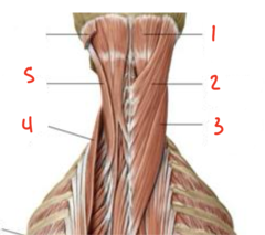
Splenius cervicis
What muscle is labeled 3?

Iliocostalis
What muscle is labeled 4?
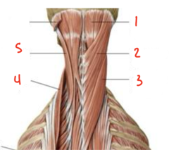
Longissimus
What muscle is labeled 5?

Splenius
What group of back muscles are labeled 1?

Suboccipital
What group of back muscles are labeled 2?
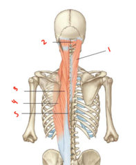
Longissimus
What back muscle is labeled 3?

Iliocostalis
What back muscle is labeled 4?
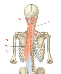
Spinalis
What back muscle is labeled 5?

Erector spinae
What group of back muscles are 3, 4, and 5 a part of?

Splenius group
Which group of deep back muscles is responsible for connecting the skull to the spine as well as adjacent vertebrae?
Extend neck, laterally bend and rotate face to same side
What is the action of the splenius group muscles?
Dorsal primary rami C2-C6
What nerve innervates the splenius group?
Occipital artery and transverse cervical artery
Which arteries supply the splenius group? (2)
Semispinalis
What group of back muscles is pointed to in blue?

Multifidi
What group of back muscles is pointed to in green?

Rotatores
What group of back muscles is pointed to in red?
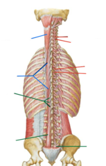
Extension of vertebral column
What is the action of the semispinalis muscle group bilaterally?
Contralateral trunk rotation
What is the action of the semispinalis muscle group individually?
Ipsilateral head rotation
What is the action of the semispinalis capitis muscle individually?
C1 and C2 vertebrae to the base of skull
What does the suboccipital triangle connect?
Extend head at atlanto-axial joint
What is the function of the suboccipital triangle?
Suboccipital nerve/C1 posterior ramus
What nerve innervates the suboccipital triangle?
Vertebral artery and C1 posterior ramus
What are the contents of the suboccipital triangle (artery and nerve)?