Exam 1
 Call Kai
 Learn
Learn Practice Test
 Spaced Repetition
 Match
Match Flashcards
 Knowt Play
1/82

There's no tags or description
Looks like no tags are added yet.
Name | Mastery | Learn | Test | Matching | Spaced | Call with Kai | Chat |
|---|
No analytics yet
Send a link to your students to track their progress
83 Terms
Heart Structure & Function
The cardiovascular system is made up of the:
Heart
Vascular system
Main purpose:
Delivers oxygen and nutrients to tissues.
Helps remove waste products.
The heart is a hollow, muscular organ that pumps blood throughout the body.
Located in the mediastinum:
Between the lungs.
Between the sternum and spinal column.
Rests on the diaphragm.
Extends downward and to the left.
Apex/Point of Maximum Impulse (PMI):
Located at the 5th intercostal space, left midclavicular line.
Layers of the Heart
Endocardium
Innermost layer.
Made of endothelial tissue.
Lines the inside of the heart and the valves.
Myocardium
Middle layer.
Thickest layer of the heart.
Made of muscle fibers.
Responsible for the pumping action of the heart.
Epicardium
Outermost layer.
Serous layer that protects the heart.
Pericardium
The pericardium is a fibrous sac surrounding the heart.
The serous pericardium has two layers:
Visceral pericardium
Covers the surface of the heart.
Parietal pericardium
Lines the pericardial sac.
The layers produce serous fluid.
Pericardial space
Space between the visceral and parietal pericardium.
Contains serous fluid.
Allows the layers to move smoothly against one another.
The parietal pericardium helps anchor the heart within the thorax.
Heart Chambers
The heart has 4 chambers:
Right atrium
Right ventricle
Left atrium
Left ventricle
Atria
Receive blood returning to the heart.
Ventricles
Receive blood from the atria.
Pump blood to the lungs and the rest of the body.
Septum
Band of tissue that separates:
Right side of the heart.
Left side of the heart.
🩸 Blood Flow Through the Heart Remember:
Body → Right side → Lungs → Left side → Body
Detailed blood flow:
Superior vena cava
Brings deoxygenated blood from the head and neck.
Inferior vena cava
Brings deoxygenated blood from the rest of the body.
Right atrium
Right ventricle
Pulmonary arteries
Carry deoxygenated blood to the lungs.
Only arteries in the adult vascular system that carry deoxygenated blood.
Lungs
Blood becomes oxygenated.
Pulmonary veins
Carry oxygenated blood back to the heart.
Only veins in the adult vascular system that carry oxygenated blood.
Left atrium
Left ventricle
Aorta
Body
Important
The left ventricle has a thicker wall than the right ventricle.
This is because the left ventricle pumps blood to the entire body, requiring more force.
Heart Valves Main function
Ensure blood flows in one direction.
Open and close as blood moves through the heart.
Two Types of Valves Atrioventricular (AV) Valves
Separate the atria from the ventricles.
Tricuspid valve
Between the right atrium and right ventricle.
Has 3 leaflets.
Mitral/Bicuspid valve
Between the left atrium and left ventricle.
Has 2 leaflets.
Semilunar Valves
Pulmonic valve
Between the right ventricle and pulmonary artery.
Aortic valve
Between the left ventricle and aorta.
Systole vs. Diastole Systole = Squeeze
Contraction phase of the cardiac cycle.
Ventricles contract and pump blood out.
Diastole = Relax
Relaxation phase.
Heart chambers fill with blood.
One complete cardiac cycle = one heartbeat.
Vascular System
The vascular system includes:
Arteries
Arterioles
Capillaries
Venules
Veins
Functions
Arteries
Transport high-pressure blood away from the heart.
Arterioles
Connect arteries to capillaries.
Capillaries
Allow:
Gas exchange.
Nutrient transfer.
Waste removal.
Venules
Connect capillaries to veins.
Veins
Act as a blood reservoir.
Carry low-pressure blood back to the heart.
Coronary Circulation
The heart muscle requires its own oxygen supply.
Blood is supplied through the:
Right coronary artery (RCA)
Left coronary artery (LCA)
Coronary arteries originate from the base of the aorta, just above the aortic valve.
Coronary perfusion occurs primarily during diastole.
Important!
Increased heart rate → shorter diastole → decreased time for coronary perfusion.
Left Coronary Artery Branches
Left main coronary artery
Left anterior descending (LAD) artery
Supplies the anterior portion of the left ventricle.
Circumflex artery
Supplies the left atrium.
Supplies the posterior-lateral portion of the left ventricle.
Right Coronary Artery Branches
Marginal artery
Supplies the lateral part of the right side of the heart.
Posterior descending artery (PDA)
Supplies the posterior part of the heart.
Cardiac Conduction System ⚡
The heart has its own electrical conduction system.
Order of electrical conduction: SA Node → AV Node → Bundle of His → Right & Left Bundle Branches → Purkinje Fibers SA Node
Located in the right atrium.
Known as the heart's natural pacemaker.
Sets the heart's rate and rhythm.
AV Node
Receives the impulse from the SA node.
Causes a slight delay.
Allows the atria to fully contract and empty into the ventricles.
Bundle of His
Conducts the impulse into the interventricular septum.
Right and Left Bundle Branches
Carry the impulse toward the right and left ventricles.
Purkinje Fibers
Final part of the conduction pathway.
Stimulation causes ventricular contraction.
Dysfunction in the conduction pathway can result in dysrhythmias.
Cardiac Output (CO) Definition
The total amount of blood ejected from one ventricle per minute.
Normal resting cardiac output
Approximately 5–6 L/min in a healthy adult.
Formula
Cardiac Output = Heart Rate × Stroke Volume
Stroke Volume
Amount of blood ejected by one ventricle with each heartbeat.
Three factors affecting stroke volume:
Preload
Afterload
Contractility
Preload
The degree of stretching of the ventricular muscle at the end of diastole.
At the end of diastole:
The ventricle contains its greatest volume of blood.
Muscle fibers experience the greatest stretch.
Preload directly affects stroke volume.
Can be estimated by measuring pulmonary capillary wedge (PCW) pressure.
Easy way to remember:
Preload = volume/stretch before contraction
Afterload
The resistance or pressure the ventricle must overcome to eject blood.
Directly related to:
Arterial blood pressure.
Diameter of blood vessels.
Smaller blood vessels = increased afterload.
Increased afterload can occur with hypertension.
Easy way to remember:
Afterload = resistance against which the heart pumps
Contractility
The force generated by the myocardium during contraction.
Influences stroke volume.
Increases contractility:
Sympathetic stimulation.
Positive inotropic medications.
Decreases contractility:
Hypoxia.
Negative inotropic medications.
Heart Sounds 🩺 Normal Heart Sounds S1 = "Lub"
Closure of:
Mitral valve
Tricuspid valve
S2 = "Dub"
Closure of:
Aortic valve
Pulmonic valve
Remember:
S1 = AV valves close S2 = Semilunar valves close
Abnormal Heart Sounds S3
Best heard with the bell of the stethoscope.
Described as a ventricular gallop.
Heard as a loud "DUB" following S2.
Indicates decreased ventricular compliance.
Can be normal in:
Children.
Adults up to age 40.
In adults older than 40, may indicate heart failure.
S4
Best heard with the bell.
Described as an atrial gallop.
Heard as a loud "LUB" immediately before S1.
Associated with decreased ventricular compliance.
May occur with:
Hypertension.
Aortic stenosis.
Coronary artery disease.
Cardiomyopathy.
Murmurs & Pericardial Friction Rub Murmur
Caused by turbulent blood flow through healthy or diseased valves.
Sounds like "whooshing."
Systolic Murmur
Heard between S1 and S2.
Diastolic Murmur
Heard between S2 and S1.
Pericardial Friction Rub
Abnormal heart sound.
Usually heard over the left sternal border.
Caused by:
Inflammation.
Infection.
Infiltration in the pericardial sac.
Sounds "grating."
Usually disappears when the underlying cause is treated.
Valvular Dysfunction
Main Function of Heart Valves
Heart valves ensure blood flows in one direction.
Valves open to allow forward blood flow.
Valves close to prevent backflow of blood.
The 4 valves are:
Tricuspid
Pulmonic
Mitral (bicuspid)
Aortic
AV valves:
Tricuspid
Mitral
Semilunar valves:
Pulmonic
Aortic
Types of Valvular Dysfunction 1. Stenosis
Stenosis = narrowing of a valve
Valve does not open completely.
Blood has difficulty moving forward through the valve.
The heart must work harder to push blood through the narrowed opening.
Can lead to:
Increased pressure behind the valve.
Chamber hypertrophy.
Decreased cardiac output.
Heart failure.
Easy way to remember:
Stenosis = "stuck closed"
2. Regurgitation (Insufficiency)
Regurgitation = valve does not close completely
Blood leaks backward through the valve.
Causes volume overload in the affected chamber.
The heart must pump extra blood to maintain adequate cardiac output.
Can eventually cause:
Chamber dilation.
Decreased cardiac output.
Pulmonary congestion.
Heart failure.
Easy way to remember:
Regurgitation = "leaky valve"
Mitral Valve Dysfunction Mitral Stenosis
Narrowing of the mitral valve.
Mitral valve is located between:
Left atrium
Left ventricle
Obstruction prevents blood from moving easily from the left atrium to the left ventricle.
Blood backs up into:
Left atrium.
Pulmonary circulation.
Possible findings
Dyspnea.
Fatigue.
Pulmonary congestion.
Crackles.
Exercise intolerance.
Increased risk for atrial dysrhythmias.
Think:
Mitral stenosis → blood backs up into the lungs
Mitral Regurgitation
Mitral valve does not close completely.
During ventricular systole:
Some blood flows backward from the left ventricle → left atrium.
Can result in:
Left atrial enlargement.
Pulmonary congestion.
Decreased forward cardiac output.
Possible findings
Fatigue.
Dyspnea.
Pulmonary congestion.
Heart murmur.
Aortic Valve Dysfunction Aortic Stenosis
Narrowing of the aortic valve.
Makes it difficult for blood to leave the left ventricle.
Left ventricle must generate more pressure to push blood through the narrowed valve.
Effects
Increased afterload.
Left ventricular hypertrophy.
Decreased cardiac output.
Classic symptoms
SAD
Syncope
Angina
Dyspnea
Important NCLEX concept
Aortic stenosis can significantly decrease blood flow to the body.
Severe stenosis may lead to:
Heart failure.
Dysrhythmias.
Syncope.
Sudden cardiac death.
Aortic Regurgitation
Aortic valve does not close completely.
Blood flows backward from the:
Aorta → left ventricle
Occurs during diastole.
Causes increased volume in the left ventricle.
Effects
Left ventricular dilation.
Increased workload.
Decreased cardiac efficiency.
Possible heart failure.
Tricuspid Valve Dysfunction Tricuspid Stenosis
Narrowing of the tricuspid valve.
Obstructs blood flow from:
Right atrium → right ventricle
Can cause blood to back up into the systemic venous circulation.
Possible findings
Jugular venous distention (JVD).
Peripheral edema.
Hepatomegaly.
Ascites.
Tricuspid Regurgitation
Tricuspid valve does not close completely.
Blood flows backward from:
Right ventricle → right atrium
Occurs during ventricular systole.
Can cause signs of right-sided heart failure:
JVD.
Peripheral edema.
Hepatomegaly.
Ascites.
Weight gain.
Pulmonic Valve Dysfunction Pulmonic Stenosis
Narrowing of the pulmonic valve.
Obstructs blood flow from:
Right ventricle → pulmonary artery
Right ventricle must work harder to pump blood to the lungs.
Possible effects
Right ventricular hypertrophy.
Decreased pulmonary blood flow.
Signs of right-sided heart failure in severe cases.
Pulmonic Regurgitation
Pulmonic valve does not close completely.
Blood flows backward from:
Pulmonary artery → right ventricle
Occurs during diastole.
Causes volume overload of the right ventricle.
Murmurs and Valvular Dysfunction
A murmur is caused by turbulent blood flow.
Often described as a "whooshing" sound.
Systolic murmurs occur between:
S1 → S2
Diastolic murmurs occur between:
S2 → S1.
Quick Rule for Murmurs Systolic
Aortic stenosis
Mitral regurgitation
Diastolic
Aortic regurgitation
Mitral stenosis
Easy Memory Trick:
"Stenosis = systolic on the semilunar valves"
Aortic stenosis
Pulmonic stenosis
"Regurgitation = diastolic on the semilunar valves"
Aortic regurgitation
Pulmonic regurgitation
For the AV valves, the pattern is reversed:
Mitral/tricuspid regurgitation = systolic
Mitral/tricuspid stenosis = diastolic
Signs and Symptoms of Valvular Disease
Depending on the valve affected, watch for:
Decreased Cardiac Output
Fatigue.
Weakness.
Exercise intolerance.
Dizziness.
Syncope.
Hypotension.
Left-Sided Effects
Dyspnea.
Orthopnea.
Crackles.
Pulmonary edema.
Right-Sided Effects
Peripheral edema.
JVD.
Hepatomegaly.
Ascites.
Weight gain.
Valvular Dysfunction
Main Function of Heart Valves
Heart valves ensure blood flows in one direction.
Valves open to allow forward blood flow.
Valves close to prevent backflow of blood.
The 4 valves are:
Tricuspid
Pulmonic
Mitral (bicuspid)
Aortic
AV valves:
Tricuspid
Mitral
Semilunar valves:
Pulmonic
Aortic
Types of Valvular Dysfunction 1. Stenosis
Stenosis = narrowing of a valve
Valve does not open completely.
Blood has difficulty moving forward through the valve.
The heart must work harder to push blood through the narrowed opening.
Can lead to:
Increased pressure behind the valve.
Chamber hypertrophy.
Decreased cardiac output.
Heart failure.
Easy way to remember:
Stenosis = "stuck closed"
2. Regurgitation (Insufficiency)
Regurgitation = valve does not close completely
Blood leaks backward through the valve.
Causes volume overload in the affected chamber.
The heart must pump extra blood to maintain adequate cardiac output.
Can eventually cause:
Chamber dilation.
Decreased cardiac output.
Pulmonary congestion.
Heart failure.
Easy way to remember:
Regurgitation = "leaky valve"
Mitral Valve Dysfunction Mitral Stenosis
Narrowing of the mitral valve.
Mitral valve is located between:
Left atrium
Left ventricle
Obstruction prevents blood from moving easily from the left atrium to the left ventricle.
Blood backs up into:
Left atrium.
Pulmonary circulation.
Possible findings
Dyspnea.
Fatigue.
Pulmonary congestion.
Crackles.
Exercise intolerance.
Increased risk for atrial dysrhythmias.
Think:
Mitral stenosis → blood backs up into the lungs
Mitral Regurgitation
Mitral valve does not close completely.
During ventricular systole:
Some blood flows backward from the left ventricle → left atrium.
Can result in:
Left atrial enlargement.
Pulmonary congestion.
Decreased forward cardiac output.
Possible findings
Fatigue.
Dyspnea.
Pulmonary congestion.
Heart murmur.
Aortic Valve Dysfunction Aortic Stenosis
Narrowing of the aortic valve.
Makes it difficult for blood to leave the left ventricle.
Left ventricle must generate more pressure to push blood through the narrowed valve.
Effects
Increased afterload.
Left ventricular hypertrophy.
Decreased cardiac output.
Classic symptoms
SAD
Syncope
Angina
Dyspnea
Important NCLEX concept
Aortic stenosis can significantly decrease blood flow to the body.
Severe stenosis may lead to:
Heart failure.
Dysrhythmias.
Syncope.
Sudden cardiac death.
Aortic Regurgitation
Aortic valve does not close completely.
Blood flows backward from the:
Aorta → left ventricle
Occurs during diastole.
Causes increased volume in the left ventricle.
Effects
Left ventricular dilation.
Increased workload.
Decreased cardiac efficiency.
Possible heart failure.
Tricuspid Valve Dysfunction Tricuspid Stenosis
Narrowing of the tricuspid valve.
Obstructs blood flow from:
Right atrium → right ventricle
Can cause blood to back up into the systemic venous circulation.
Possible findings
Jugular venous distention (JVD).
Peripheral edema.
Hepatomegaly.
Ascites.
Tricuspid Regurgitation
Tricuspid valve does not close completely.
Blood flows backward from:
Right ventricle → right atrium
Occurs during ventricular systole.
Can cause signs of right-sided heart failure:
JVD.
Peripheral edema.
Hepatomegaly.
Ascites.
Weight gain.
Pulmonic Valve Dysfunction Pulmonic Stenosis
Narrowing of the pulmonic valve.
Obstructs blood flow from:
Right ventricle → pulmonary artery
Right ventricle must work harder to pump blood to the lungs.
Possible effects
Right ventricular hypertrophy.
Decreased pulmonary blood flow.
Signs of right-sided heart failure in severe cases.
Pulmonic Regurgitation
Pulmonic valve does not close completely.
Blood flows backward from:
Pulmonary artery → right ventricle
Occurs during diastole.
Causes volume overload of the right ventricle.
Murmurs and Valvular Dysfunction
A murmur is caused by turbulent blood flow.
Often described as a "whooshing" sound.
Systolic murmurs occur between:
S1 → S2
Diastolic murmurs occur between:
S2 → S1.
Quick Rule for Murmurs Systolic
Aortic stenosis
Mitral regurgitation
Diastolic
Aortic regurgitation
Mitral stenosis
Easy Memory Trick:
"Stenosis = systolic on the semilunar valves"
Aortic stenosis
Pulmonic stenosis
"Regurgitation = diastolic on the semilunar valves"
Aortic regurgitation
Pulmonic regurgitation
For the AV valves, the pattern is reversed:
Mitral/tricuspid regurgitation = systolic
Mitral/tricuspid stenosis = diastolic
Signs and Symptoms of Valvular Disease
Depending on the valve affected, watch for:
Decreased Cardiac Output
Fatigue.
Weakness.
Exercise intolerance.
Dizziness.
Syncope.
Hypotension.
Left-Sided Effects
Dyspnea.
Orthopnea.
Crackles.
Pulmonary edema.
Right-Sided Effects
Peripheral edema.
JVD.
Hepatomegaly.
Ascites.
Weight gain.
Cardiomyopathy
Cardiomyopathy is a chronic cardiac condition that affects the heart's ability to pump blood efficiently.
There are 4 main types:
Dilated cardiomyopathy (DCM)
Hypertrophic cardiomyopathy (HCM)
Restrictive cardiomyopathy (RCM)
Arrhythmogenic cardiomyopathy (ACM)
The direct cause is often unknown, but multiple contributing factors may increase the risk.
Cardiomyopathy is closely associated with heart failure.
Types of Cardiomyopathy 1. Dilated Cardiomyopathy (DCM) Pathophysiology
Most common type of cardiomyopathy.
Progressive cardiac disorder.
The ventricles become dilated/stretched out.
Ventricular muscle fibers become weakened.
The heart cannot contract effectively.
Ineffective contractions lead to:
↓ Cardiac output
Heart failure
Think:
Dilated = stretched → weak squeeze → ↓ cardiac output
2. Hypertrophic Cardiomyopathy (HCM) Pathophysiology
Caused by hypertrophy/thickening of:
Left ventricular walls
Interventricular septum
Thickened ventricles do not allow adequate blood refilling.
Results in:
↓ Left ventricular filling
↓ Cardiac output
Think:
Hypertrophic = thick → can't fill
3. Restrictive Cardiomyopathy (RCM) Pathophysiology
Least common type of cardiomyopathy.
Typically has a poor prognosis.
Ventricles become stiff.
Stiff ventricles cannot fill adequately during diastole.
Results in:
↓ Diastolic ventricular filling
↓ Cardiac output
Think:
Restrictive = rigid/stiff → can't fill
4. Arrhythmogenic Cardiomyopathy (ACM) Pathophysiology
Ventricular muscle fibers are replaced with:
Fibrous tissue
Fatty tissue
The abnormal tissue causes electrical instability.
Can lead to lethal dysrhythmias:
Ventricular tachycardia (VT)
Ventricular fibrillation (VF)
Can result in sudden cardiac death.
Think:
Arrhythmogenic = abnormal tissue → abnormal electrical activity → lethal dysrhythmias
⭐ Quick Comparison
Type | Main Problem | Effect |
|---|---|---|
DCM | Ventricles are stretched/dilated | Weak contractions |
HCM | Ventricular walls/septum are thickened | Decreased filling |
RCM | Ventricles are stiff | Decreased diastolic filling |
ACM | Muscle replaced with fatty/fibrous tissue | Lethal dysrhythmias |
Etiology / Causes DCM
Not age-specific.
More typically seen in male clients.
Possible causes:
Viral infections.
Autoimmune disorders.
Myocarditis.
Sarcoidosis.
Malnutrition.
Endocrine disorders.
Inflammatory processes.
Alcohol use.
HCM
Often has a genetic predisposition.
Associated with mutations to cardiac contractile muscle proteins.
Can occur in athletes.
RCM
Cause is usually unknown.
May be linked to:
Amyloidosis.
Sarcoidosis.
ACM
Associated with a genetic defect.
Impact on Overall Health Psychosocial Effects
Cardiomyopathy requires ongoing:
Lifestyle changes.
Management.
Modifications to daily life.
Clients may have an increased risk of depression due to loss of independence.
Medications such as diuretics and beta blockers may affect:
Sexual libido.
Mood.
Urinary continence.
The financial burden of multiple medications may contribute to feeling overwhelmed.
Safety Considerations ⚠
Clients with arrhythmogenic or restrictive cardiomyopathy have an increased risk of sudden cardiac death.
Risk is particularly important with competitive recreational sports.
Other safety considerations are similar to those for heart failure.
Considerations for Older Adults
Cognitive deficits may interfere with the client's ability to:
Follow self-care recommendations.
Manage cardiomyopathy effectively.
Clinical Manifestations DCM
Common findings:
Chest discomfort.
Peripheral edema.
Progressively increasing fatigue.
Exertional dyspnea.
HCM
May be asymptomatic.
Possible manifestations:
Fatigue.
Syncope.
Chest discomfort.
Palpitations.
Exhaustion.
Dyspnea.
Increased risk of sudden cardiac death, including in clients without manifestations.
RCM
Manifestations progress over time.
Symptoms are similar to heart failure manifestations seen with:
DCM
HCM
ACM
Manifestations may include:
Symptoms similar to other cardiomyopathies.
Ascites
Increased JVD
Lethal dysrhythmias:
VT
VF
Diagnostic Tests 🔬 Chest X-Ray
Used to identify cardiac enlargement.
Blood Tests
CBC
CMP
Monitor for abnormalities involving:
Liver function.
Thyroid function.
Renal function.
Cardiac Markers Troponin I & Troponin T
Heart muscle-specific biomarkers.
Elevation indicates myocardial injury.
NCLEX Question:
What does an elevated troponin I indicate?
➡ Myocardial injury
BNP
A hormone released when ventricular contraction has weakened due to muscle fiber stretching from excessive fluid overload.
ECG Findings
An ECG may show:
Irregular heart rhythm.
Widened QRS complexes.
Bundle branch block.
Abnormal P waves.
Abnormal T waves.
Absence of P waves.
Ventricular tachycardia.
Ventricular fibrillation.
Echocardiogram
Used to evaluate:
Thickness of the ventricles.
Ventricular function.
Movement of the heart valves.
Role of the Nurse 👩⚕ ICD Safety
Some clients may have an implantable cardioverter-defibrillator (ICD).
ICD function:
Monitors for abnormal cardiac rhythms.
Detects life-threatening rhythms such as:
VT
VF
Delivers an electrical shock when indicated.
Nursing Assessment:
Ask the client:
Has the ICD delivered a shock?
How often has it delivered shocks?
Report this information to the healthcare provider.
Individual Assessment
Obtain a complete:
Medical history.
Family history.
Assess for factors that may predispose the client to cardiomyopathy.
Determine whether the client can perform ADLs without increased:
Fatigue.
Dyspnea.
Palpitations.
Client Education
Teach the importance of:
Follow-up appointments.
Frequent vital sign monitoring.
Monitoring medication effectiveness.
Lifestyle Modifications
Reduce dietary sodium.
Restrict fluids to approximately 2 L/day.
Eliminate or decrease:
Nicotine use.
Alcohol use.
Avoid strenuous activity.
Participate in low-impact exercise once cleared by the healthcare provider.
Pace activities.
Take rest periods as needed.
Monitor daily for:
Weight changes.
Lower extremity edema.
Nursing Process 1. Recognize Cues — Assessment
Assess for manifestations of heart failure:
Lower extremity edema.
JVD.
Pulmonary congestion.
Additional assessments:
Palpate pulses for irregularities that may indicate A-fib.
Assess for:
Liver distention.
Ascites.
Auscultate for:
Heart murmurs.
Extra heart sounds.
Irregular rhythms.
2. Analyze Cues
Gather all relevant client data.
Develop a plan of care.
Provide support and education.
Reinforce:
Medication adherence.
Lifestyle modifications.
Goal: improve the client's quality of life.
3. Priority of Care ⭐ Main priority:
Improve cardiac output and quality of life.
4. Generate Solutions
Collaborate with the interprofessional healthcare team to plan:
Medication management.
Dietary restrictions.
Activity restrictions.
Education about monitoring manifestations.
5. Take Actions
Treatment may include:
Lifestyle modifications.
Medications.
Surgical interventions.
6. Evaluate Outcomes
Clients require frequent follow-up.
Treatments are complex.
Regular monitoring is needed to prevent potentially life-threatening complications.
Evaluate the effectiveness of the plan of care.
Treatments & Therapies Nonpharmacological Interventions
Teach clients to:
Reduce sodium intake.
Reduce fluid intake.
Maintain or reduce weight as appropriate.
Avoid:
Alcohol.
Smoking.
Illicit drug use.
Discontinue strenuous activities.
Participate in low-impact exercise.
Consider genetic testing and counseling.
Pharmacological Interventions 💊 General Treatment
For all four types:
Diuretics
Manage fluid overload.
Vasodilators
Help decrease manifestations of fluid overload.
Example: Nitroglycerin
DCM Medications
ACE inhibitors
Enalapril.
Lisinopril.
ARBs
Losartan.
Valsartan.
Beta blockers
Metoprolol.
Mineralocorticoid antagonists
Eplerenone.
Spironolactone.
HCM Medications
Beta blocker.
Calcium channel blocker:
Verapamil
Used to reduce:
Chest discomfort/pressure.
Dyspnea.
RCM Medications ⚠
The heart may require a higher heart rate to maintain adequate cardiac output.
Use beta blockers and calcium channel blockers cautiously because they can decrease the heart rate.
Surgical & Invasive Interventions DCM and RCM
For end-stage heart failure:
Heart Transplantation
Can improve long-term survival and heart function.
Finding a compatible donor can take a long time.
LVAD — Left Ventricular Assist Device
Surgically implanted cardiac device.
Helps maintain:
Heart function.
Cardiac output.
May be used:
While waiting for a heart transplant.
As a long-term treatment option.
Following LVAD placement:
Clients typically remain in the ICU for 4–5 days while IV medications are weaned.
HCM Procedures Septal Myectomy
Invasive procedure similar to open-heart surgery.
Removes or reduces portions of the hypertrophied ventricular septum.
Allows increased capacity for diastolic filling.
Alcohol Septal Ablation
Less invasive procedure.
Performed with cardiac catheterization.
Medical alcohol is injected into the thickened septal tissue.
Eliminates blood flow to the targeted area.
Tissue death reduces the thickness of the septum.
ICD
May reduce the high risk of sudden cardiac death.
Battery life: approximately 6–12 years.
Clients should be monitored by the healthcare provider at least every 6 months.
Heart Failure
Heart failure (HF) is a complex, progressive clinical syndrome.
Occurs due to abnormalities in the:
Structure of the heart
Function of the heart
These abnormalities lead to:
↓ Cardiac output (CO)
↑ Pressure within the heart
Can occur:
At rest.
During stress.
Heart failure occurs when the heart cannot meet the needs of the systemic circulatory system.
This happens when:
Ventricles do not fill properly.
Blood is not effectively ejected into the systemic circulation.
Conditions that contribute to HF:
Hypertension.
Coronary artery disease (CAD).
Uncontrolled arrhythmias.
Myocardial infarction (MI).
Valvular disease.
Key Concept 🧠
Heart failure = filling problem, pumping problem, or both.
Etiology / Causes Most Common Causes
CAD
MI
Diabetes mellitus
Structural Causes
Uncontrolled arrhythmias.
Hypertension.
Myocarditis.
Congenital heart disease.
Other Causes
Heart failure can also occur due to:
Thyrotoxicosis.
Anemia.
Thiamine deficiency.
Pregnancy.
Risk Factors
Family history of heart disease.
Chronic pulmonary disease.
CAD.
Hypertension.
Chronic infection or inflammation.
Diabetes mellitus.
Obesity.
Metabolic diseases.
Alcohol abuse.
Treatment with cardiotoxic agents, such as:
Anthracyclines
Common Comorbidities
More than half of clients with heart failure also have another comorbid condition.
Cardiac and Noncardiac Comorbidities
Obesity.
Atrial fibrillation (A-fib).
Chronic kidney disease (CKD).
Diabetes mellitus.
Hypertension.
Anemia.
Iron deficiency.
Frailty.
Impact on Overall Health Physiological Effects
Common manifestations can include:
Shortness of breath.
Fatigue.
Weakness.
Difficulty sleeping.
Lower extremity edema.
Psychosocial Effects
Diminished quality of life.
Difficulty completing ADLs independently.
Activity intolerance.
Increased dependence on caregivers.
May contribute to:
Depression.
Hopelessness.
Helplessness.
Safety Considerations ⚠
Heart failure is progressive, but manifestations can worsen quickly.
Clients need to know:
How to monitor their manifestations.
When to report changes.
Oxygen Safety
If the client uses oxygen:
Follow proper oxygen administration.
Post no-smoking signs.
Keep oxygen at least 6 feet from open flames.
Use electrical equipment safely.
Considerations for Older Adults
Incidence of heart failure increases with age.
Adults age 65 and older should be educated about:
Manifestations of developing heart failure.
Modifiable risk factors.
Strategies to reduce risk.
Types of Heart Failure Left-Sided Heart Failure 🫁
Primarily causes blood and fluid to back up toward the lungs.
Manifestations
Shortness of breath.
Hypotension with tachycardia.
Orthopnea.
Exertional dyspnea.
Paroxysmal nocturnal dyspnea (PND).
Lower extremity edema.
Weight gain.
Increased abdominal girth.
Pulmonary congestion.
Rales.
Productive cough.
White or pink blood-tinged sputum.
Displaced point of maximum impulse (PMI).
Memory Trick:
LEFT = LUNGS
Right-Sided Heart Failure 💧
Associated with systemic fluid buildup.
Manifestations
JVD.
Chest discomfort.
S3 heart sound.
Heart murmurs.
Palpitations.
Ascites.
Peripheral edema.
Generalized swelling.
Enlarged liver.
Enlarged spleen.
Memory Trick:
RIGHT = REST of the body
Biventricular Heart Failure
Involves both ventricles.
Client experiences manifestations of both:
Left-sided HF.
Right-sided HF.
Common findings
Fluid buildup.
Dyspnea.
Pulmonary and systemic congestion.
⭐ Left vs. Right Heart Failure
Left-Sided HF | Right-Sided HF |
|---|---|
Pulmonary congestion | Systemic congestion |
Dyspnea | JVD |
Orthopnea | Peripheral edema |
PND | Ascites |
Rales | Hepatomegaly |
Pink/white blood-tinged sputum | Generalized swelling |
Framingham Diagnostic Criteria
To diagnose heart failure, the client must meet:
2 major criteria
OR
1 major criterion + 2 minor criteria
Major Criteria
Acute pulmonary edema.
Cardiomegaly.
Hepatojugular reflux.
Neck vein distention.
Paroxysmal nocturnal dyspnea.
Orthopnea.
Pulmonary rales.
Third heart sound (S3 gallop).
Weight loss of 4.5 kg or more in 5 days in response to treatment.
Central venous pressure greater than 16 cm of water.
Radiographic cardiomegaly.
Minor Criteria
Ankle edema.
Dyspnea on exertion.
Hepatomegaly.
Nocturnal cough.
Pleural effusion.
Tachycardia:
Heart rate greater than 120/min.
Decrease in vital capacity by one-third of the maximum recorded value.
NYHA Functional Classification
Used to classify the severity of heart failure based on manifestations during physical activity.
Class I
No limitation.
Able to perform normal activities.
No manifestations.
Class II
Slight limitation with normal activity.
Mild manifestations with activity.
Comfortable at rest.
Class III
Moderate limitation.
Moderate manifestations with decreased activity.
Comfortable only at rest.
Class IV
Severe limitation.
Manifestations with minimal activity.
Manifestations may be present at rest.
Possible Manifestations
Chest pain.
Shortness of breath.
Fatigue.
Palpitations.
Fainting.
Easy Memory 🧠
I = no symptoms
II = slight limitation
III = moderate limitation
IV = symptoms at rest
Diagnostic Tests & Labs 🔬 Complete Laboratory Profile
Used to evaluate:
Renal function.
Liver function.
Anemia.
Iron deficiency.
Possible cause and severity of HF.
BNP — B-Type Natriuretic Peptide
Used to help determine whether dyspnea is:
Cardiac-related
Noncardiac
Can also monitor effectiveness of treatment.
BNP levels correspond with the NYHA classification.
BNP obtained before discharge can help predict:
Readmission risk.
Risk of death.
NCLEX Pearl ⭐
Dyspnea + suspected heart failure → check BNP
Echocardiogram
Used to assess:
Systolic dysfunction.
Diastolic dysfunction.
Wall motion abnormalities.
Heart valve function.
Systolic Dysfunction
Left ventricle does not contract normally.
Not enough blood is pushed into circulation.
Diastolic Dysfunction
Left ventricle does not relax normally.
Ventricle does not fill properly during relaxation.
Easy Memory:
Systolic = squeeze problem Diastolic = filling/relaxation problem
Chest X-Ray
Used to assess:
Heart size.
Degree of pulmonary congestion.
Findings suggestive of HF:
Pulmonary vascular congestion.
Enlarged heart.
Fluid in the base of the lungs.
Other Tests ECG
Helps identify underlying causes.
Can identify rhythm abnormalities.
Cardiac Catheterization
May be performed if HF is related to an acute MI.
May be followed by PCI to restore blood flow.
Stress Test
Used to help determine the underlying cause.
Role of the Nurse 👩⚕ Environmental Assessment
During home visits, assess:
Ability to perform ADLs.
Meals the client is preparing.
Safe use of oxygen.
Ability to ask questions and understand teaching.
Individualized Assessment
Assess each client's:
Manifestations.
Functional ability.
Ambulation.
Mobility.
Cognition.
Ability to remain independent safely.
Assess understanding of:
Manifestations to report.
How and when to take medications.
Importance of daily weights.
Daily Weight Monitoring ⚖
A weight gain may indicate fluid retention and worsening heart failure.
Report:
More than 2–3 lb in 1 day
OR
More than 5 lb in 1 week
How to Weigh
Use the same scale.
Weigh at the same time every day.
Ideally weigh:
In the morning.
Before eating.
Client Education Monitor Daily For:
Increased shortness of breath.
Swelling.
Weight gain.
Other worsening manifestations.
Lifestyle Changes
Smoking cessation.
Limit alcohol.
Reduce sodium intake to less than 3 g/day.
Approximately 2 L fluid restriction/day, especially with:
Hyponatremia.
Advanced symptoms.
Take medications as prescribed.
Attend regular follow-up appointments.
Maintain recommended vaccinations, including:
Influenza.
Pneumococcal.
Nursing Process 1. Recognize Cues — Assessment
Obtain a complete health history:
Current and past manifestations.
Other medical conditions.
Ability to perform ADLs.
Physical Assessment
Assess:
General appearance.
Lung sounds.
Heart sounds.
JVD.
Peripheral edema.
Full set of vital signs.
Review:
Laboratory results.
Diagnostic testing.
2. Analyze Cues
Look for signs of worsening HF:
Weight gain.
Decreased ability to perform activities.
Edema.
Rales.
Evidence of fluid overload.
Elevated or abnormal BNP findings.
3. Priority of Care ⭐ Main Priority:
Reduce fluid overload and improve respiratory and cardiac status.
4. Generate Solutions
Collaborate with the interprofessional team.
Goals include:
Control and monitor manifestations.
Medication adherence.
Dietary adherence.
Daily weight monitoring.
Improve quality of life.
Reduce morbidity and mortality.
Reduce hospital readmissions.
5. Take Actions
If the client has respiratory distress:
Elevate the head of the bed to High-Fowler's.
Administer O₂ as prescribed.
Encourage anxiety-relieving techniques:
Pursed-lip breathing.
Guided imagery.
Relaxation.
Encourage coughing and deep breathing every 2 hours.
Reassess lung sounds at least every 4 hours, or more frequently as indicated.
Treatment may include:
Medications.
Lifestyle modifications.
Surgical interventions.
6. Evaluate Outcomes
Evaluate whether treatment resulted in:
Maintained weight.
Decreased edema.
Improved quality of life.
Decreased hospital readmissions.
Continue frequent monitoring of manifestations.
Nonpharmacological Treatment
Treatment focuses on:
Correcting the underlying cause when possible.
Relieving manifestations.
Slowing disease progression.
Improving quality of life.
Lifestyle Changes
Stop smoking.
Reduce or maintain weight.
Monitor fluid intake.
Use alcohol and caffeine in moderation.
Follow a heart-healthy diet.
Remain physically active as appropriate.
Manage stress.
Control blood pressure.
Pharmacological Treatment 💊
Heart failure treatment may involve multiple medications.
Medication Classes
SGLT-2 inhibitors
Reduce HF hospitalizations.
Reduce cardiovascular mortality.
ACE inhibitors
Examples:
Captopril.
Enalapril (Vasotec).
Angiotensin II receptor blockers (ARBs)
Angiotensin receptor-neprilysin inhibitors (ARNIs)
Beta blockers
Example: Metoprolol
Aldosterone antagonists
Example: Spironolactone
Hydralazine and isosorbide dinitrate
Diuretics
Other Medications
May include:
Anticoagulants
For clients with A-fib.
Example: Warfarin.
Statins
For elevated cholesterol or history of MI.
Digoxin
Surgical & Invasive Interventions Cardiac Catheterization + PCI
Used when HF is caused by an acute MI.
Purpose:
Restore blood flow to cardiac muscle.
Decrease the extent of myocardial damage.
Cardiac Resynchronization Therapy (CRT) Ventricular Desynchrony
Right and left ventricles do not contract and relax at the same time.
Different areas of the ventricles may contract at different times.
Can contribute to dysrhythmias.
CRT with Biventricular Pacemaker
Used to synchronize ventricular contractions.
Pacemaker is inserted below the collarbone.
Three wires are threaded into the ventricles.
The device detects and corrects rhythm problems.
Implantable Cardioverter-Defibrillator (ICD) ⚡
Clients with LVEF less than 35% are at high risk for dysrhythmias.
An ICD may be inserted to help prevent sudden cardiac death.
Device is placed beneath the skin below the collarbone.
Two wires are threaded into:
Right atrium.
Right ventricle.
Detects dysrhythmias.
Sends an electrical impulse to correct the abnormal rhythm.
ICD Discharge Teaching
For approximately 4–6 weeks, follow prescribed activity restrictions.
Incision Care
Monitor for infection:
Redness.
Drainage.
Increased pain.
Keep incision dry for 4–5 days.
After that:
Shower as instructed.
Gently pat incision dry.
Avoid:
Tub baths.
Swimming.
Hot tubs.
Continue avoiding these until the incision is completely healed.
Activity Restrictions
Wear comfortable clothing that does not rub the incision.
Do not lift more than 10–15 lb.
Avoid:
Twisting.
Pushing.
Pulling for 2–3 weeks.
Do not raise the affected arm above the shoulder for 4–6 weeks.
Keep follow-up appointments for regular device monitoring.
diuretics
Overview Indications for medications affecting urinary output
Management of blood pressure.
Removal of excess/edematous fluid related to:
Heart failure.
Kidney disease.
Liver disease.
Prevention of kidney failure in certain situations.
Main Types of Diuretics
High-ceiling loop diuretics
Thiazide diuretics
Potassium-sparing diuretics
Osmotic diuretics
1. High-Ceiling Loop Diuretics 💧 Prototype
Furosemide (Lasix)
Other Medications
Ethacrynic acid
Bumetanide
Torsemide
Expected Pharmacological Action
Work in the loop of Henle.
Block reabsorption of:
Sodium.
Chloride.
Prevent reabsorption of water.
Cause extensive diuresis, even with severe renal impairment.
Increase excretion of:
Water.
Potassium.
Sodium.
Chloride.
Magnesium.
Calcium.
Memory Trick 🧠
Loop diuretics = lose lots of fluid and electrolytes.
Therapeutic Uses
Used when there is an emergent need for rapid fluid removal.
Pulmonary edema caused by heart failure.
Edema caused by:
Liver disease.
Cardiac disease.
Kidney disease.
Conditions that do not respond to other diuretics.
Hypertension:
Torsemide.
Unlabeled use: Hypercalcemia
Routes
Oral
IV
IM
Loop Diuretic Complications ⚠ 1. Dehydration Manifestations
Dry mouth.
Increased thirst.
Oliguria.
Lethargy.
Weight loss.
Nursing Actions
Monitor for dehydration.
Monitor daily weight.
Report urine output less than 30 mL/hr.
Notify the provider.
Medication may need to be withheld.
Therapy is generally started with a low dose.
2. Hypotension / Orthostatic Hypotension Nursing Actions
Monitor blood pressure.
Teach the client to:
Change positions slowly.
Rise slowly from lying or sitting.
Client Education
If light-headedness or dizziness occurs:
Sit down.
Lie down.
3. Ototoxicity 👂 Important!
Usually transient with furosemide.
Can be irreversible with ethacrynic acid.
Nursing Actions
Avoid combining with other ototoxic medications when possible.
Example:
Aminoglycoside antibiotics such as gentamicin.
Client Education
Report:
Tinnitus.
Ringing.
Buzzing.
Vertigo.
Feeling of fullness in the ears.
4. Hypokalemia Potassium level:
K⁺ less than 3.5 mEq/L
Manifestations
Nausea.
Vomiting.
Fatigue.
Leg cramps.
General weakness.
Nursing Actions
Monitor:
Potassium.
Cardiac status.
Report K⁺ less than 3.5 mEq/L.
Monitor ECG if potassium drops below 3.5 mEq/L.
Client may require a potassium supplement.
Client Education
Eat potassium-rich foods:
Bananas.
Potatoes.
Dried fruits.
Nuts.
Spinach.
Citrus fruits.
5. Other Electrolyte Imbalances
Loop diuretics can cause:
Hyponatremia
Hypomagnesemia
Hypochloremia
Hypocalcemia
Nursing Actions
Monitor electrolyte levels.
Report abnormal results.
6. Other Adverse Effects
Hyperglycemia.
Hyperuricemia.
↓ HDL cholesterol.
↑ LDL cholesterol.
↑ Triglycerides.
Nursing Actions
Monitor:
Blood glucose.
Uric acid.
Lipid levels.
Loop Diuretics: Contraindications & Precautions Contraindicated
Anuria = no urine output.
Use Cautiously With
Severe liver disease.
Diabetes mellitus.
Dehydration.
Electrolyte depletion.
Gout.
Use caution with clients taking:
Digoxin.
Lithium.
Ototoxic medications.
NSAIDs.
Antihypertensives.
Important
Hypoproteinemia can increase the risk of ototoxicity.
Loop Diuretic Drug Interactions Digoxin + Hypokalemia ⚠
Hypokalemia increases the risk for digoxin toxicity.
Can lead to ventricular dysrhythmias.
Nursing Actions
Monitor:
Cardiac status.
Potassium levels.
Digoxin levels.
Important
Potassium-sparing diuretics may be given with loop diuretics to reduce the risk of hypokalemia.
Antihypertensives
Can cause an additive hypotensive effect.
Nursing Action
Monitor blood pressure.
Lithium
Hyponatremia caused by loop diuretics can increase lithium levels.
Can result in lithium toxicity.
Nursing Action
Monitor lithium levels.
NSAIDs
Decrease blood flow to the kidneys.
Can decrease the effectiveness of the diuretic.
Nursing Action
Monitor for:
Decreased urine output.
Decreased diuretic effectiveness.
Loop Diuretic Nursing Administration
Obtain baseline:
Orthostatic blood pressure.
Weight.
Electrolytes.
Location and extent of edema.
Monitor:
Blood pressure.
I&O.
Daily weight.
Daily Weight
Weigh at the same time each day.
Use the same amount of clothing.
Usually weigh upon awakening.
Administration Timing ⏰
Avoid giving late in the day to prevent nocturia.
Usual dosing:
0800
1400
IV Furosemide
May be given:
IV bolus.
Continuous IV infusion.
IV bolus should be administered at 20 mg/min or slower.
Slow administration helps prevent:
Abrupt hypotension.
Hypovolemia.
Safety
Initiate fall precautions, especially for older adults.
Monitor for pain in:
Chest.
Calves.
Pelvis.
Notify the provider if these occur.
Loop Diuretic Effectiveness
Therapy is effective when there is:
↓ Pulmonary edema.
↓ Peripheral edema.
Weight loss.
↓ Blood pressure.
↑ Urine output.
↓ Calcium level.
2. Thiazide Diuretics 💊 Prototype
Hydrochlorothiazide (HCTZ)
Other Medications
Chlorothiazide.
Methyclothiazide.
Thiazide-Type Diuretics
Indapamide.
Chlorthalidone.
Metolazone.
Expected Pharmacological Action
Work in the early distal convoluted tubule.
Block reabsorption of:
Sodium.
Chloride.
Prevent water reabsorption.
Promote diuresis when renal function is not impaired.
Thiazide Therapeutic Uses
Often the first choice for essential hypertension.
Mild to moderate edema caused by:
Heart failure.
Liver disease.
Kidney disease.
Often combined with other antihypertensive medications.
Reduce urine production in diabetes insipidus.
Promote calcium reabsorption.
Can reduce the risk of postmenopausal osteoporosis.
Memory Trick 🧠
Thiazides hold onto calcium.
Thiazide Complications 1. Dehydration & Hyponatremia Manifestations
Dry mouth.
Increased thirst.
Minimal urine output.
Weight loss.
Nursing Actions
Monitor:
Electrolytes.
Weight.
Urine output.
Report urine output less than 30 mL/hr.
Stop medication and notify the provider as directed.
2. Hypokalemia & Hypochloremia Monitor
Cardiac status.
Potassium levels.
Report
K⁺ less than 3.5 mEq/L.
Client Education
Consume potassium-rich foods.
Watch for:
Nausea.
Vomiting.
Weakness.
Fatigue.
Leg cramps.
3. Hyperglycemia Nursing Action
Monitor blood glucose.
4. Other Effects
Hyperuricemia.
Hypomagnesemia.
Increased lipid levels.
Monitor:
Uric acid.
Magnesium.
Total cholesterol.
HDL.
LDL.
Triglycerides.
Low Magnesium Manifestations
Weakness.
Muscle twitching.
Tremors.
Thiazide Contraindications & Precautions Contraindicated
Renal impairment.
Lactation.
Pregnancy
Use cautiously due to risk of:
Jaundice in the newborn.
Thrombocytopenia in the newborn.
Use Cautiously With
Cardiovascular disease.
Diabetes mellitus.
Hypokalemia.
Hyperlipidemia.
Hypomagnesemia.
Gout.
Use caution with:
Digoxin.
Lithium.
Antihypertensives.
Important
Thiazides do not cause hearing loss.
They can be combined with ototoxic medications.
Thiazide Nursing Administration
Chlorothiazide: Oral or IV.
All other thiazides: Oral.
Obtain baseline:
Orthostatic BP.
Weight.
Electrolytes.
Location and extent of edema.
Monitor:
Potassium.
Blood pressure.
I&O.
Daily weight.
Alternate-Day Dosing
Can help decrease electrolyte imbalances.
If K⁺ is less than 3.5 mEq/L:
Monitor ECG.
Notify the provider.
Potassium supplementation may be needed.
Thiazide Client Education Timing
Take first dose in the morning.
If prescribed twice daily:
Take second dose by 1400.
Prevents:
Nocturia
Daily Weight
Weigh at the same time every day.
Wear the same amount of clothing.
Notify the provider for a weight gain of more than 3 lb in 1 day.
Fluids
Maintain approximately 1,500 mL/day, unless contraindicated.
GI Upset
Take with or after meals.
Other Teaching
Rise slowly.
Monitor BP.
Monitor blood glucose if diabetic.
Report:
Significant weight loss.
Dizziness.
Lightheadedness.
GI distress.
General weakness.
Thiazide Effectiveness
Expected outcomes:
↓ Blood pressure.
↓ Edema.
↑ Urine output.
↓ Urine output in diabetes insipidus.
Preserved bone integrity in postmenopausal clients.
3. Potassium-Sparing Diuretics 🥔 Prototype
Spironolactone
Other Medications
Triamterene.
Amiloride.
Expected Pharmacological Action
Block the action of aldosterone.
Aldosterone normally promotes sodium and water retention.
Potassium-sparing diuretics:
Retain potassium.
Increase sodium excretion.
Increase water excretion.
Memory Trick 🧠
Potassium-sparing = save the K⁺!
Therapeutic Uses
Often combined with:
Loop diuretics.
Thiazide diuretics.
Helps reduce the risk of hypokalemia.
Used to treat:
Hypertension.
Edema.
Heart failure.
Used for primary hyperaldosteronism.
Onset
Therapeutic effects can take 48–72 hours.
Route
Oral.
Potassium-Sparing Complications 1. Hyperkalemia ⚠ High potassium:
Greater than 5 mEq/L
Manifestations
Weakness.
Fatigue.
Dyspnea.
Dysrhythmias.
Nursing Actions
Monitor potassium.
Initiate cardiac monitoring if K⁺ is greater than 5 mEq/L.
Monitor electrolytes.
If hyperkalemia occurs:
Discontinue medication.
Restrict potassium in diet.
May give a potassium-excreting diuretic.
May administer IV glucose and insulin to shift potassium into cells.
Do NOT Combine With
Potassium supplements.
Other potassium-sparing diuretics.
Use caution with:
ACE inhibitors.
ARBs.
Direct renin inhibitors.
These can all increase potassium levels.
2. Endocrine Effects
Spironolactone can cause:
Deepened voice.
Impotence.
Menstrual irregularities.
Gynecomastia.
Hirsutism.
Client Education
Observe for these effects.
Notify the provider if they occur.
3. Drowsiness & Metabolic Acidosis Nursing Actions
Monitor for:
Drowsiness.
Restlessness.
Client Education
Avoid activities requiring alertness until the medication's effects are known.
Potassium-Sparing Contraindications
Do not administer to clients who:
Have hyperkalemia.
Take potassium supplements.
Take another potassium-sparing diuretic.
Have severe kidney failure.
Have anuria.
Pregnancy
Associated with fetal harm.
Wear gloves when handling medication.
Use Cautiously With
Kidney disease.
Liver disease.
Electrolyte imbalances.
Metabolic acidosis.
Potassium-Sparing Drug Interactions
The following increase the risk of hyperkalemia:
ACE inhibitors.
ARBs.
Direct renin inhibitors.
Potassium supplements.
Potassium-containing salt substitutes.
Other potassium-sparing diuretics.
Nursing Actions
Monitor K⁺.
Notify the provider if K⁺ is greater than 5.0 mEq/L.
Avoid concurrent use when indicated.
Potassium-Sparing Nursing Administration
Obtain baseline data.
Monitor:
Daily weight.
Blood pressure.
I&O.
ECG periodically.
Potassium levels.
Potassium-Sparing Client Education Avoid
Salt substitutes containing potassium.
Excess potassium-rich foods, such as:
Oranges.
Bananas.
Potatoes.
Dates.
Monitor
Blood pressure.
Weight.
Keep a log.
Triamterene
Can cause bluish-colored urine.
Report:
Cramps.
Diarrhea.
Thirst.
Altered menstruation.
Deepened voice.
Potassium-Sparing Effectiveness
Expected outcomes:
Potassium maintained between:
3.5–5.0 mEq/L
Weight loss.
↓ Blood pressure.
↓ Edema.
4. Osmotic Diuretics 🧠👁💧 Prototype
Mannitol
Expected Pharmacological Action
Raises serum osmolality.
Draws fluid into the bloodstream.
Reduces:
Intracranial pressure (ICP)
Intraocular pressure (IOP)
Memory Trick 🧠
Mannitol = pulls water out of tissues and into the bloodstream.
Therapeutic Uses Kidney Protection
Can help prevent kidney failure in:
Hypovolemic shock.
Severe hypotension.
Mannitol:
Is not reabsorbed.
Remains in the nephron.
Draws water into the nephron.
Helps preserve urine flow.
Other Uses
Decreases ICP caused by cerebral edema.
Decreases IOP.
Promotes sodium retention and water excretion in clients with:
Hyponatremia.
Fluid volume excess.
Used during the oliguria phase of acute kidney injury.
Mannitol Complications ⚠ 1. Heart Failure & Pulmonary Edema
Watch for:
Dyspnea.
Weakness.
Fatigue.
Distended neck veins.
Weight gain.
Nursing Action
Stop the medication immediately.
Notify the provider.
2. Rebound Increased ICP
Monitor for:
Change in level of consciousness.
Changes in pupils.
Headache.
Nausea.
Vomiting.
3. Fluid & Electrolyte Imbalances
Can cause:
Electrolyte abnormalities.
Metabolic acidosis.
Metabolic Acidosis Manifestations
Drowsiness.
Restlessness.
Nursing Actions
Monitor laboratory values.
Monitor for manifestations of metabolic acidosis.
Mannitol Contraindications
Contraindicated in clients with:
Active intracranial bleeding.
Anuria.
Severe pulmonary edema.
Severe dehydration.
Renal failure.
Use Extreme Caution With
Heart failure.
Pregnancy.
Breastfeeding.
Renal insufficiency.
Electrolyte imbalances.
Mannitol Drug Interactions Lithium
Mannitol increases renal excretion of lithium.
Nursing Action
Monitor lithium levels.
Cardiac Glycosides
Increased risk of hypokalemia.
Nursing Actions
Monitor potassium.
Monitor ECG.
Mannitol Administration Route
Administer by continuous IV infusion.
Important: Crystals! ⚠
To prevent administration of microscopic crystals:
Use a filter needle when drawing from the vial.
Use a filter in the IV tubing.
Mannitol Nursing Assessment
Monitor:
Daily weight.
I&O.
Blood electrolytes.
Blood pressure.
Dehydration.
Increased edema.
Potassium.
If K⁺ falls below 3.5 mEq/L:
Monitor ECG.
Notify the provider.
Monitor for increased ICP:
Change in LOC.
Pupil changes.
Headache.
Nausea/vomiting.
Monitor for metabolic acidosis:
Drowsiness.
Restlessness.
Mannitol Client Education
Get up slowly to prevent postural hypotension.
Monitor blood pressure.
If dizzy or faint:
Sit down.
Lie down.
Report:
Significant weight loss.
Lightheadedness.
Dizziness.
GI distress.
General weakness.
These can indicate:
Hypokalemia.
Hypovolemia.
Mannitol Effectiveness
Expected therapeutic outcomes:
Kidney Function
Urine output at least 30 mL/hr.
Creatinine:
Males: 0.6–1.3 mg/dL
Females: 0.5–1.1 mg/dL
BUN:
10–20 mg/dL
Other indicators:
↓ Intracranial pressure.
↓ Intraocular pressure.
⭐ Quick Comparison of Diuretics
Type | Prototype | Main Site/Action | Potassium Effect | High-Yield Use |
|---|---|---|---|---|
Loop | Furosemide | Loop of Henle; massive diuresis | ↓ K⁺ | Pulmonary edema |
Thiazide | Hydrochlorothiazide | Distal convoluted tubule | ↓ K⁺ | First-line for HTN |
Potassium-sparing | Spironolactone | Blocks aldosterone | ↑ K⁺ / saves K⁺ | HF + prevents hypokalemia |
Osmotic | Mannitol | Pulls water into circulation/nephron | Monitor electrolytes | ↓ ICP and ↓ IOP |
🧠 NCLEX High-Yield Points Furosemide
Think: “Fluid out, potassium out.”
Monitor for:
Dehydration.
Hypotension.
Hypokalemia.
Ototoxicity.
Important
Report urine output < 30 mL/hr.
Give early in the day.
IV furosemide: 20 mg/min or slower.
Avoid combining with other ototoxic medications.
Hydrochlorothiazide
Think: “Thiazides = first-line hypertension + save calcium.”
Monitor for:
Dehydration.
Hyponatremia.
Hypokalemia.
Hyperglycemia.
Hyperuricemia.
Hypomagnesemia.
Take in the morning.
Second dose by 1400.
Can reduce the risk of postmenopausal osteoporosis because it promotes calcium reabsorption.
Spironolactone
Think: “Spironolactone spares potassium.”
Monitor for:
Hyperkalemia.
Dysrhythmias.
Endocrine effects.
Avoid:
Potassium supplements.
Potassium-containing salt substitutes.
Excess potassium-rich foods.
Possible endocrine effects:
Gynecomastia.
Impotence.
Menstrual irregularities.
Hirsutism.
Deepened voice.
Mannitol
Think: “Mannitol moves fluid out of the brain and eyes.”
Used to:
↓ ICP.
↓ IOP.
Promote urine flow/prevent kidney failure in specific situations.
Important:
IV continuous infusion.
Use a filter needle and filter tubing.
Monitor for:
Pulmonary edema.
Heart failure.
Rebound increased ICP.
Fluid/electrolyte imbalances.
Complementary Therapy Interactions 🌿
Ginkgo biloba + thiazide diuretic
Can cause hypertension.
Licorice + antihypertensives
Can contribute to hypokalemia.
cardiac glycosides
Heart failure (HF) occurs when the heart cannot pump enough blood to meet the body's circulatory needs.
Results in:
↓ Cardiac output (CO)
↓ Tissue perfusion
Fatigue
Weakness
Shortness of breath
Activity intolerance
Important Factors Affected
Heart rate (HR)
Stroke volume (SV)
Preload
Afterload
Cardiac output (CO)
Types of Heart Failure
Left-sided HF
Mainly causes pulmonary manifestations
Dyspnea
Cough
Oliguria
Pulmonary congestion
Right-sided HF
Mainly causes systemic venous congestion
Peripheral edema
Jugular venous distention (JVD)
Weight gain
Common Medications for Heart Failure
Diuretics
ACE inhibitors
ARBs
Beta-adrenergic blockers
Cardiac glycosides may be used when other medications do not adequately control manifestations.
Cardiac Glycosides Prototype Medication: Digoxin Main Actions
Digoxin has two major effects:
1. Positive Inotropic Effect = Stronger Contractions 💪
↑ Force of myocardial contraction
↑ Efficiency of the heart as a pump
↑ Stroke volume
↑ Cardiac output
Remember:
Positive inotropic = positive force
2. Negative Chronotropic Effect = Slower Heart Rate ❤
↓ SA node depolarization
↓ Heart rate
Slows conduction through the heart
Gives ventricles more time to fill
Overall Result
↓ HR + ↑ contraction strength = ↑ SV and ↑ CO
Therapeutic Uses of Digoxin
Second-line treatment for heart failure
Can be used for atrial fibrillation
Helps reduce manifestations
Does not prolong life
Digoxin Complications & Toxicity 🚨 1. Cardiac Effects
Dysrhythmias
Bradycardia
Cardiotoxicity
Risk Factors for Digoxin-Induced Dysrhythmias
Hypokalemia
↑ Digoxin levels
Heart disease
Older adult clients
Nursing Actions
Monitor:
Heart rate
Heart rhythm
ECG
Potassium level
Digoxin level
Potassium Goal
K⁺: 3.5–5.0 mEq/L
Important NCLEX Connection
Low potassium = increased risk of digoxin toxicity
Low K⁺ → Digoxin toxicity ↑
Digoxin Therapeutic Level
Expected therapeutic range: 0.8–2.0 ng/mL
Obtain blood level:
At least 6–8 hr after the last dose
Preferably just before the next dose
Remember
Do not rely only on the digoxin level.
Assess the client for manifestations of toxicity.
Consider:
Symptoms
Digoxin level
Potassium level
Cardiac rhythm
Digoxin Toxicity 🚨 GI Manifestations
Often the earliest sign:
Anorexia
Nausea
Vomiting
Abdominal pain
CNS Manifestations
Fatigue
Weakness
Visual Changes 👀
Blurred vision
Yellow-green vision
White or yellow-green halos around objects
Easy Memory Trick
Digoxin toxicity = GI + Vision + Heart
GI: anorexia, nausea, vomiting
Vision: yellow/green halos, blurred vision
Heart: bradycardia and dysrhythmias
Nursing Administration: Digoxin Check Apical Pulse Before Giving
Notify the provider if:
Adult HR < 60/min
Child HR < 70/min
Infant HR < 90/min
Administration
Administer at the same time every day
Do not double doses if a dose is missed.
Avoid OTC medications unless approved by the provider.
IV Digoxin
Infuse over at least 5 minutes
For clients with pulmonary edema:
Infuse over 10–15 minutes
Monitor for dysrhythmias.
Client Teaching for Digoxin
Monitor pulse rate and rhythm.
Take at the same time every day.
Do not double a missed dose.
Report:
Anorexia
Nausea/vomiting
Fatigue
Weakness
Blurred vision
Yellow-green halos
Irregular pulse
If Taking a Potassium-Wasting Diuretic
Examples:
Furosemide
Hydrochlorothiazide
Monitor potassium closely.
May need potassium supplements.
May be prescribed a potassium-sparing diuretic such as spironolactone.
Consume potassium-rich foods if appropriate:
Bananas
Potatoes
Green leafy vegetables
Digoxin Contraindications & Precautions Contraindicated In
Ventricular fibrillation
Ventricular tachycardia
Second-degree heart block
Third-degree heart block
Use Cautiously In
Hypokalemia
Partial AV block
Advanced heart failure
Impaired kidney function
Digoxin Drug Interactions Loop & Thiazide Diuretics
Examples:
Furosemide
Hydrochlorothiazide
Effect
Can cause hypokalemia
Hypokalemia ↑ risk for digoxin dysrhythmias and toxicity
Nursing Action
Maintain K⁺ between 3.5–5.0 mEq/L
ACE Inhibitors & ARBs
Can cause hyperkalemia
May decrease the therapeutic effects of digoxin
Nursing Action
Monitor potassium.
Use caution with:
Potassium supplements
Potassium-sparing diuretics
Sympathomimetics
Example:
Dopamine
Effect
Increased inotropic action
↑ Risk for tachydysrhythmias
Nursing Action
Monitor ECG.
Monitor pulse.
Report palpitations.
Quinidine
↑ Risk for digoxin toxicity
Displaces digoxin from binding sites.
↓ Kidney excretion of digoxin
Nursing Action
Avoid concurrent use.
Verapamil
↑ Plasma levels of digoxin
Can increase risk of toxicity
Nursing Action
Digoxin dose may need to be decreased.
Concurrent use is usually avoided.
Antacids
↓ Absorption of digoxin
Can decrease effectiveness
Client Teaching
Talk to the provider before taking antacids.
Management of Digoxin Toxicity 🚨
Stop digoxin.
Stop potassium-wasting diuretics.
Monitor potassium.
If K⁺ < 3.5 mEq/L:
Administer potassium as prescribed.
Do not give additional potassium if:
K⁺ > 5.0 mEq/L
AV block is present
Treat dysrhythmias with:
Phenytoin
Lidocaine
Treat bradycardia with:
Atropine
Severe toxicity may require:
Activated charcoal
Cholestyramine
Digoxin immune Fab
Adrenergic Agonists Common Medications Catecholamines
Epinephrine
Dopamine
Dobutamine
Isoproterenol
Norepinephrine
Noncatecholamines
Albuterol
Ephedrine
Adrenergic Receptors Alpha₁ Receptors Effects
Vasoconstriction
↑ Blood pressure
Mydriasis
Location
Blood vessels of:
Skin
Viscera
Mucous membranes
Veins
Beta₁ Receptors ❤ Effects
↑ Heart rate
↑ Myocardial contractility
↑ AV node conduction
↑ Cardiac output
Also
Stimulates renin release from kidneys
Memory Trick
Beta 1 = 1 heart ❤
Beta₂ Receptors 🫁 Effects
Vasodilation
Bronchodilation
Uterine relaxation
Glycogenolysis → ↑ blood glucose
Memory Trick
Beta 2 = 2 lungs 🫁
Dopamine Receptors Effects
Dilate renal blood vessels
↑ Renal perfusion
Can help maintain urine output
Epinephrine Alpha₁ Effects
Vasoconstriction
↑ Blood pressure
Uses
Anaphylactic shock
Slow absorption of local anesthetics
Control superficial bleeding
Decrease nasal congestion
Beta₁ Effects
↑ HR
↑ Contractility
↑ AV node conduction
↑ CO
↑ Tissue perfusion
Uses
AV block
Heart failure
Shock
Cardiac arrest
Beta₂ Effects
Bronchodilation
Use
Asthma
Dopamine Low Dose = Dopamine Receptors 🫘 Effects
Renal vasodilation
↑ Renal perfusion
↑ Urine output
Uses
Shock
Heart failure
Acute kidney injury
Moderate Dose = Beta₁ Effects ❤
↑ HR
↑ Contractility
↑ AV node conduction
Maintains renal vasodilation
Uses
Shock
Heart failure
High Dose = Dopamine + Beta₁ + Alpha₁ Effects
↑ HR
↑ Contractility
Vasoconstriction
Mydriasis
Renal vasoconstriction
Uses
Shock
Heart failure
Easy Way to Remember
Low = kidneys Medium = heart High = blood vessels
Dobutamine Primarily Beta₁ Effects
↑ Heart rate
↑ Contractility
↑ Cardiac output
↑ AV node conduction
Main Use
Heart failure
Complications of Adrenergic Agonists 🚨 Hypertensive Crisis
Caused by Alpha₁ stimulation.
Can lead to cerebral hemorrhage.
Nursing Actions
Continuous cardiac monitoring
Continuous blood pressure monitoring
Report significant vital sign changes.
Cardiac Complications
Dysrhythmias
Tachycardia
Increased myocardial workload
Increased oxygen demand
Angina
Nursing Actions
Continuous ECG monitoring
Monitor HR.
Monitor for chest pain.
Monitor urine output.
Report dysrhythmias.
Extravasation → Tissue Necrosis Especially With
Epinephrine
High-dose dopamine
Nursing Actions
Monitor IV site closely.
Stop infusion if irritation occurs.
Prefer a central line for high-dose dopamine if possible.
Treatment
Phentolamine
Alpha blocker
Injected subcutaneously into the extravasation area
Counteracts vasoconstriction
Contraindications & Precautions Dopamine
Contraindicated in:
Tachydysrhythmias
Ventricular fibrillation
Use cautiously with:
Hypovolemia
Angina
History of MI
Hypertension
Diabetes
Dobutamine
Use cautiously with:
Hypovolemia
Angina
History of MI
Hypertension
Diabetes
Epinephrine
Use cautiously with:
Hyperthyroidism
Angina
Cardiac dysrhythmias
Hypertension
Adrenergic Agonist Interactions MAOIs
Prolong effects of epinephrine.
Can cause cardiotoxicity with dopamine and dobutamine.
Avoid concurrent use.
Tricyclic Antidepressants
Intensify and prolong effects.
May require lower doses.
General Anesthetics
Can increase sensitivity of the heart.
↑ Risk for dysrhythmias.
Nursing Action
Continuous ECG monitoring
Alpha Blockers
Example:
Phentolamine
Used for:
Epinephrine toxicity
Extravasation of epinephrine or dopamine
Beta Blockers
Example:
Propranolol
Can be used to treat:
Chest pain
Dysrhythmias
Nursing Administration: Adrenergic Agonists
Administer by continuous IV infusion.
Use an IV pump.
Titrate dosage based on blood pressure response.
Continuously monitor:
ECG
Blood pressure
Heart rate
Urine output
Peripheral perfusion
Cardiac output
Pulmonary capillary wedge pressure
Central venous pressure
Report
Chest pain
Tachycardia
Dysrhythmias
Clients With Diabetes
Monitor for hyperglycemia with:
Epinephrine
Isoproterenol
Albuterol
Effectiveness of Adrenergic Agonists
Improved perfusion is evidenced by:
Urine output ≥ 30 mL/hr
Improved mental status
Systolic BP ≥ 90 mm Hg
Angiotensin Receptor-Neprilysin Inhibitor (ARNI) Prototype Medication
Sacubitril/Valsartan
Therapeutic Use
Used for NYHA Class II–IV heart failure
Used in clients with reduced ejection fraction
Used to replace an ACE inhibitor or ARB
Sacubitril/Valsartan Complications
Angioedema
Hyperkalemia
Hypotension
Cough
Dizziness
Renal failure
Nursing Actions
Monitor:
Renal function
Potassium level
Blood pressure
ARNI Contraindications & Precautions Avoid Use
During pregnancy
While breastfeeding
Severe hepatic impairment
Do Not Combine With
ACE inhibitors
ARBs
Potassium-sparing diuretics
NSAIDs
Lithium
Also should not be used by clients with diabetes who are taking:
Aliskiren
ARNI Administration
Oral tablet
Monitor:
Kidney function
Potassium
Expected Effectiveness
Improved control of heart failure manifestations
⭐ High-Yield NCLEX Summary Digoxin
Positive inotrope → ↑ contraction strength
Negative chronotrope → ↓ HR
Check apical pulse for 1 full minute
Hold and notify provider for adult HR < 60/min
Therapeutic level: 0.8–2.0 ng/mL
K⁺ 3.5–5.0 mEq/L
Hypokalemia = ↑ digoxin toxicity
Toxicity:
Anorexia
Nausea/vomiting
Fatigue/weakness
Blurred vision
Yellow-green halos
Bradycardia/dysrhythmias
Severe toxicity antidote: Digoxin immune Fab
Dopamine
Low dose → kidneys → ↑ urine output
Moderate dose → Beta₁ → heart stimulation
High dose → Alpha₁ → vasoconstriction
Extravasation
Adrenergic agonist extravasation → Phentolamine
Sacubitril/Valsartan
ARNI
Used for HF with reduced EF
Monitor K⁺ and renal function
Can cause:
Hypotension
Hyperkalemia
Angioedema
Do not combine with ACE inhibitors or ARBs
Meds that increase contractility
cardiac glycosides
positive inotrope (increase squeeze), negative chronotrope (decrease HR)
increase force of myocardial contraction
improve cardiac output
slow AV conduction
ex: digoxin (se: decreased K+ toxicity risk → halos, arrythmias, nausea. check K+ levels, HR, dopamine, dobutamine
meds that decrease contractility
beta blockers (go low and slow)
decrease HR + decrease contractility
block adrenaline
negative chronotrope
ex: metoprolol, carvedilol (All HF patients should be discharged with Beta blockers, check HR and BP)
meds that decrease preload
diuretics
decrease fluid in system (“reduce circulating blood volume”, lowers pulmonary congestion in turn
decrease preload
ex: furosemide, bumex, spironolactone
loop: watch K+ for hypokalemia
spironolactone: watch for hyperkalemia
meds that decrease afterload (think BP)
ace inhibitors, ARBs, ARNIs, direct vasodilators (nitro)
decrease resistance to overcome
block ace enzyme from converting angiotensin I to angiotensin II which is a vasoconstrictor
increase vasodilation
decrease cardiac workload
ex: lisinopril, acinopril
ACEs: assess BP and K+, worry about hyperkalemia, angioedema, cough
HF
heart fails to pump due to damage to muscle or valves
difference between a TTE and TEE
TTE: thoracic echo, noninvasive. looks at functionality of heart, for HF, look at EF
TEE: esophageal echo, invasive, looks at backside of heart
what special considerations are needed for a TEE
make NPO (risk for aspiration), consent form (invasive procedure, from provider), sedation/anesthesia (cardiac/O2 monitor), numbing meds (test gag reflex before eat/drink)
what is a normal EF
55-70%
dry weight for daily weights
daily weight, in morning, after pee, same amount of clothes, record it
when should HF pt call doctor with daily weights
all if gain 2-3 lbs overnight or 5lbs in a week
how does RAAS system make HF so much worse
sympathetic NS activation
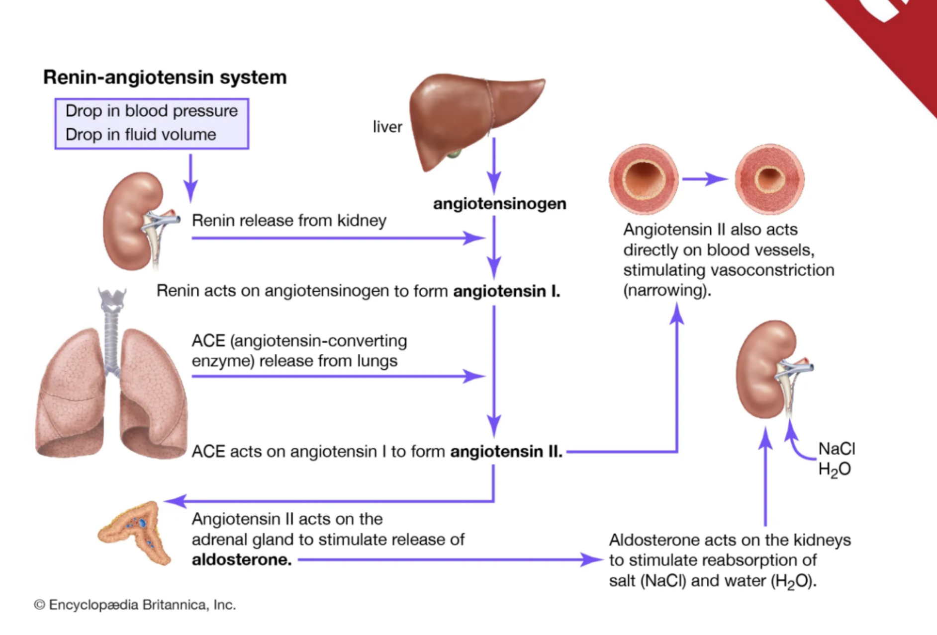
What is heart failure?
Heart failure occurs when damage to the heart muscle or valves impairs the heart's ability to function as an effective pump.
The heart functions as what?
A pump.
“squeeze” “forward flow”
goal= pump blood
damage = decreased forward flow efficiency
What can cause heart failure?
Damage to the heart muscle or valves.
What valve diseases are associated with heart failure?
Mitral and/or tricuspid valve disease.
What cardiomyopathies are associated with heart failure?
Hypertrophic cardiomyopathy and dilated cardiomyopathy.
How can the heart compensate for increased workload?
The heart can hypertrophy or dilate. (cardiac remodeling)
Hypertrophic means what?
Stiff and thick.
Dilated means what?
Thin and stretched. and weak
risk factors for HF
CAD: feeds heart, plaque buildup in arteries = increase workload
HTN: systemic vascular resistance = afterload, force backs up to heart = HF
DM
metabolic syndrome: big abdominal girth, BMI, and triglycerides
obesity
smoking: vasoconstriction
high sodium dietary intake: not alone it doesnt hurt you
chemo: vasotoxic
illicit drugs
cardiac output
The amount of blood ejected from the left ventricle each minute.
What is the formula for cardiac output? CO = Heart Rate × Stroke Volume. (SV: blood ejected w/ each contraction)
What is stroke volume? The amount of blood ejected with each contraction.
What three factors affect stroke volume? Preload, afterload, and contractility.
preload: meds that effect: diuretics (furosemide) blocks sodium reuptake, synonymous with fluid volume
afterload: systemic vascular resistance
contractility: the force, how well the pump actually pumps
what things decrease the effects of CO
HF
renal disease (increase fluid volume)
dehydration
hypovolemia
fluid retention equation
1L of fluid = 1 kg = 2.2 lbs
daily weights
call dr is gain 2-3 Ibs overnight or 5 Ibs in a weeks
Left vs right side HF
start showing s/s at 6 L thats why daily weights are important
L = lungs: pulmonary congestion, crackles (bases=little, higher=more), blood tinged sputum, confusion, orthopnea, exertional dyspnea, fatigue
pulmonary edema: increase workload on capillary vessels, coughing blood tinges pink frothy sputum
R = site (what you see): ascites (fluid in belly), enlarged liver and spleen, distended jugular veins, dependent edema (sacral, legs, feeling full)
babies: poor feeding, irritability
NY heart association functionality classification scale
Class I? No symptoms even with activity.
Class II? Symptoms are present with ordinary exertion, such as ADLs or grocery shopping.
Class III? Symptoms are present with minimal exertion.
Class IV? Symptoms are present at rest
What happens when cardiac output decreases?
The body activates compensatory mechanisms.
What nervous system is activated as compensation for decreased cardiac output? The sympathetic nervous system.
What hormonal system is activated as compensation for decreased cardiac output? The renin-angiotensin-aldosterone system (RAAS).
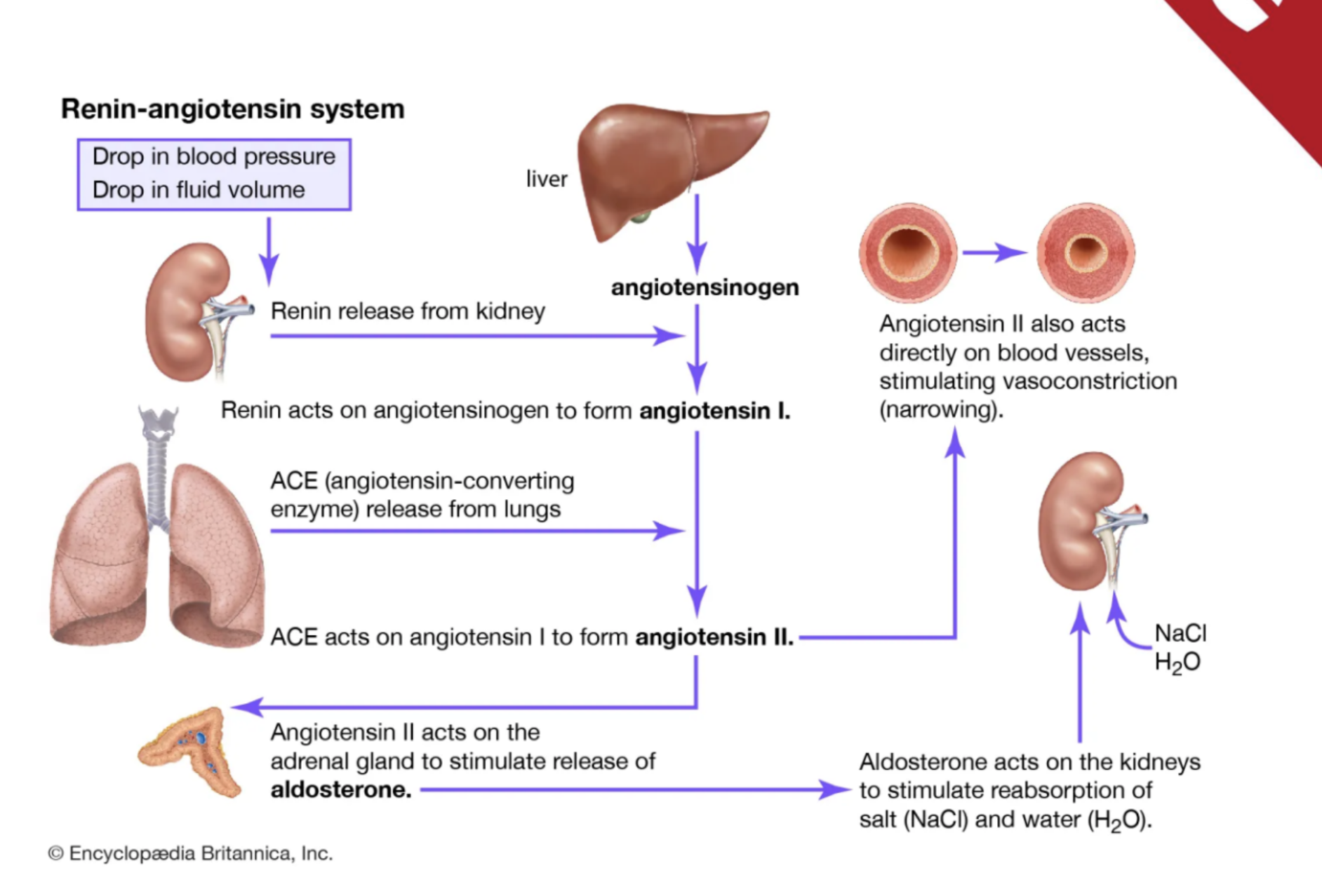
What BNP level is considered high in these notes?
Greater than 400 pg/mL.
hormone released in response to cardiac muscle stretching
greater the number = greater the HF exacerbation
What does a transthoracic echocardiogram (TTE) assess?
Functionality or problems with the heart's blood flow, chamber walls, and valves.
non invasive
What is another name for a transthoracic echocardiogram? An echo.
TEE: transesophageal echo: echo that is invasive and looks at backside of heart
What is ejection fraction (EF)?
The total volume percentage the heart ejects with each beat.
What is a typical healthy ejection fraction? 55% to 70%.
What is an important education topic for clients with heart failure?
Daily weights and intake and output (I&O) (2-3 Ibs a night or 5Ibs a week → call doctor, med adherance, sodium (1.5 g restriction)
Why are daily weights important for clients with heart failure? They are related to fluid balance and accurate intake and output. if intake is higher than output than you are retaining fluid
What should be considered when teaching clients with heart failure about food? The nutrition content of foods, especially factors relevant to heart failure management such as sodium intake.
ECG Interpretation
Overview
An ECG (electrocardiogram) records the electrical activity of the heart.
ECG interpretation can help identify:
Dysrhythmias
Myocardial infarction (MI)
Cardiomyopathy
Conduction abnormalities
Other cardiac conditions
ECGs may be interpreted by specially trained:
Nurses
Physicians
Monitor technicians
Many clinicians interpreting ECGs also have ACLS training.
If an abnormal rhythm is identified on 3- or 5-lead telemetry, a 12-lead ECG should be obtained for more accurate diagnosis.
An 18-lead ECG may be used to identify abnormalities not seen on standard ECGs, such as right-sided heart abnormalities.
Safety Considerations
Determine whether the client has any allergies.
Helps prevent allergic reactions.
Verify the client's identity according to facility protocol.
Ensures the correct procedure is performed on the correct client.
Use standard and infection-control precautions.
Helps prevent transmission of infectious organisms.
Additional precautions may be needed based on client history and facility policy.
Equipment/Supplies
Rhythm strip at least 6 seconds long
Calipers
Ink pen
Calculator
General Steps Before ECG Interpretation 1. Review the Client's Medical Record
Assess for a history of:
Chest pain or pressure
Palpitations
Dysrhythmias
Conduction abnormalities
Electrolyte abnormalities
Previous cardiac surgery
Previous cardiac catheterization
Coronary artery disease
Heart failure
Syncope/fainting
Previous cardiac arrest
Recent changes in heart rhythm or rate
Reviewing the client's history helps identify:
Why the ECG is being performed
Which abnormalities may be expected
2. Understand ECG Graph Paper
ECG paper measures time horizontally and amplitude vertically.
1 small box = 0.04 seconds
1 large box = 0.20 seconds
1 large box = 5 small boxes
5 large boxes = 1 second
25 small boxes = 1 second
3. Know Normal ECG Components
Review normal:
P waves
QRS complexes
T waves
PR intervals
QT intervals
Knowing expected characteristics helps identify abnormalities.
PQRST Complex
P wave
Represents atrial depolarization.
Atrial repolarization
Usually hidden within the QRS complex.
QRS complex
Represents ventricular depolarization.
T wave
Represents ventricular repolarization.
ECG Interpretation Steps Step 1: Gather Supplies
Bring all necessary equipment to the clinical area.
Ensure the nurse is prepared before beginning interpretation.
Step 2: Obtain a Rhythm Strip
Obtain a 6- to 12-second telemetry strip, according to facility policy.
A standard time period allows calculation of:
Heart rate
Rhythm regularity
Step 3: Calculate the Heart Rate
For the 6-second method:
Count the number of QRS complexes in 6 seconds.
Multiply by 10.
Normal adult heart rate: 60-100 beats/minute
Use calipers for accurate ECG measurements.
Purpose
Identify:
Bradycardia: HR < 60 bpm
Tachycardia: HR > 100 bpm
Step 4: Measure the PR Interval
Measure from the beginning of the P wave to the beginning of the QRS complex.
Normal PR interval: 0.12-0.20 seconds
Equivalent to 120-200 milliseconds.
Abnormal PR Intervals
Prolonged: > 0.20 seconds
May indicate first-degree AV block.
May occur with second-degree type I AV block.
Shortened: < 0.12 seconds
May indicate a junctional dysrhythmia.
PR interval cannot be accurately measured if:
No P waves are present.
The PR interval varies between beats.
Step 5: Measure the QRS Complex
Measure the width of the QRS complex.
Normal QRS duration: < 0.12 seconds.
A wide or abnormal QRS may indicate:
Bundle branch block (BBB)
Ventricular origin of the beat
Step 6: Measure the QT Interval
Measure from the beginning of the QRS complex to the end of the T wave.
Normal QT interval: approximately 0.36-0.44 seconds.
QT interval varies with:
Heart rate
Age
Gender
QT interval:
Increases with slower heart rates
Decreases with faster heart rates
With a normal heart rate, the QT interval generally should not be more than half of the R-R interval.
Clinical Significance of a Prolonged QT
May result from:
Medication side effects
Electrolyte abnormalities
Other cardiac abnormalities
Increases the risk for potentially fatal dysrhythmias.
Expected Normal ECG Findings
Heart rate: 60-100 bpm
A P wave precedes each QRS complex
PR interval: 0.12-0.20 seconds
QRS complex: < 0.12 seconds
QT interval: generally 0.36-0.44 seconds, but varies with heart rate
Step 8: Determine Rhythm Regularity
Compare the R-R intervals using calipers.
Regular rhythm: R-R intervals are consistent.
Irregular rhythm: R-R intervals vary.
Regular with occasional irregular beats: rhythm is mostly regular with occasional variations.
Step 9: Methods for Calculating Ventricular Rate 6-Second Method
Count QRS complexes in 6 seconds.
Multiply by 10.
300 Method
Count the number of large boxes between R waves.
Divide that number into 300.
Use only when the rhythm is regular.
1500 Method
Count the number of small boxes between R waves.
Divide that number into 1500.
Small-Box Time Method
Count small boxes in one R-R interval.
Multiply by 0.04 seconds.
Use the calculated interval to determine the heart rate.
Step 10: Evaluate the P Waves
If no P waves are present, the rhythm is not sinus.
For a sinus rhythm, P waves should:
Be present
Occur before each QRS complex
Have a similar shape
Typically be upright in lead II
Step 11: Evaluate the PR Interval
Determine whether PR intervals are:
Constant
Prolonged
Shortened
Variable
Measure using the small boxes between the beginning of the P wave and beginning of the QRS complex.
Multiply the number of small boxes by 0.04 seconds.
Document the PR interval on the rhythm strip when measurable.
Common Supraventricular/Sinus Rhythms Sinus Bradycardia
Rhythm: Regular
Rate: < 60 bpm
PR interval: Normal
QRS: Normal
Sinus Tachycardia
Rhythm: Regular
Rate: > 100 bpm
PR interval: Normal
QRS: Normal
Sinus Arrhythmia
Rhythm: Irregular
Rate: Usually normal
Often varies with respiration
QRS: Normal
Sinus Arrest
Rhythm: Irregular
P waves: Normal when present
PR interval: Normal
QRS: Normal
May have occasional missed beats.
Sick Sinus Syndrome
Rhythm: Irregular
Rate: May alternate between fast and slow
PR interval: Variable
QRS: Usually normal
Premature Atrial Contractions (PACs)
Rhythm: Irregular
Rate: Usually normal
PR interval: Usually normal
QRS: Normal
P wave:
Occurs early
May have an abnormal appearance
Paroxysmal Atrial Tachycardia
Rhythm: Regular
Rate: 150-250 bpm
PR interval: May be abnormal
QRS: Usually normal
P and T waves may be abnormal.
Atrial Flutter
Rhythm: Usually regular
Atrial rate: Approximately 300 bpm
Ventricular rate: May be fast or slow
PR interval: Usually unable to measure
QRS: Usually normal
Characteristic finding:
Sawtooth waves
Clear atrial-to-ventricular conduction ratio
T waves may be difficult to distinguish
Atrial Fibrillation
Rhythm: Irregular
Rate: Variable
PR interval: Unable to measure
QRS: Usually normal
Characteristic finding:
Chaotic or quivering atrial activity
Variable ventricular response
Wolff-Parkinson-White (WPW) Syndrome
Rhythm: Usually regular
Rate: May be normal
PR interval: Short, < 0.10 seconds
QRS: Wide, > 0.10 seconds
Associated with increased risk for serious dysrhythmias, including torsades de pointes.
Step 12: Evaluate the QRS Complex
Determine whether QRS complexes are:
Normal in shape
Abnormal in shape
Narrow
Wide
Normal width: < 0.12 seconds
A wide/abnormal QRS can suggest:
Bundle branch block
Ventricular-origin beat
Check for associated P waves to help determine the origin of the rhythm.
Ventricular Rhythms Premature Ventricular Contractions (PVCs)
Rhythm: Irregular
Rate: Normal or fast
PR interval:
Usually normal for normal beats
Not applicable to the PVC itself
QRS:
Wide and abnormal during the ventricular beat
PVC patterns include:
Bigeminy: every second beat is a PVC
Trigeminy: every third beat is a PVC
Idioventricular Rhythm (Agonal Rhythm)
Rhythm: Regular
Ventricular rate: < 40 bpm
May be accelerated
P waves: Usually absent
QRS: Wide and abnormal
Occurs when no other functional conduction system is controlling the heart.
Ventricular Tachycardia (VT)
Rhythm: Regular or irregular
Rate: > 100 bpm
P waves: Usually absent
PR interval: Not measurable
QRS: Wide and abnormal
Other findings:
Beats originate in the ventricles
T waves may be inverted
Torsades de Pointes
Rhythm: Irregular
Rate: 150-250 bpm
P waves: Absent
QRS:
Wide
Variable morphology
Characteristic appearance:
QRS complexes appear to twist or rotate around the baseline
Ventricular Fibrillation (VF)
Rhythm: No organized rhythm
Rate: None
P waves: Absent
QRS: Absent
Characterized by chaotic electrical activity and quivering of the ventricles.
Asystole
Rhythm: No rhythm
Rate: None
P waves: Absent
QRS: Absent
ECG appears nearly as a flat line with no significant electrical activity.
Pulseless Electrical Activity (PEA)
Rhythm: May appear regular or irregular
ECG may show normal electrical activity.
No effective mechanical contractions or pulse are present despite electrical activity.
Step 13: Evaluate T Waves
Determine whether T waves are:
Present
Consistent in appearance
Round and smooth
Similar in amplitude
Compare T-wave direction to the QRS complex.
T waves are typically upright in leads II and III.
Abnormal T waves may indicate changes affecting ventricular repolarization.
Step 14: Evaluate the QT Interval
Measure from the beginning of the QRS/Q wave to the end of the T wave.
Count the number of small boxes.
Multiply by 0.04 seconds.
Determine whether the QT interval is:
Normal
Prolonged/wide
Document the QT interval on the ECG strip.
Step 15: Interpret the Rhythm
Interpret the ECG based on:
Rate
Rhythm regularity
Presence and appearance of P waves
PR interval
QRS width and morphology
T-wave characteristics
QT interval
Use these findings to identify possible:
Electrical abnormalities
Conduction problems
Structural cardiac abnormalities
Step 16: Ensure Client Safety
Before leaving the client:
Place the call light within reach.
Place personal items within easy reach.
Lower the bed to the lowest position.
Ensure the bed brakes are locked.
Address any individual client safety needs.
Client Considerations
Always consider the client's cardiac history when interpreting an ECG.
ECG interpretation and communication may be urgent, especially when the client:
Is experiencing chest pain
Is being evaluated for cardiac disease
Has a new abnormal rhythm
Is in a code situation
Complete interpretation promptly and communicate urgent findings immediately.
Documentation
Document completion of ECG interpretation in the client's chart.
Notify the provider ASAP of abnormal results.
Communicate critical findings immediately.
Educate the client and family about results within the RN's scope of practice.
Quick Normal ECG Values to Memorize
Heart rate: 60-100 bpm
PR interval: 0.12-0.20 seconds
QRS duration: < 0.12 seconds
QT interval: approximately 0.36-0.44 seconds
Small box: 0.04 seconds
Large box: 0.20 seconds
5 large boxes: 1 second
25 small boxes: 1 second
lead placement and heart rythms
Overview
ECG/EKG = noninvasive, painless test that records the heart’s electrical activity.
Electrodes are attached to the client’s skin and connected to an ECG machine.
Procedure typically takes 5–10 minutes.
ECG tracing can be used to:
Evaluate heart rhythm.
Evaluate pacemaker effectiveness.
Identify signs of structural abnormalities.
Identify ischemia.
18-lead ECG provides additional views not obtained with a 12- or 15-lead ECG.
Especially useful for diagnosing:
Right-sided myocardial infarction (MI).
Posterior MI.
Delegation
The nurse may delegate ECG performance to assistive personnel (AP) if:
The client has already been assessed by a licensed nurse or provider.
The client’s condition is stable.
The AP has been trained and competent in the skill.
AP should report all findings to the nurse.
Do NOT delegate if:
Client’s condition is unknown.
Client is unstable.
AP has not been trained in the skill.
Safety Considerations Check for Adhesive Allergies
Determine whether the client has an allergy or sensitivity to adhesives.
Helps prevent an allergic reaction from ECG electrodes.
Verify Client Identity
Use facility protocol to ensure the correct procedure is performed on the correct client.
Infection Control
Use standard precautions.
Perform hand hygiene.
Use gloves/PPE as indicated.
Follow additional infection-control precautions based on:
Client history.
Isolation requirements.
Facility policy.
Equipment and Supplies
ECG machine — records the heart’s electrical activity.
Alcohol swabs — cleanse skin and improve electrode adhesion.
Clippers/scissors — remove excess hair if necessary.
Use an electronic clipper, not a razor.
ECG electrodes — adhere to the skin and transmit electrical signals.
Adhesive remover wipes/disposable wet wipes — remove adhesive residue.
Nonsterile gloves and other PPE — infection prevention.
Approved disinfectant wipes — clean ECG machine after use.
General Steps Before the Procedure 1. Review the Client’s Medical Record and MAR
Review:
Allergies.
Medical history.
Age and gender.
Medications.
Previous vital signs.
Pertinent laboratory values.
Provider’s prescription/order.
Previous ECG results.
Facility policy and procedure manual.
Important considerations:
Identify contraindications or adhesive allergies.
Consider anatomical differences, such as an amputated limb.
Verify the provider’s order and purpose of the ECG.
Review facility policy before performing an unfamiliar skill.
2. Check the ECG Machine
Ensure the machine is operational before entering the client’s room.
Check for malfunction before beginning the procedure.
3. Gather Supplies
Bring all necessary equipment to the bedside.
Being prepared helps prevent interruptions during the procedure.
Step-by-Step Procedure 1. Provide Privacy
Close the door or curtain.
Protect the client’s:
Confidentiality.
Physical privacy.
Dignity.
2. Introduce Yourself
Identify yourself to the client.
Helps establish a therapeutic nurse-client relationship.
3. Perform Hand Hygiene and Apply PPE
Perform hand hygiene before client contact.
Apply nonsterile gloves/PPE as indicated.
4. Identify the Client
Use two unique identifiers according to facility policy.
5. Confirm Allergies
Double-check allergy status, especially adhesive allergies.
6. Educate the Client
Explain:
What will happen.
Why the ECG is being performed.
Allow the client to ask questions.
Verify understanding.
Education can help decrease anxiety.
7. Position the Client
Position comfortably, preferably at approximately 45 degrees.
Raise the bed/stretcher to a comfortable working height.
Keep the ECG machine within reach.
8. Ensure Proper Grounding
Verify the third grounding prong is intact.
Ensure the machine is properly plugged in.
Helps prevent:
Electrical malfunction.
Electrical shock.
9. Enter Client Information
Turn on the ECG machine.
Enter required client and nurse information according to:
Facility policy.
Machine instructions.
Ensures correct identification and proper transmission of results.
10. Expose Necessary Areas
Assist the client with adjusting clothing to expose:
Chest.
Arms.
Lower limbs.
Keep the pelvic area and thighs covered.
Ensure the room is warm enough.
Shivering can cause artifact on the ECG.
11. Prepare the Skin
Clean electrode sites gently with alcohol wipes.
Remove excess hair with an electronic clipper if needed.
Promotes good electrode adhesion and accurate electrical conduction.
ECG Electrode Placement Limb Leads
RA (Right Arm): Right shoulder or arm.
LA (Left Arm): Left shoulder or arm.
RL (Right Leg): Right leg.
LL (Left Leg): Left leg.
Chest/Precordial Leads
V1: 4th intercostal space, right sternal border.
V2: 4th intercostal space, left sternal border.
V3: Midway between V2 and V4.
V4: 5th intercostal space, left midclavicular line.
V5: 5th intercostal space, between V4 and V6.
V6: 5th intercostal space, just left of the spine.
Special Considerations
Follow facility policy for variations in placement.
Some facilities use the wrist for arm leads.
If the client has an amputation:
Place the electrode on the remaining limb or stump.
Key point: Correct electrode placement is essential for an accurate ECG reading.
Recording the ECG 13. Connect the Cables
If not already connected:
Connect ECG cables to the machine.
Attach each cable to the correct electrode.
Allows electrical signals to travel to the ECG machine.
14. Instruct the Client to Remain Still
Tell the client to:
Relax shoulders.
Relax legs.
Remain still.
Muscle movement and tension can cause artifact.
15. Activate and Record the ECG
Verify:
Machine is turned on.
Correct client information is entered.
Follow machine directions to record the ECG.
Print a copy and/or transfer results to the electronic medical record (EMR) according to facility policy.
16. Review the Tracing Before Removing Electrodes
Check for:
Artifact.
Poor-quality tracing.
An adequate ECG recording.
Repeat the ECG if necessary before disconnecting the client.
After the ECG 17. Remove Electrodes
Disconnect cords from electrodes.
Remove electrodes gently.
Use adhesive remover if needed.
Remove any remaining adhesive residue.
Purpose: Prevent skin irritation or skin breakdown.
18. Clean Equipment
Disinfect the ECG machine with a facility-approved disinfectant.
Dispose of:
Used electrodes.
Used wipes.
Store the machine according to facility policy.
19. Provide Post-Procedure Education
Teach the client/family:
What the next steps are after the ECG.
An ECG does not measure symptoms such as:
Chest pain.
Dyspnea/shortness of breath.
The client should continue reporting symptoms to the healthcare provider.
Outpatients should continue current treatment recommendations until the provider communicates the ECG results.
20. Ensure Client Safety
Before leaving:
Position client safely.
Address individual safety needs.
Reduce risk of:
Falls.
Injury.
21. Remove PPE and Perform Hand Hygiene
Follow facility infection-control policy.
22. Notify the Provider
Notify the ordering provider that the ECG is complete.
Report preliminary results provided by the ECG machine as appropriate.
STAT report abnormal results to ensure prompt treatment.
Unexpected Outcomes
Notify the provider of any unexpected outcomes or abnormal findings.
Prompt communication helps ensure timely treatment.
Documentation
Document accurately in the client’s MAR and medical record according to facility policy.
Include as indicated:
Date and time ECG was completed.
Type of ECG performed.
Example: 18-lead instead of 12- or 15-lead.
Client’s response to the procedure.
Any unexpected outcomes.
Education provided to the client and family.
Provider notification that the ECG was completed.
Provider notification of unexpected/abnormal outcomes.
ECG Interpretation & Non-Ventricular Dysrhythmia Notes Electrocardiogram (ECG) Overview
ECG: Noninvasive test that records the heart's electrical activity.
ECG helps identify:
Abnormal electrical conduction
Areas of decreased or absent cardiac perfusion
Dysrhythmias
Myocardial ischemia/injury
ECG does not visualize actual cardiac structures.
Cardiac Electrical Activity
Depolarization: Electrical stimulation that causes cardiac muscle contraction.
Repolarization: Electrical recovery/relaxation as the heart prepares for the next impulse.
Steps for ECG Analysis
Calculate heart rate
Determine if rhythm is regular or irregular
Assess P waves
Measure PR interval
Measure QRS duration
Assess ST segment
Observe T waves
Measure QT interval
Interpret the rhythm
ECG Waveform Components P Wave
Represents atrial depolarization.
Normally:
One P wave before each QRS complex
P waves should have a consistent shape
PR Interval
Measured from the beginning of the P wave to the beginning of the QRS complex.
Represents conduction of the impulse through the atria and AV node.
Normal: 0.12–0.20 seconds
Should be consistent.
QRS Complex
Represents ventricular depolarization.
Normal duration: approximately 0.06–0.10 seconds.
A QRS duration >0.12 seconds may indicate abnormal ventricular conduction.
ST Segment
Located between the QRS complex and T wave.
Should remain near the isoelectric baseline.
Elevation or depression may indicate:
Altered cardiac perfusion/ischemia
Electrolyte abnormalities
T Wave
Represents ventricular repolarization.
Peaked T waves: May indicate hyperkalemia.
Inverted T waves: May indicate:
Ischemia
Altered cardiac perfusion
Pulmonary embolism
QT Interval
Measured from the beginning of the QRS complex to the end of the T wave.
Represents the total time for ventricular depolarization and repolarization.
Prolonged QT increases the risk for dangerous dysrhythmias.
ECG Grid Paper & Heart Rate Calculation ECG Paper Measurements
1 small box = 0.04 seconds
1 large box = 0.20 seconds
5 small boxes = 1 large box
A typical 6-second ECG strip = 30 large boxes
300 Method
Used for regular rhythms.
Count the number of large boxes between two R waves.
300 ÷ number of large boxes = heart rate
6-Second Method
Count the number of QRS complexes in 6 seconds.
Multiply by 10.
Useful for obtaining an average heart rate.
Determining Regularity
R-to-R intervals: Determine ventricular rhythm.
P-to-P intervals: Determine atrial rhythm.
Equal spacing = regular.
Unequal spacing = irregular.
Expected Normal ECG Criteria
Heart rate: 60–100/min
Rhythm: Regular
P waves: One P wave for every QRS
PR interval: 0.12–0.20 sec
QRS: 0.06–0.10 sec
ST segment: No significant elevation or depression
QT interval:
Prepuberty: <0.45 sec
Postpubertal males: <0.46 sec
Postpubertal females: <0.47 sec
Normal Sinus Rhythm (NSR) Pathophysiology
Electrical impulse follows the normal conduction pathway:
SA node
AV node
Bundle of His
Right and left bundle branches
Purkinje fibers
Impulse originates in the SA node and travels normally through the heart.
ECG Criteria
Rate: 60–100/min
Rhythm: Regular
P waves: One consistently shaped P wave before every QRS
PR interval: 0.12–0.20 sec
QRS: 0.06–0.10 sec
Sinus Bradycardia Definition
SA node fires at a rate <60 bpm.
All other ECG characteristics are generally the same as NSR.
ECG Criteria
Rate: <60/min
Rhythm: Regular
P waves: One consistent P wave before every QRS
PR interval: 0.12–0.20 sec
QRS: 0.06–0.10 sec
Causes/Risk Factors
Well-conditioned athletes
Normal aging
Myocardial infarction
Sleep apnea
Increased intracranial pressure
Hypothyroidism
Eating disorders/decreased metabolic needs
Hypoxia
Increased vagal stimulation:
Coughing
Gagging
Straining during bowel movements
Certain infections
Medications
Medications That Can Cause Bradycardia
Parasympathomimetics
Beta blockers: Metoprolol
Digoxin
Calcium channel blockers: Diltiazem
Amiodarone
Lithium
Clinical Manifestations
May be asymptomatic
If symptomatic:
Dizziness
Lightheadedness
Syncope/near syncope
Chest pain
Altered mental status
Dyspnea
Diaphoresis
Exercise intolerance/fatigue
Diagnostics
ECG
Electrolytes, including magnesium and calcium
Thyroid hormone testing
Troponin
Drug/toxicology screening if indicated
Nursing Priorities
First priority: Determine if the client is stable or unstable.
Assess for decreased cardiac output.
Monitor:
Heart rate
Blood pressure
ECG
Mental status
Chest pain
Oxygenation
Implement fall precautions if symptomatic.
Encourage slow position changes.
Review and potentially hold medications that contribute to bradycardia as directed.
Treatment
Asymptomatic: Usually observation and treatment of the underlying cause.
Unstable/symptomatic:
Atropine 1 mg IV
May repeat every 3–5 minutes
Maximum total dose: 3 mg
If atropine is ineffective:
Prepare for transcutaneous pacing
A permanent pacemaker may be needed for persistent underlying conduction problems.
NCLEX Key Point
Symptomatic bradycardia = assess perfusion and hemodynamic stability first.
Sinus Tachycardia Definition
SA node fires at a rate >100 bpm.
Usually a response to an underlying physiological or psychological stressor.
Prolonged tachycardia can decrease cardiac output by reducing ventricular filling time.
ECG Criteria
Rate: >100 to <150 bpm
Rhythm: Regular
P waves: One consistent P wave before every QRS
PR interval: 0.12–0.20 sec
QRS: 0.06–0.10 sec
Causes/Risk Factors
Fever
Pain
Anxiety/stress
Hypovolemia/fluid volume loss
Fluid volume excess
MI
Shock
Hyperthyroidism
Anemia
Physical exertion
Hypoxia
Sepsis
Medications:
Atropine
Catecholamines
Theophylline
Stimulants:
Cocaine
Amphetamines
Caffeine
Nicotine
Clinical Manifestations
May be asymptomatic
If symptomatic:
Palpitations
Chest pain
Dyspnea
Dizziness/lightheadedness
Elevated temperature
Signs of Instability
Hypotension
Increased respiratory rate
Shortness of breath
Chest pain
Altered level of consciousness
Decreased urine output
Signs of dehydration
Nursing Priorities
Identify and treat the underlying cause.
Assess for hemodynamic instability.
Monitor:
ECG
Vital signs
Perfusion
Urine output
Mental status
Implement fall precautions if dizziness is present.
Treatment
Treat the underlying cause:
Fever → reduce temperature
Pain → manage pain
Hypovolemia → replace fluids as prescribed
Anxiety/stress → reduce stressor
Depending on the clinical situation, treatment may include:
Medications such as beta blockers
Adenosine in specific tachyarrhythmia evaluation/treatment situations
Catheter ablation for persistent cases
Client Education
Reduce/avoid stimulants that increase heart rate.
Reduce caffeine and nicotine.
Avoid illicit drugs.
Use stress-management techniques:
Controlled breathing
Meditation
Relaxation
NCLEX Key Point
Sinus tachycardia is usually a symptom of another problem—find and treat the cause.
Premature Ventricular Contractions (PVCs) Definition/Pathophysiology
Early ventricular beat caused by an irritable ventricular focus.
Occurs before the next expected normal SA node impulse.
PVC QRS complexes are typically wide and abnormal.
ECG Findings
Underlying rhythm may be normal but slightly irregular.
Premature beat occurs early.
Wide QRS >0.12 sec
P wave associated with the PVC may be absent or hidden.
PR interval may be unmeasurable.
PVC Patterns
Unifocal: PVCs have the same shape → originate from one ventricular site.
Multifocal: PVCs have different shapes → originate from multiple sites.
Bigeminy: Every other beat is a PVC.
Trigeminy: Every third beat is a PVC.
Quadrigeminy: Every fourth beat is a PVC.
Causes/Risk Factors
May occur in healthy individuals and be benign.
MI/cardiac muscle damage
Hypertension
Cardiomyopathy
Chronic lung disease/COPD
Pulmonary hypertension
Sleep apnea
Thyroid disorders
Hypokalemia
Hypomagnesemia
Caffeine sensitivity
Nicotine
Alcohol
Illicit drugs
Clinical Manifestations
Often asymptomatic
Palpitations
Feeling like the heart “skipped a beat”
Lightheadedness
Chest pain
Shortness of breath
Diagnostics
ECG
Electrolytes:
Potassium
Magnesium
Thyroid testing
Holter/ambulatory monitor: Often worn 24–48 hours
Echocardiogram to assess structural heart disease
Nursing Priorities
Assess:
Frequency and pattern of PVCs
Peripheral pulses
Symptoms
Cardiac output/perfusion
Monitor ECG and vital signs.
Identify and correct triggers.
Correct electrolyte imbalances as prescribed.
Implement fall precautions if dizzy or lightheaded.
Treatment
Asymptomatic/benign PVCs:
Often require no specific treatment
Manage triggers
Frequent/persistent symptomatic PVCs:
Beta blockers: Metoprolol, carvedilol
Antiarrhythmics may be prescribed
Persistent PVCs may be treated with catheter ablation.
NCLEX Key Point
Frequent PVCs + low potassium or magnesium → prioritize correcting the electrolyte imbalance and monitoring for worsening dysrhythmias.
Premature Atrial Contractions (PACs) Definition/Pathophysiology
Early beat originating from an irritable atrial focus.
An atrial impulse fires before the next expected SA node impulse.
Usually considered benign.
ECG Findings
Underlying rhythm is usually normal.
Premature/early P wave may be:
Hidden
Difficult to identify
Different in shape from normal P waves
PR interval may be:
Shortened (<0.12 sec)
Unmeasurable
Causes/Risk Factors
Often unknown
MI
Hypertension
Diabetes mellitus
Heart failure
Certain medications:
Digoxin
Beta blockers
Chemotherapy medications
Some antidepressants
Stress/anxiety
Fatigue with excessive exercise
Smoking
Alcohol use
Clinical Manifestations
Often asymptomatic
Palpitations
Fluttering sensation in chest
Shortness of breath with activity
Diagnostics
ECG
Echocardiogram if cardiac abnormalities are suspected
Electrolyte testing may be helpful
Nursing Priorities
Identify triggers.
Obtain history of:
Chronic conditions
Medications
Stress
Smoking
Alcohol use
Assess pulse and heart sounds.
Reassure the client that PACs are usually benign.
Treatment
Usually no treatment for infrequent PACs.
Reduce or eliminate triggering factors.
Frequent PACs may be treated with low-dose beta blockers.
Client Education
Reduce stress.
Avoid smoking and alcohol.
Monitor exercise-related fatigue.
Contact the provider if palpitations or dyspnea interfere with ADLs.
NCLEX Key Point
PAC = premature beat from the ATRIA; usually benign and treated by identifying/reducing triggers.
First-Degree AV (Heart) Block Definition/Pathophysiology
Delay in conduction through the AV node.
The impulse is slowed but not blocked.
Every atrial impulse is eventually conducted to the ventricles.
Key ECG Finding
PR interval >0.20 seconds
PR interval is consistently prolonged.
Other conduction is generally normal.
Easy Memory Tip
First-degree block = “First, the PR is long.”
Causes/Risk Factors
Increased age
MI/coronary artery disease
Hypokalemia
Hypomagnesemia
Increased vagal tone
Certain infections
Autoimmune disorders
Athletes with high vagal tone
Medications that slow AV conduction
Medications That Can Prolong AV Conduction
Sodium channel blockers
Beta blockers
Calcium channel blockers
Amiodarone
Digoxin
Magnesium
Clinical Manifestations
Usually asymptomatic
Possible:
Dizziness
Shortness of breath with activity
Fatigue
Diagnostics
ECG:
PR interval >0.20 sec
PR interval remains consistent
Electrolyte testing may help identify the cause.
Drug/toxicology testing may be indicated.
Nursing Priorities
Review medication list.
Monitor ECG for:
Worsening PR prolongation
Progression to higher-degree heart block
Assess for decreased cardiac output.
Hold medications that prolong the PR interval if indicated by the provider/clinical orders.
Implement fall precautions if symptomatic.
Treatment
Asymptomatic: Usually observation and follow-up.
Treat the underlying cause.
If significant bradycardia or progression occurs, treatment may include:
Atropine
Isoproterenol
Pacemaker, if necessary
NCLEX Key Point
First-degree AV block = PR interval >0.20 sec and consistent; usually asymptomatic.
Quick Comparison Chart
Rhythm | Rate | Rhythm | Main ECG Clue |
|---|---|---|---|
Normal Sinus Rhythm | 60–100 | Regular | Normal P-QRS relationship |
Sinus Bradycardia | <60 | Regular | Normal rhythm, slow rate |
Sinus Tachycardia | >100 to <150 | Regular | Normal rhythm, fast rate |
PVC | Variable | May be irregular | Early, wide QRS >0.12 sec |
PAC | Usually normal | May be irregular | Early/abnormal P wave |
1st-Degree AV Block | Usually normal | Regular | PR >0.20 sec, consistent |
High-Yield Memory Tricks
P wave = Atria depolarize
QRS = Ventricles depolarize
T wave = Ventricles repolarize
PR >0.20 = First-degree AV block
Wide premature QRS = PVC
Early abnormal P wave = PAC
Slow but otherwise normal = Sinus bradycardia
Fast but otherwise normal = Sinus tachycardia
Sinus rhythms originate in the SA node
Atrial Fibrillation (A-fib) Pathophysiology
A-fib is a common cardiac dysrhythmia.
Occurs when the SA node is not firing appropriately.
Multiple areas in the atria send electrical impulses.
Electrical activity becomes:
Rapid
Chaotic
Irregular
The atria cannot effectively contract.
Blood is not effectively moved from the atria into the ventricles.
Results in:
Decreased cardiac output
Ineffective cardiac contractions
Irregular blood flow
Blood can pool and form blood clots.
Embolized clots can cause damage to:
Brain → stroke
Lungs
Kidneys
Other organs
Etiology & Risk Factors
Diabetes
Hyperthyroidism
Obstructive sleep apnea
Smoking
Excessive alcohol intake
Sedentary lifestyle/minimal exercise
Comorbidities associated with A-fib
Recovery from cardiac surgery
Common Comorbidities
Hypertension
Heart failure
Diabetes mellitus
Overweight/obesity
COPD
Thyroid disease
Kidney disease
History of stroke
Major Complications
Stroke
Heart failure
Increased morbidity and mortality
Epidemiology
Most common type of treated cardiac dysrhythmia.
More common in:
Adults older than 65 years
Individuals who are overweight or obese
Causes a significant number of hospitalizations.
Impact on Daily Life
May decrease ability to perform usual activities.
Possible problems include:
Dizziness
Palpitations
Exercise intolerance
Fatigue
Can decrease overall quality of life.
Safety Considerations
Increased risk for:
Stroke
Systemic emboli
Heart failure
Falls due to dizziness/lightheadedness
Long-term anticoagulant therapy increases the risk for:
Spontaneous bleeding
Excessive bleeding after injury
Clinical Presentation A-fib May Be Asymptomatic
May only be identified by an irregular pulse.
Symptomatic Findings
Irregular apical pulse
Increased heart rate
Hypotension
Palpitations
Chest discomfort
Shortness of breath:
At rest
With activity
Exertional fatigue
Anxiety
Dizziness
Lightheadedness
Syncope
Weight gain
Increased urination
Laboratory & Diagnostic Testing Blood Tests
TSH and thyroxine (T4)
Assess for hyperthyroidism.
Other possible tests:
CBC
Creatinine
Glucose
Clients taking certain oral anticoagulants, such as warfarin, may require PT/INR monitoring.
A-fib ECG Findings ⭐
Rhythm: Irregular
P waves:
Absent
Replaced by indiscernible/chaotic atrial activity
Ventricular rate may be:
60-100/min: controlled ventricular response
100-200/min: increased ventricular rate
Rapid ventricular response (RVR): ventricular rate > 100/min
Quick Memory Tip
A-fib = “Irregularly irregular” + no identifiable P waves.
Echocardiogram & TEE
Echocardiogram evaluates:
Size of the atria and ventricles
Heart valve function
Transesophageal echocardiogram (TEE):
Detects blood clots, especially in the left atrium.
May be performed before cardioversion.
Nursing Assessment: A-fib Recognize Cues
Obtain a thorough medical history.
Ask about:
Palpitations
Difficulty breathing
Dizziness/lightheadedness
Ability to complete usual ADLs
Auscultate the apical pulse for a full 60 seconds.
Obtain a manual blood pressure when indicated.
Palpate pulses for a full 60 seconds because the rhythm is irregular.
Assess for a pulse deficit.
Pulse Deficit
Compare:
Apical pulse
Radial pulse
A-fib may cause the two pulse rates to differ.
Pulse deficit = apical pulse − radial pulse
A result of 0 = no pulse deficit.
A pulse deficit greater than 0 indicates:
Inadequate cardiac output
Potentially decreased organ perfusion
Analyze Cues
Identify:
Irregular ECG rhythm
Clinical manifestations of decreased cardiac output
Assess for signs of embolized clots:
Changes in level of consciousness
Extremity pain
Stroke symptoms
Shortness of breath
Priority Nursing Goals
Improve cardiac perfusion.
Control heart rate.
Control or restore heart rhythm.
Prevent:
Blood clots
Stroke
Other embolic complications
Treatments for A-fib Major Goals
Restore and maintain normal sinus rhythm (NSR) when appropriate.
Control ventricular rate.
Prevent thromboembolic complications, especially stroke.
Medications Antiarrhythmics
Used to help restore and maintain NSR.
Monitor for:
Bradycardia
Prolonged QT interval
Rate-Control Medications
Beta blockers
Calcium channel blockers
Amiodarone
Digoxin
Effects
Slow the ventricular rate.
Decrease conduction through the AV node.
Nursing Consideration
Assess the apical heart rate before administration.
Amiodarone
Often used when A-fib is more difficult to control.
Important adverse effects:
Pulmonary fibrosis
Liver damage
Obtain baseline:
Pulmonary function tests (PFTs)
Liver enzyme testing
Anticoagulation Therapy Purpose
Prevent:
Blood clot formation
Systemic emboli
Stroke
Major Nursing Concern
Bleeding
Bleeding Precautions
Use a soft-bristled toothbrush.
Use an electric razor or safety razor.
Avoid contact sports.
Avoid high-risk activities that could cause injury.
Monitor for spontaneous or excessive bleeding.
Warfarin (Coumadin) Monitoring
Requires PT/INR monitoring.
Frequent monitoring is required initially.
Once stable, monitoring may occur less frequently.
Important Teaching
Maintain a consistent intake of vitamin K.
Do not suddenly increase or decrease vitamin K intake.
Vitamin K changes can alter warfarin's effectiveness.
High Vitamin K Foods
Brussels sprouts
Leafy greens:
Beet greens
Collard greens
Mustard greens
Turnip greens
Kale
Spinach
Moderate Vitamin K Foods
Asparagus
Broccoli
Cabbage
Carrots
Cauliflower
Celery
Green beans
Lettuce
Mixed vegetables
Okra
Peas
Pickles
Low Vitamin K Foods
Avocados
Bananas
Corn
Fruit
Garbanzo beans
Green/red peppers
Potatoes
Tomatoes
Reversal of Warfarin
Vitamin K
Fresh frozen plasma (FFP) may also be used depending on the clinical situation.
Medications/Substances That Affect Warfarin Can Increase INR → ↑ Bleeding Risk
Acetaminophen
Allopurinol
Many antibiotics
Cephalosporins
Doxycycline
Fluoroquinolones
Macrolides
Metronidazole
Penicillins
Antifungals
Some chemotherapy medications
Testosterone
Can Decrease INR → ↑ Clotting Risk
Dicloxacillin
Nafcillin
Rifampin
Some antiseizure medications:
Carbamazepine
Phenobarbital
Phenytoin
Azathioprine
Ritonavir
Sucralfate
St. John's wort
Vitamin K
Direct Oral Anticoagulants (DOACs) Examples
Apixaban (Eliquis)
Dabigatran (Pradaxa)
Rivaroxaban (Xarelto)
Advantages
Generally do not require routine PT/INR testing.
Usually do not require frequent dose adjustments.
Important Teaching
Take exactly as prescribed.
Do not miss doses.
Missed doses increase the risk for:
Systemic emboli
Stroke
Reversal Agents
Dabigatran → Idarucizumab (Praxbind)
Apixaban & rivaroxaban → Andexanet alfa
Herbal Supplements & Anticoagulants Decrease Anticoagulant Effect → ↑ Clotting Risk
Coenzyme Q-10
Ginseng
Licorice
St. John's wort
Increase Anticoagulant Effect → ↑ Bleeding Risk
Danshen
Evening primrose
Ginkgo biloba
Saw palmetto
Synchronized Electrical Cardioversion Purpose
Delivers an electrical shock to reset the heart rhythm to NSR.
Key Points
Shock is synchronized with the QRS complex/R wave.
Energy typically ranges from 50-200 joules.
If unsuccessful:
Another attempt may be made at a higher energy level.
Conscious sedation is used.
Nursing Priorities
Maintain an open airway.
Monitor:
Respiratory status
Vital signs
Cardiac rhythm
Anticoagulation may be used before and continued after cardioversion to reduce clot risk.
Catheter Ablation
May be considered if medications or cardioversion are ineffective.
An electrophysiology (EP) study is performed to:
Map the heart's electrical system
Identify abnormal conduction pathways
A catheter delivers:
Radiofrequency energy (heat) OR
Cryotherapy (cold)
Creates scar tissue that blocks abnormal electrical conduction.
A-fib Client Education Report Immediately
Palpitations
Chest pain
Irregular heart rate
Dizziness
Increased fatigue
Shortness of breath
Lifestyle Modifications
Maintain a healthy weight.
Limit alcohol.
Stop smoking.
Manage:
Sleep apnea
Blood glucose
Blood pressure
Weight
Avoid unapproved stimulants:
Excess caffeine
OTC decongestants
Illicit stimulants
Herbal supplements
❤ Atrial Flutter Pathophysiology
A supraventricular dysrhythmia.
Less common than A-fib.
Atria beat at a:
Regular
Very rapid rate
Atrial rate: approximately 240-400 bpm.
Multiple rapid electrical impulses occur in the atria.
Impulses occur too rapidly for the AV node to process normally.
Risk Factors
Recent MI
Postoperative cardiac surgery
Diabetes
Hyperthyroidism
Obstructive sleep apnea
Obesity
Alcohol use
Smoking
Cardiomyopathy
Pericarditis
Common Comorbidities
Similar to A-fib:
Hypertension
Heart failure
Diabetes
Obesity
COPD
Thyroid disease
Kidney disease
Stroke
Clinical Presentation
Similar to A-fib:
Hypotension
Lightheadedness
Dizziness
Palpitations
Chest discomfort
Shortness of breath
Fatigue
Syncope
Atrial Flutter ECG Findings ⭐
Rhythm: Usually regular
P waves:
Absent
Replaced by characteristic sawtooth/flutter waves
Ventricular rate: typically 60-100/min
Atrial rate: approximately 240-340/min
Quick Memory Tip
Atrial flutter = “Sawtooth waves.”
Diagnostic Tests
Blood tests may evaluate for:
Electrolyte abnormalities
Diabetes
Thyroid disease
Kidney disease
Liver dysfunction
Echocardiogram:
Evaluates chamber size and function.
TEE:
Detects left atrial blood clots before cardioversion.
Nursing Priorities for Atrial Flutter Assessment
Obtain medical history and determine onset of symptoms.
Monitor:
ECG
Vital signs
Oxygen saturation
Signs of decreased cardiac output
Signs the Client May Not Be Tolerating the Rhythm
Hypotension
Decreased cardiac output
Ventricular rate > 150/min
Priority Goals
Improve cardiac perfusion.
Restore normal rhythm.
Prevent:
Blood clots
Hypotension
Bradycardia
Tachycardia-related complications
Treatment of Atrial Flutter
Similar to A-fib:
Anticoagulants
Antiarrhythmics
Beta blockers
Calcium channel blockers
Amiodarone
Digoxin
Synchronized cardioversion
Catheter ablation
Client education and lifestyle modifications are similar to A-fib.
❤ Supraventricular Tachycardia (SVT) Pathophysiology
SVT is a category of narrow-complex tachycardias.
Electrical impulses originate above the ventricles/AV node.
Increased atrial excitability causes a rapid heart rate.
PSVT
Paroxysmal SVT (PSVT) is also called AV nodal reentrant tachycardia (AVNRT).
Usually:
Starts abruptly
Stops suddenly
May occur without warning
Etiology & Triggers
Stress
Smoking
Alcohol
Caffeine
Other stimulants
Heart disease
Heart failure
Wolff-Parkinson-White syndrome
Pregnancy
Chronic lung disease
Associated Conditions
Coronary artery disease
Hypertension
Diabetes
A-fib
Heart failure
Cardiomyopathy
Hyperthyroidism
Renal disease
Clinical Presentation Common Manifestations
Sudden onset of:
Palpitations
Chest discomfort
Dizziness
Lightheadedness
Shortness of breath
Possible:
Hypotension
Syncope
Diaphoresis
Fatigue
Anxiety
Safety Concern
Dizziness and syncope increase the risk for falls and injury.
SVT/PSVT Diagnostic Testing Blood Tests
No blood test confirms SVT or PSVT.
Blood work may identify underlying causes:
Electrolytes
CBC
Thyroid function
Other chronic medical conditions
SVT ECG Findings ⭐
Rhythm: Regular
Heart rate: approximately 100-220 bpm
QRS: Narrow, < 0.12 seconds
P waves:
Often difficult or impossible to identify
PSVT:
Similar narrow QRS
Heart rate often around 160 bpm
Quick Memory Tip
SVT = Sudden, Very fast, Thin (narrow) QRS.
Nursing Process: SVT/PSVT Recognize Cues
Obtain a thorough medical history.
Assess ability to complete usual activities.
Monitor:
ECG
Heart rate
Blood pressure
Peripheral pulses
Assess for:
Diaphoresis
Fatigue
Syncope
Anxiety
Hypotension
Priority
Maintain hemodynamic stability.
Convert the rhythm back to a normal rhythm when indicated.
Prevent complications from:
Hypoxia
Dyspnea
Hypotension
SVT Treatment 1. Vagal Maneuvers
May slow conduction through the AV node.
Can help terminate certain SVTs or reveal the underlying rhythm.
Examples
Valsalva maneuver
Forceful coughing
Gagging
Carotid massage performed by an appropriate provider
Valsalva Maneuver
Hold breath for approximately 15 seconds.
Bear down as if having a bowel movement.
Nursing Care
Monitor:
Vital signs
Cardiac rhythm
Assess before, during, and after the maneuver.
2. Adenosine Used When
Vagal maneuvers are ineffective.
Key Points
Has a very short duration of action.
Must be administered rapidly.
Continuous cardiac monitoring is required.
Keep:
Defibrillator
Resuscitation equipment
Emergency supplies readily available.
Common Administration
First dose: 6 mg rapid IV push over 1-3 seconds
Follow immediately with a rapid normal saline flush.
If ineffective:
12 mg may be given, followed by a rapid saline flush.
Purpose
Temporarily slows AV nodal conduction.
May allow identification of the underlying rhythm.
3. Additional Medications
If vagal maneuvers and adenosine are ineffective, medications may include:
Diltiazem
Esmolol
Metoprolol
Goal
Slow the heart rate.
Improve cardiac perfusion and hemodynamic stability.
⭐ Quick Comparison: A-fib vs. Atrial Flutter vs. SVT
Feature | A-fib | Atrial Flutter | SVT/PSVT |
|---|---|---|---|
Rhythm | Irregularly irregular | Usually regular | Regular |
P waves | Absent/chaotic | Sawtooth flutter waves | Often not visible |
Atrial activity | Chaotic | Rapid and organized | Rapid electrical activity |
Rate | Ventricular rate variable | Atrial rate 240-340+ | Usually 100-220 bpm |
QRS | Usually normal/narrow | Usually normal/narrow | Narrow <0.12 sec |
Major concern | Stroke/clot formation | Clots & decreased cardiac output | Hemodynamic instability |
Key treatment | Rate/rhythm control + anticoagulation | Similar to A-fib | Vagal maneuvers → adenosine |
NCLEX High-Yield Takeaways ⭐
A-fib = irregularly irregular + no distinct P waves + stroke risk.
A-fib with HR >100 = rapid ventricular response (RVR).
Atrial flutter = sawtooth waves.
SVT = regular, rapid, narrow QRS tachycardia.
For SVT, vagal maneuvers are often attempted first if appropriate.
If vagal maneuvers are ineffective, adenosine may be used.
Clients with A-fib/flutter often require anticoagulation to prevent thromboembolic stroke.
Always monitor for bleeding in clients receiving anticoagulants.
Warfarin requires consistent vitamin K intake and PT/INR monitoring.
Adenosine is administered rapidly with continuous cardiac monitoring.
HTN
Overview
Hypertension (HTN) = persistently elevated blood pressure (BP).
One of the most important modifiable risk factors for cardiovascular disease (CVD).
As BP increases, risk increases for:
Myocardial infarction (MI)
Heart failure (HF)
Stroke
Renal disease
Vision loss
Often requires a combination of:
Lifestyle modifications
Antihypertensive medications
Patient education is essential for long-term BP control.
Social determinants of health can significantly affect BP control:
Limited access to health care
Poverty
Chronic stress
Limited resources/support
Normal Regulation of Blood Pressure Blood Pressure Basics
BP = force exerted by blood against vessel walls.
BP must be adequate to maintain tissue perfusion at rest and during activity.
Main determinants of BP:
Cardiac output (CO)
Systemic vascular resistance (SVR)
Cardiac Output
CO = Stroke Volume (SV) × Heart Rate (HR)
Stroke volume = amount of blood ejected from the left ventricle with each beat.
↑ HR and/or ↑ SV → ↑ CO → ↑ BP.
Systemic Vascular Resistance
SVR = resistance opposing blood flow through blood vessels.
Mainly determined by the radius of small arteries and arterioles.
Vasoconstriction → ↑ SVR → ↑ BP.
Vasodilation → ↓ SVR → ↓ BP.
Even a small change in arteriole diameter can cause a major change in SVR.
Mechanisms of BP Regulation
BP regulation involves:
Short-term mechanisms: seconds to hours
Long-term mechanisms: days to weeks
Short-Term Regulation
Sympathetic nervous system (SNS)
Baroreceptors
Vascular endothelium
Long-Term Regulation
Kidneys
Hormones
Renin-angiotensin-aldosterone system (RAAS)
Control of blood volume and sodium
Sympathetic Nervous System (SNS)
Responds within seconds to changes in BP.
SNS activation causes:
↑ HR
↑ Cardiac contractility
Peripheral vasoconstriction
↑ Renin release
Overall result: ↑ CO + ↑ SVR = ↑ BP
Parasympathetic Nervous System (PNS)
Mainly decreases BP by:
Decreasing HR through the vagus nerve
↓ HR → ↓ CO → ↓ BP
Baroreceptors
Located in:
Carotid arteries
Aortic arch
Sensitive to stretch caused by BP changes.
When BP Increases
Baroreceptors send inhibitory signals to the SNS.
Results:
↓ HR
↓ Contractility
Peripheral vasodilation
↓ BP
When BP Decreases
SNS is activated.
Results:
Vasoconstriction
↑ HR
↑ Contractility
↑ BP
Important
In long-standing hypertension, baroreceptors adapt to the higher BP.
The elevated BP becomes the patient's new “normal.”
SNS Receptors Affecting BP α1 Receptors
Location:
Vascular smooth muscle
Heart
Activation causes:
Vasoconstriction
↑ Contractility
α2 Receptors
Activation:
Inhibits norepinephrine release
Can cause vasoconstriction in vascular smooth muscle
β1 Receptors
Location:
Heart
Juxtaglomerular cells of kidneys
Activation causes:
↑ HR (positive chronotropic effect)
↑ Contractility (positive inotropic effect)
↑ Conduction speed (positive dromotropic effect)
↑ Renin secretion
β2 Receptors
Located in smooth muscle of:
Coronary arteries
Lungs/bronchi
Skeletal muscle blood vessels
Activation causes vasodilation.
Dopamine Receptors
Primarily located in renal blood vessels.
Activation causes vasodilation.
Vascular Endothelium
Single-cell layer lining blood vessels.
Important functions:
Regulates platelet adhesion
Regulates coagulation
Immune function
Fluid balance
Vascular tone
Endothelial Substances
Nitric oxide (NO) → vasodilation
Prostacyclin → vasodilation
Endothelin (ET) → potent vasoconstriction
Endothelial Dysfunction
Smoking and diabetes can damage endothelial function.
Can lead to:
Excessive vasoconstriction or dilation
Early CVD changes
Hypertension
Renal System and BP Sodium and Water
Kidneys regulate BP by controlling:
Sodium excretion
Extracellular fluid (ECF) volume
Sodium retention → water retention.
Water retention → ↑ blood volume.
↑ Blood volume → ↑ venous return → ↑ SV → ↑ CO → ↑ BP.
Renin-Angiotensin-Aldosterone System (RAAS) Renin Release
Kidneys release renin in response to:
SNS stimulation
↓ Renal blood flow
↓ Serum sodium
RAAS Sequence
Renin converts angiotensinogen → angiotensin I
ACE converts angiotensin I → angiotensin II (A-II)
Effects of Angiotensin II
Potent vasoconstrictor → ↑ SVR → immediate ↑ BP.
Stimulates adrenal cortex to release aldosterone.
Causes vascular remodeling and tissue growth.
Contributes to:
Primary hypertension
Atherosclerosis
Renal disease
Cardiac hypertrophy
Aldosterone
Causes sodium and water retention.
↑ Blood volume → ↑ CO → ↑ BP.
Natriuretic Peptides
ANP and BNP are released by heart cells.
Oppose ADH and aldosterone.
Cause:
Natriuresis = sodium excretion
Diuresis = water excretion
Result:
↓ Blood volume
↓ BP
Antidiuretic Hormone (ADH)
Released from the posterior pituitary.
Triggered by:
↑ Blood sodium
↑ Osmolarity
Promotes water reabsorption by the kidneys.
↑ Blood volume → ↑ CO → ↑ BP.
Epinephrine and BP
Released by the adrenal medulla during SNS stimulation.
Causes:
↑ HR
↑ Myocardial contractility
↑ CO
Effects vary by receptor:
β2 receptors → vasodilation
α1 receptors → vasoconstriction
BP Classification
Category | Systolic BP | Diastolic BP |
|---|---|---|
Normal | <120 | <80 |
Elevated | 120–129 | <80 |
Stage 1 HTN | 130–139 | or 80–89 |
Stage 2 HTN | ≥140 | or ≥90 |
Key Points
If SBP and DBP fall into different categories, use the higher category.
Example: 115/86 = Stage 1 hypertension.
Diagnosis should be based on:
2 or more readings
Both arms
2 separate occasions
SBP tends to increase with age.
DBP usually rises until about age 55, then declines.
Etiology of Hypertension Primary Hypertension
Also called:
Essential hypertension
Idiopathic hypertension
No identifiable cause
Accounts for 90%–95% of cases.
Contributing Factors
Endothelial dysfunction
↑ SNS activity
Sodium-retaining hormones
High sodium intake
Overweight/obesity
Age
Family history/genetics
Diabetes
Tobacco use
Excess alcohol use
Secondary Hypertension
Hypertension with a specific identifiable cause.
Accounts for 5%–10% of cases.
Often suspected when hypertension:
Develops suddenly
Is severe
Is difficult to control
Common Causes
Renal disease/renal artery stenosis
Sleep apnea
Endocrine disorders:
Pheochromocytoma
Cushing syndrome
Thyroid disease
Pregnancy-induced hypertension
Coarctation of the aorta
Neurologic disorders:
Brain tumors
Stroke
TBI
Cirrhosis
Drugs:
Oral contraceptives
Estrogen therapy
Corticosteroids
NSAIDs
Sympathetic stimulants
Pathophysiology of Primary Hypertension
BP increases when CO or SVR increases.
Early hypertension:
↑ Blood volume
↑ CO
As hypertension progresses:
SVR progressively increases
CO may return to normal
Hemodynamic hallmark of chronic hypertension = persistently increased SVR.
Major Risk Factors for Primary Hypertension Nonmodifiable
Increasing age
Family history
Genetics
Ethnicity
Modifiable
Excess sodium intake
Obesity/central obesity
Sedentary lifestyle
Excess alcohol use
Tobacco use
Stress
Diabetes/insulin resistance
Increased serum lipids
Sodium and Hypertension
High sodium intake contributes to hypertension, especially in salt-sensitive individuals.
Sodium retention → water retention → ↑ blood volume → ↑ BP.
Sodium sensitivity has a strong genetic component.
Effects may be more significant in:
Black individuals
Middle-aged adults
Older adults
Stress and SNS Activity
Stressors include:
Anger
Fear
Pain
Persistent stress can cause prolonged SNS activation.
Results:
Vasoconstriction
↑ HR
↑ Renin release
RAAS activation
↑ BP
Insulin Resistance and Hyperinsulinemia
Common in primary hypertension.
High insulin levels can:
↑ SNS activity
Impair NO-mediated vasodilation
Promote vascular hypertrophy
↑ Renal sodium reabsorption
Contributes to hypertension and CVD.
Clinical Manifestations “Silent Killer”
Hypertension is often asymptomatic until severe or target-organ damage develops.
Possible Symptoms of Severe Hypertension
Fatigue
Dizziness
Palpitations
Angina
Dyspnea
Important
Headaches and nosebleeds are not reliable routine symptoms of hypertension.
In a hypertensive crisis, patients may experience:
Severe headache
Dyspnea
Nosebleeds
Target Organ Damage / Complications Heart
Coronary artery disease (CAD)
Angina
MI
Left ventricular hypertrophy (LVH)
Heart failure
Brain
TIA
Stroke
Hypertensive encephalopathy
Kidneys
Nephrosclerosis
CKD
Renal failure
Eyes
Retinal damage
Blurred vision
Retinal hemorrhages
Vision loss
Peripheral Vessels
Peripheral vascular disease (PVD)
Aortic aneurysm
Aortic dissection
Intermittent claudication
Other
Erectile dysfunction
Hypertensive Heart Disease Coronary Artery Disease
Hypertension damages coronary endothelium.
Promotes atherosclerosis.
Can lead to:
Angina
MI
CAD
Left Ventricular Hypertrophy
Sustained high BP → increased cardiac workload.
Left ventricle thickens to compensate.
Eventually:
↑ Myocardial oxygen demand
Progressive cardiac dysfunction
Increased risk for HF
Heart Failure
Occurs when compensatory mechanisms fail.
↓ Contractility → ↓ SV and CO.
Symptoms:
Dyspnea on exertion
Paroxysmal nocturnal dyspnea
Fatigue
Cerebrovascular Complications
Hypertension is a major risk factor for:
Cerebral atherosclerosis
TIA
Stroke
Even mild hypertension significantly increases stroke risk.
Adequate BP control decreases stroke risk.
Hypertensive Encephalopathy
Occurs with a marked rise in BP.
Cerebral autoregulation fails.
Results:
Cerebral vasodilation
Increased capillary permeability
Cerebral edema
↑ Intracranial pressure
Can rapidly cause severe brain damage or death.
Nephrosclerosis
Hypertension is a leading cause of CKD.
Narrowed renal blood vessels cause:
Ischemia
Tubular atrophy
Glomerular destruction
Nephron loss
Renal failure
Signs of Renal Damage
Albuminuria
Proteinuria
Microscopic hematuria
↑ Creatinine
↑ BUN
Nocturia may be an early symptom
Diagnostic Assessment Basic Studies
History and physical examination
Ophthalmic examination
Fasting blood glucose
Urinalysis
Basic metabolic panel
eGFR
CBC
Lipid profile
Uric acid
Calcium and magnesium
12-lead ECG
Possible Additional Tests
24-hour creatinine clearance
Echocardiography
Liver function tests
TSH
Purpose of Testing
Identify secondary causes.
Detect target-organ damage.
Assess cardiovascular risk.
Obtain baseline values before therapy.
ECG Can Detect
LVH
Cardiac ischemia
Previous MI
Ambulatory Blood Pressure Monitoring (ABPM) White Coat Hypertension
BP is high in the clinical setting but normal outside the clinic.
ABPM
Automated BP monitoring over 12–24 hours.
Patient wears:
BP cuff
Small monitoring device
Patient should:
Keep arm still during readings
Keep a diary of activities affecting BP
Other Uses
Suspected medication resistance
Symptoms of hypotension during therapy
Episodic hypertension
Suspected SNS dysfunction
Normal Diurnal Pattern
BP is usually:
Highest in early morning
Lower during the day
Lowest during sleep
Normal nighttime BP usually decreases by 10% or more.
Nondippers
No normal nighttime BP decrease.
Increased CVD risk.
Reverse Dippers
Nighttime SBP increases.
Highest cardiovascular risk.
Lifestyle Modifications Main Goals
Lower BP.
Reduce overall CVD risk.
Lifestyle changes are recommended for all patients with elevated BP or hypertension.
Key Health Behaviors
Manage BP
Control cholesterol
Control glucose
Be physically active
Eat a healthy diet
Lose weight if needed
Stop smoking
Weight Loss
Excess weight increases risk for hypertension and CVD.
Even moderate weight loss can lower BP.
Rule of thumb:
1 kg of weight loss ≈ 1 mm Hg BP reduction
Recommended approach:
Calorie restriction
Moderate physical activity
Nutrition Recommended Diet Patterns
DASH diet
Mediterranean diet
Plant-based dietary patterns
Emphasize
Fruits
Vegetables
Nuts
Legumes
Lean proteins
Fish
Sodium Restriction
General goal: ≤2300 mg/day.
Intake <1500 mg/day may lower BP even further.
“Salty Six”
Major high-sodium food groups:
Bread products
Lunch/cured meats
Pizza
Soup
Sandwiches
Poultry
Teaching
Do not add extra salt.
Read labels on:
Packaged foods
Prepared foods
OTC medications
Health products with hidden sodium
Benefits
May control BP without medication in some patients.
May allow lower medication doses.
Can decrease risk of diuretic-related hypokalemia.
Potassium and Calcium
Adequate dietary potassium and calcium are associated with lower BP.
Encourage obtaining these nutrients from food sources.
Calcium supplements are not recommended solely to lower BP.
Alcohol
Excess alcohol use increases BP and CVD/stroke risk.
Limit intake to:
Men: no more than 2 drinks/day
Women and lighter-weight men: no more than 1 drink/day
Physical Activity
Recommended:
150 minutes/week moderate exercise, OR
75 minutes/week vigorous exercise
Even some activity is better than none.
Muscle-strengthening exercises:
At least 2 days/week
Older adults should also perform:
Flexibility exercises
Balance exercises
Benefits
Can reduce SBP by approximately 4–9 mm Hg.
Helps with:
Weight control
Relaxation
Overall cardiovascular health
Nursing Teaching
Increase activity gradually.
Choose activity that is:
Safe
Enjoyable
Affordable
Easy to fit into daily life
Patients with serious CVD may need evaluation before starting an exercise program.
Tobacco Use
Nicotine causes:
Vasoconstriction
↑ BP
Smoking greatly increases CVD risk.
Hypertension + smoking = especially high CVD risk.
Strongly encourage smoking/tobacco cessation.
Social and Psychosocial Risk Factors
Consider:
Socioeconomic status
Food access
Housing
Education
Health care access
Work/family stress
Support systems
Exposure to violence
These factors can affect the cardiovascular system through:
SNS activation
Stress hormones
Poor access to healthy lifestyle resources
Possible referrals:
Counseling
Social work
Community support
Stress-management programs
Exercise programs
Drug Therapy General BP Goals
Many adults with hypertension and CVD/risk factors have a treatment goal of 130/80 mm Hg.
Treatment goals should be individualized based on:
Age
Comorbidities
Functional status
Patient preferences
Older Adults
For many ambulatory adults ≥65:
Goal SBP is <130 mm Hg
More individualized treatment is needed for those with:
Multiple comorbidities
Limited life expectancy
Residence in skilled care settings
Main Actions of Antihypertensive Drugs
Antihypertensive medications generally work by:
Decreasing circulating blood volume
Decreasing systemic vascular resistance
First-Line Medications
For Stage 1 hypertension:
Nonpharmacologic treatment + usually 1 medication.
Preferred First-Line Classes
Thiazide diuretic
Calcium channel blocker (CCB)
ACE inhibitor
ARB
For Stage 2 hypertension:
Nonpharmacologic treatment + 2 medications from different classes.
Adrenergic-Inhibiting Drugs Central-Acting α-Agonists
Examples:
Clonidine
Guanfacine
Methyldopa
Effects
↓ Sympathetic outflow
↓ Peripheral sympathetic tone
Vasodilation
↓ SVR
↓ BP
Nursing Considerations
Do not stop clonidine suddenly.
Sudden discontinuation can cause:
Rebound hypertension
Tachycardia
Headache
Tremors
Can cause dry mouth.
Alcohol/sedatives increase sedation.
Methyldopa may cause daytime sedation.
α1-Adrenergic Blockers
Examples:
Doxazosin
Prazosin
Terazosin
Effects
Peripheral vasodilation
↓ SVR
↓ BP
Nursing Consideration
Give at bedtime, especially the first dose.
Major risk: orthostatic hypotension and syncope.
Doxazosin Alert
First dose can cause a significant BP drop.
Syncope may occur 30–90 minutes after the first dose.
Severe hypotension can occur with PDE-5 inhibitors.
β-Blockers Cardioselective β1-Blockers
Examples:
Atenolol
Metoprolol
Esmolol
Bisoprolol
Effects
↓ HR
↓ Contractility
↓ CO
↓ Renin secretion
↓ BP
Nursing Considerations
Monitor:
HR
BP
Use caution in diabetes:
May mask tachycardia associated with hypoglycemia.
Often beneficial for patients with:
Previous MI
HF
Esmolol is IV only.
Nonselective β-Blockers
Examples:
Propranolol
Nadolol
Pindolol
Important
Block β1 and β2 receptors.
Can cause bronchospasm.
Use caution in patients with asthma.
Mixed α- and β-Blockers
Examples:
Carvedilol
Labetalol
Effects
↓ HR
↓ CO
Peripheral vasodilation
↓ SVR
↓ BP
Nursing Considerations
IV forms may be used during hypertensive crisis.
Monitor for severe orthostatic hypotension.
ACE Inhibitors
Examples:
Lisinopril
Enalapril
Benazepril
Captopril
Ramipril
Mechanism
Inhibit ACE.
↓ Conversion of angiotensin I → angiotensin II.
↓ Vasoconstriction.
↓ Sodium and water retention.
Key Nursing Considerations
Monitor serum creatinine.
Can cause dry, hacking cough due to bradykinin.
Aspirin and NSAIDs may decrease effectiveness.
Adding a diuretic can enhance the effect.
Avoid combining with potassium-sparing diuretics due to hyperkalemia risk.
Captopril may be used orally for hypertensive crisis.
ARBs
Examples:
Losartan
Valsartan
Candesartan
Irbesartan
Mechanism
Block angiotensin II from binding to its receptors.
Cause:
Vasodilation
↑ Sodium and water excretion
Key Points
Full effect may take 3–6 weeks.
Do not increase bradykinin.
Can be used as an alternative for patients who develop an ACE inhibitor cough.
Do not combine ACE inhibitors and ARBs in patients with kidney disease because of adverse renal effects.
Calcium Channel Blockers (CCBs) Non-Dihydropyridines
Examples:
Diltiazem
Verapamil
Effects
Vasodilation
↓ HR
Slows AV conduction
Nursing Considerations
Use cautiously in HF.
Avoid with:
2nd-degree AV block
3rd-degree AV block
Left ventricular systolic dysfunction
Avoid grapefruit juice with certain CCBs.
Dihydropyridines
Examples:
Amlodipine
Nicardipine
Nifedipine
Clevidipine
Effects
Strong peripheral vasodilation
↓ SVR
↓ BP
Important
Short-acting sublingual nifedipine is unsafe and ineffective in hypertensive emergencies.
IV nicardipine/clevidipine may be used for hypertensive crisis.
Direct Vasodilators Hydralazine
Direct arterial vasodilator.
↓ SVR and BP.
May be IV for hypertensive crisis.
Not typically used alone.
Contraindicated in CAD.
Minoxidil
Powerful arterial vasodilator.
Reserved for severe/resistant hypertension, especially with renal failure.
Fenoldopam
Activates dopamine receptors.
Causes systemic and renal vasodilation.
IV only for hypertensive crisis.
Nitroglycerin
Relaxes arterial and venous smooth muscle.
Low dose → mainly venodilation.
Higher dose → arterial dilation.
IV may be used in hypertensive crisis with myocardial ischemia.
Sodium Nitroprusside
Direct arterial vasodilator.
IV infusion for hypertensive crisis.
Requires close BP monitoring.
Protect solution from light.
Prolonged use can lead to cyanide/thiocyanate toxicity.
Diuretics Main Effects
Promote sodium and water excretion.
↓ Plasma volume.
↓ BP.
Thiazide Diuretics
Examples:
Hydrochlorothiazide
Chlorthalidone
Indapamide
Metolazone
Effects
↑ Sodium and chloride excretion
Initially ↓ ECF volume
Long-term ↓ SVR
Nursing Considerations
Monitor for:
Orthostatic hypotension
Hypokalemia
Metabolic alkalosis
Hypokalemia can increase digoxin toxicity.
Teach about potassium-rich foods.
NSAIDs can decrease the diuretic/antihypertensive effect.
Loop Diuretics
Examples:
Furosemide
Bumetanide
Torsemide
Key Points
More potent than thiazides.
Effective despite renal insufficiency.
Monitor:
Electrolytes
Orthostatic hypotension
Potassium-Sparing Diuretics
Examples:
Amiloride
Triamterene
Nursing Considerations
Monitor for hyperkalemia.
Avoid potassium supplements.
Contraindicated in renal failure.
Aldosterone Receptor Blockers
Examples:
Spironolactone
Eplerenone
Effects
Block aldosterone.
Decrease sodium retention.
Spare potassium.
Nursing Considerations
Monitor for:
Hyperkalemia
Orthostatic hypotension
Use caution with ACE inhibitors and ARBs.
Renin Inhibitor Aliskiren
Directly inhibits renin.
Reduces formation of angiotensin I.
Can cause angioedema.
Do not use during pregnancy.
Resistant Hypertension
Failure to reach goal BP despite full doses of an appropriate 3-drug regimen that includes a diuretic.
Associated with a significantly increased risk of:
MI
Stroke
Other complications
Management
Assess:
Medication adherence
Contributing risk factors
Secondary causes
Drug interactions
Alternative medications may be needed.
Renal denervation may be considered in some patients.
Nursing Management Priority Clinical Problems
Altered BP
Body weight problems
Difficulty coping
Health maintenance alteration
Impaired cardiac function
Impaired sexual function
Inadequate tissue perfusion
Nutritional problems
Overall Goals
The patient should:
Achieve and maintain goal BP.
Experience minimal treatment side effects.
Understand and manage their condition.
Reduce risk of target-organ damage.
Nursing Assessment Assess
BP trends
Age and sex considerations
Diet:
Sodium intake
Alcohol intake
Weight patterns
Tobacco use
Physical activity
Stress
Family history of:
Hypertension
CVD
Stroke
Renal disease
Diabetes
All medications:
Prescription
OTC
Recreational drugs
Previous history of high BP and treatment
Accurate BP Measurement Proper Technique
Use the correct cuff size.
Place cuff on the bare upper arm.
Align cuff bladder with the brachial artery.
Keep the arm at heart level.
Support the arm during measurement.
Cuff Size
Too small → falsely high BP
Too large → falsely low BP
Both Arms
Measure BP in both arms initially.
If readings differ:
Document the difference.
Use the arm with the higher BP for future measurements.
If Using the Forearm
Document the site.
Forearm and upper-arm BP readings are not interchangeable.
Orthostatic BP Assessment Assess In
Older adults
Patients taking antihypertensive medications
Patients with:
Lightheadedness
Dizziness
Syncope
Procedure
Rest supine for 3 minutes.
Measure BP and pulse.
Have patient sit; repeat measurements.
Have patient stand; measure BP and pulse.
Repeat after standing for 3 minutes.
Orthostatic Hypotension
Drop in SBP ≥20 mm Hg, OR
Drop in DBP ≥10 mm Hg, AND/OR
HR increase of ≥20 beats/min
Symptoms such as dizziness or lightheadedness are abnormal.
Common Causes
Dehydration
Disease-related impaired vasoconstriction
Antihypertensive medications
Auscultatory Gap
A temporary disappearance of Korotkoff sounds between the first sound and later beats.
If the cuff is not inflated high enough:
SBP may be underestimated.
Important for accurate BP measurement.
Resistant Hypertension Causes of Pseudoresistant Hypertension
Improper BP measurement
Wrong BP cuff size
Incorrect technique
Inadequate medication doses
Inappropriate drug therapy
Poor medication adherence
Side effects
Financial concerns
White coat syndrome
Causes Related to Volume Overload
Drug-induced
Corticosteroids
Cyclosporine
Tacrolimus (Prograf)
Erythropoietin
NSAIDs
Oral contraceptives
Sympathomimetics
Decongestants
Diet pills
Excess sodium intake
Herbal supplements
Ma huang
Bitter orange
Inadequate diuretic therapy
Illegal drugs
Cocaine
Amphetamines
Licorice
Kidney disease causing fluid/volume retention
Associated Conditions
Excessive alcohol use
Increasing obesity
Obstructive sleep apnea (OSA)
Home Blood Pressure Monitoring
Verify the home BP machine is validated for accuracy.
Encourage measurements at the same time each day for consistency.
Compare home and office BP readings to determine average BP.
Home readings may be:
Lower than office readings
Better predictors of cardiovascular disease (CVD) risk
Patients should understand:
Their target/goal BP
When to contact their health care provider (HCP)
Why regular monitoring is important
Patient Teaching for Home BP Monitoring
Use an automatic/oscillometric BP monitor with an upper-arm or wrist cuff.
Bring the monitor to the office to verify:
Correct cuff size
Device accuracy
Correct patient technique
Measure BP:
In the nondominant arm, OR
In the arm with the higher BP if there is a known difference
Check BP:
First thing in the morning, preferably before medications
At night before bed
Record all readings and bring the record to office visits.
For medication changes or clinical decisions:
Take readings as instructed for 1 week
Stable patients with normal BP:
Check morning and evening readings for at least 1 week every 3 months
Community Hypertension Screening
Used to identify people with elevated BP.
Give each person:
A written, numeric BP reading
An explanation of what the reading means
Information about the need for further evaluation if indicated
Nursing Assessment: Hypertension Subjective Data Health History
Assess for:
Duration and previous evaluation of hypertension
Cardiovascular disease
Cerebrovascular disease
Renal disease
Thyroid disease
Diabetes
Pituitary disorders
Obesity
Dyslipidemia
Menopause or hormone replacement therapy
Medications
Ask about:
Prescription medications
OTC medications
Recreational drugs
Herbal products
Current antihypertensive medications
Health Management
Assess for:
Family history of hypertension or CVD
Tobacco use
Alcohol use
Sedentary lifestyle
Health literacy
Readiness to make lifestyle changes
Nutrition
Assess:
Sodium intake
Fat intake
Weight gain or loss
Elimination
Nocturia
Activity/Exercise
Assess for:
Fatigue
Dyspnea on exertion
Palpitations
Exertional chest pain
Intermittent claudication
Muscle cramps
Usual exercise pattern
Neurologic/Perceptual
Assess for:
Dizziness
Blurred vision
Paresthesias
Sexual Health
Assess for:
Erectile dysfunction
Decreased libido
Stress
Identify stressful life events.
Objective Assessment Findings Cardiovascular
Consistently elevated BP
Orthostatic changes in BP and HR
Significant BP difference between arms
Abnormal heart sounds
Laterally displaced apical pulse
Decreased or absent peripheral pulses
Carotid, renal, or femoral bruits
Peripheral edema
Gastrointestinal/Body Habitus
Obesity
BMI ≥ 30 kg/m²
Abnormal waist-to-hip ratio
Neurologic
Mental status changes
Possible Diagnostic Findings
Abnormal electrolytes, especially potassium
Increased:
BUN
Creatinine
Glucose
Cholesterol
Triglycerides
Proteinuria
Albuminuria
Microscopic hematuria
ECG findings:
Ischemic heart disease
Left ventricular hypertrophy (LVH)
Echocardiogram:
Structural heart disease
LVH
Funduscopic examination:
Arteriovenous nicking
Retinal hemorrhages
Papilledema
Accurate Blood Pressure Measurement Before Measurement
No:
Smoking
Exercise
Caffeine
Avoid these for 30 minutes before BP measurement.
Proper Technique
Seat patient with:
Legs uncrossed
Feet flat on floor
Back supported
Bare the arm and support it at heart level.
Allow the patient to rest quietly for 5 minutes.
Patient should:
Relax
Not talk during measurement
Use the correct cuff size.
Initially measure BP in both arms.
Use the arm with the higher BP for future measurements.
Do Not Use an Extremity With
Deep venous thrombosis (DVT)
Arteriovenous fistula or graft
PICC line
Lymphedema
Limb ischemia
Oscillometric/Automatic Devices
Preferred for routine BP measurement.
Accuracy may be limited in patients with:
Severe hypertension
Hypotension
Dysrhythmias such as atrial fibrillation
Atrial Fibrillation
HR variations can cause BP variations.
Take 3 separate measurements over several minutes to confirm BP.
Manual/Auscultatory Measurement
Palpate radial pulse and inflate cuff until the pulse disappears.
Inflate an additional 20–30 mm Hg above that point.
Deflate at 2–3 mm Hg/second.
SBP: first of 2 or more Korotkoff sounds.
DBP: point when Korotkoff sounds disappear.
Document:
BP reading
Patient's BP goal
Follow-up recommendations
Antihypertensive Medication Teaching Common Reasons for Nonadherence
Unpleasant side effects
Side effects may improve with time.
Severity of side effects may depend on the dose.
The medication or dose may need to be changed.
Teach patients to report side effects to the HCP.
Important Medication Instructions
Know:
Medication names
Actions
Dosages
Side effects
Take medications at regular, convenient times.
Do NOT stop antihypertensive medications abruptly.
Can cause severe rebound hypertension.
Do NOT double doses after a missed dose.
Do not change the dose based on BP readings without consulting the HCP.
Do not take someone else's medication.
Diuretics
May cause:
Dry mouth
Frequent urination
Helpful interventions:
Sugarless gum or hard candy for dry mouth
Take earlier in the day to reduce nighttime urination
Orthostatic Hypotension Causes
Common side effect of antihypertensive medications.
Results from altered autonomic regulation of BP during position changes.
Symptoms
Dizziness
Lightheadedness
Faintness when standing
Teaching
Rise slowly.
Sit on the side of the bed for several minutes before standing.
Stand slowly.
Begin walking only if no dizziness occurs.
Avoid standing still for prolonged periods.
Perform leg exercises to increase venous return.
Sleep with the head of the bed elevated if instructed.
If dizziness occurs:
Sit or lie down immediately.
Sexual Side Effects
Many antihypertensive medications can cause:
Decreased libido
Erectile dysfunction
Sexual side effects may lead patients to stop medications.
Nurses should:
Address the topic sensitively
Encourage discussion with the HCP
Explain that the medication may be causing the problem
Changing the medication or dose may improve symptoms.
Patient Adherence Common Causes of Nonadherence
Inadequate patient education
Low health literacy
Unpleasant side effects
BP returns to normal, so patient thinks medication is no longer needed
High medication costs
Lack of insurance
Lack of social support
Lifestyle or cultural barriers
Nursing Interventions
Determine the reason for nonadherence.
Assess:
Diet
Activity
Lifestyle
Social support
Develop a plan with the patient and caregiver.
Make the plan compatible with:
Patient habits
Cultural beliefs
Lifestyle
Encourage active patient participation.
Consider:
More affordable medications
Involving caregivers
Combination medications to reduce pill burden and cost
Key Point
Primary hypertension is chronic and cannot be cured.
It can be controlled with:
Medication
Diet changes
Physical activity
Lifestyle modifications
Regular follow-up
Nursing Management of Hypertension Nurse Responsibilities
Develop hypertension screening programs.
Assess risk factors.
Develop risk-modification plans.
Teach about:
Lifestyle changes
Medications
Home BP monitoring
Monitor for adverse medication effects.
Evaluate treatment effectiveness.
Ensure accurate BP measurements.
Monitor for complications:
Coronary artery disease (CAD)
Heart failure (HF)
Cerebrovascular disease/stroke
Peripheral vascular disease (PVD)
Renal disease
Assess hypertensive crisis for target-organ damage.
Manage hypertensive urgency/emergency as ordered.
Supervise assistive personnel measuring BP.
Report significantly high or low BP promptly.
Obtain postural BP measurements as directed.
Refer to:
Dietitian
Exercise programs/physical therapy
Stress management programs
Expected Outcomes
The patient should:
Achieve and maintain their individualized goal BP.
Understand and follow the treatment plan.
Have minimal medication side effects.
General Patient Teaching
Know and understand BP readings and goals.
Monitor BP at home.
Contact the HCP if BP exceeds prescribed high or low limits.
Hypertension is often asymptomatic.
Symptoms such as nosebleeds do not reliably indicate BP level.
Long-term treatment is necessary.
Lifestyle modifications include:
Weight management
Sodium reduction
Smoking cessation
Regular physical activity
Controlled hypertension usually has a good prognosis.
Untreated hypertension can lead to:
Stroke
MI/heart attack
Other target-organ damage
High-Risk OTC Medications/Substances
Use caution with:
High-sodium antacids
NSAIDs
Appetite suppressants
Cold and sinus medications
Older Adult Considerations Why Hypertension Is Common
Age-related changes include:
Loss of arterial elasticity/atherosclerosis
Increased myocardial stiffness
Increased peripheral vascular resistance
Decreased adrenergic receptor sensitivity
Blunted baroreceptor reflexes
Decreased renal function
Decreased renin response
Nursing Considerations
Older adults are more likely to experience:
White coat hypertension
Orthostatic hypotension
Acute kidney injury
Measure BP and HR:
Supine
Sitting
Standing
Start medications at low doses.
Increase doses slowly.
Assess closely for:
Falls
Dizziness
Orthostatic hypotension
Renal dysfunction
Postprandial Hypotension
BP may drop after meals.
Greatest decrease is often about 1 hour after eating.
BP usually returns toward baseline within 3–4 hours.
NSAID Warning
NSAIDs may:
Cause loss of BP control.
Worsen heart failure.
Increase renal adverse effects.
Increase risk of hyperkalemia when combined with:
ACE inhibitors
ARBs
Aldosterone antagonists
Hypertensive Crisis Definition
SBP >180 mm Hg and/or DBP >120 mm Hg
BP may be as high as >220/140 mm Hg.
Two Types Hypertensive Urgency
Severe BP elevation
NO acute target-organ damage
Usually managed with:
Oral medications
Outpatient follow-up, often within 24 hours
Hypertensive Emergency
Severe BP elevation WITH acute or progressive target-organ damage
Requires:
Hospitalization
IV antihypertensive medications
Intensive monitoring
Possible Target-Organ Damage
Encephalopathy
Intracranial or subarachnoid hemorrhage
Heart failure
MI
Renal failure
Aortic dissection
Retinopathy
Causes of Hypertensive Crisis
Acute aortic dissection
Cocaine use
Amphetamine use
Exacerbation of chronic hypertension
Head injury
MAOIs combined with tyramine-containing foods
Pheochromocytoma
Preeclampsia/eclampsia
Abrupt withdrawal of antihypertensive medications:
Clonidine
Beta-blockers
Clinical Manifestations of Hypertensive Crisis Neurologic
Severe headache
Confusion
Seizures
Altered level of consciousness
Coma
Blurred vision
Nausea/vomiting
Cardiac/Respiratory
Chest pain
Dyspnea
Unstable angina
MI
Pulmonary edema
Other
Nosebleeds
Renal insufficiency or renal failure
Sudden severe chest and back pain with decreased/absent pulses may indicate aortic dissection.
Hypertensive Encephalopathy
Caused by a sudden severe increase in BP.
May cause:
Severe headache
Nausea
Vomiting
Seizures
Confusion
Coma
Results from increased cerebral capillary permeability.
Can cause:
Cerebral edema
Impaired cerebral function
Retinal findings may include:
Exudates
Hemorrhages
Papilledema
Emergency Management Immediate Interventions
Obtain baseline:
Vital signs
O₂ saturation
Begin:
Continuous BP monitoring
ECG monitoring
Auscultate:
Heart sounds
Breath sounds
Insert IV access.
Obtain baseline blood work.
Administer O₂ as indicated.
Administer IV antihypertensives as prescribed.
Ongoing Monitoring
Monitor:
BP
HR
Level of consciousness
Neurologic status
Heart and breath sounds
ECG/rhythm
O₂ saturation
Titrate medication according to ordered:
MAP
SBP
Assess response to treatment.
Measure urine output hourly.
Maintain bed rest as indicated.
Provide reassurance and emotional support.
Explain procedures and interventions to the patient and caregiver.
MAP and BP Reduction MAP Formula
MAP = (SBP + 2 × DBP) ÷ 3
Treatment Goal
Initially decrease MAP by no more than 20%–25%, OR
Lower MAP to approximately 110–115 mm Hg as ordered.
Critical Safety Point
Do NOT lower BP too quickly.
Rapid BP reduction can decrease:
Cerebral perfusion
Coronary perfusion
Renal perfusion
This can cause:
Stroke
MI
Renal failure
Special Situations Aortic Dissection
SBP may need to be lowered rapidly to <100–120 mm Hg, if tolerated.
Acute Ischemic Stroke
BP management differs depending on:
Thrombolytic eligibility
Need to maintain cerebral perfusion
Elevated BP immediately after a stroke may be a compensatory response.
IV Medications for Hypertensive Emergency Vasodilators
Sodium nitroprusside
Fenoldopam
Nicardipine
Adrenergic Inhibitors
Phentolamine
Labetalol
Esmolol
Calcium Channel Blocker
Clevidipine (Cleviprex)
Sodium Nitroprusside
Highly effective IV medication for hypertensive emergencies.
IV Antihypertensive Monitoring
IV drugs act within seconds to minutes.
Initially assess BP and HR every 2–3 minutes.
Use:
Arterial line, OR
Automated noninvasive BP monitoring
Titrate medications according to ordered MAP or SBP.
Monitor ECG for:
Dysrhythmias
Ischemia
MI
Use extreme caution with:
CAD
Cerebrovascular disease
Measure urine output hourly to assess renal perfusion.
Bed rest may be required.
Standing or getting up may cause cerebral ischemia or syncope.
Hypertensive Urgency Management
Usually does not require hospitalization.
Often treated with oral antihypertensive medications.
Common oral medications:
Captopril
Labetalol
Clonidine (Catapres)
Amlodipine (Norvasc)
Follow-up is typically needed within 24 hours.
Determine and address the underlying cause to prevent recurrence.
Nonpharmacologic Measures
Allow patient to sit quietly for 20–30 minutes.
Reduce environmental stimuli:
Noise
Stress
Encourage the patient to discuss fears and concerns.
Answer questions and provide reassurance.
BP may decrease significantly with rest and a calm environment.
Drug Alerts Labetalol
Do NOT stop abruptly.
Abrupt withdrawal can cause:
Angina
Heart failure
Clonidine (Catapres)
Change positions slowly.
May cause:
Orthostatic hypotension
Drowsiness
Avoid hazardous activities if drowsy.
Do NOT stop abruptly.
Can cause rebound hypertension.
heart rhythms
Atrial Fibrillation (A-fib) Pathophysiology
A-fib is a common cardiac dysrhythmia.
Occurs when the SA node is not firing appropriately.
Multiple areas in the atria send electrical impulses.
Electrical activity becomes:
Rapid
Chaotic
Irregular
The atria cannot effectively contract.
Blood is not effectively moved from the atria into the ventricles.
Results in:
Decreased cardiac output
Ineffective cardiac contractions
Irregular blood flow
Blood can pool and form blood clots.
Embolized clots can cause damage to:
Brain → stroke
Lungs
Kidneys
Other organs
Etiology & Risk Factors
Diabetes
Hyperthyroidism
Obstructive sleep apnea
Smoking
Excessive alcohol intake
Sedentary lifestyle/minimal exercise
Comorbidities associated with A-fib
Recovery from cardiac surgery
Common Comorbidities
Hypertension
Heart failure
Diabetes mellitus
Overweight/obesity
COPD
Thyroid disease
Kidney disease
History of stroke
Major Complications
Stroke
Heart failure
Increased morbidity and mortality
Epidemiology
Most common type of treated cardiac dysrhythmia.
More common in:
Adults older than 65 years
Individuals who are overweight or obese
Causes a significant number of hospitalizations.
Impact on Daily Life
May decrease ability to perform usual activities.
Possible problems include:
Dizziness
Palpitations
Exercise intolerance
Fatigue
Can decrease overall quality of life.
Safety Considerations
Increased risk for:
Stroke
Systemic emboli
Heart failure
Falls due to dizziness/lightheadedness
Long-term anticoagulant therapy increases the risk for:
Spontaneous bleeding
Excessive bleeding after injury
Clinical Presentation A-fib May Be Asymptomatic
May only be identified by an irregular pulse.
Symptomatic Findings
Irregular apical pulse
Increased heart rate
Hypotension
Palpitations
Chest discomfort
Shortness of breath:
At rest
With activity
Exertional fatigue
Anxiety
Dizziness
Lightheadedness
Syncope
Weight gain
Increased urination
Laboratory & Diagnostic Testing Blood Tests
TSH and thyroxine (T4)
Assess for hyperthyroidism.
Other possible tests:
CBC
Creatinine
Glucose
Clients taking certain oral anticoagulants, such as warfarin, may require PT/INR monitoring.
A-fib ECG Findings ⭐
Rhythm: Irregular
P waves:
Absent
Replaced by indiscernible/chaotic atrial activity
Ventricular rate may be:
60-100/min: controlled ventricular response
100-200/min: increased ventricular rate
Rapid ventricular response (RVR): ventricular rate > 100/min
Quick Memory Tip
A-fib = “Irregularly irregular” + no identifiable P waves.
Echocardiogram & TEE
Echocardiogram evaluates:
Size of the atria and ventricles
Heart valve function
Transesophageal echocardiogram (TEE):
Detects blood clots, especially in the left atrium.
May be performed before cardioversion.
Nursing Assessment: A-fib Recognize Cues
Obtain a thorough medical history.
Ask about:
Palpitations
Difficulty breathing
Dizziness/lightheadedness
Ability to complete usual ADLs
Auscultate the apical pulse for a full 60 seconds.
Obtain a manual blood pressure when indicated.
Palpate pulses for a full 60 seconds because the rhythm is irregular.
Assess for a pulse deficit.
Pulse Deficit
Compare:
Apical pulse
Radial pulse
A-fib may cause the two pulse rates to differ.
Pulse deficit = apical pulse − radial pulse
A result of 0 = no pulse deficit.
A pulse deficit greater than 0 indicates:
Inadequate cardiac output
Potentially decreased organ perfusion
Analyze Cues
Identify:
Irregular ECG rhythm
Clinical manifestations of decreased cardiac output
Assess for signs of embolized clots:
Changes in level of consciousness
Extremity pain
Stroke symptoms
Shortness of breath
Priority Nursing Goals
Improve cardiac perfusion.
Control heart rate.
Control or restore heart rhythm.
Prevent:
Blood clots
Stroke
Other embolic complications
Treatments for A-fib Major Goals
Restore and maintain normal sinus rhythm (NSR) when appropriate.
Control ventricular rate.
Prevent thromboembolic complications, especially stroke.
Medications Antiarrhythmics
Used to help restore and maintain NSR.
Monitor for:
Bradycardia
Prolonged QT interval
Rate-Control Medications
Beta blockers
Calcium channel blockers
Amiodarone
Digoxin
Effects
Slow the ventricular rate.
Decrease conduction through the AV node.
Nursing Consideration
Assess the apical heart rate before administration.
Amiodarone
Often used when A-fib is more difficult to control.
Important adverse effects:
Pulmonary fibrosis
Liver damage
Obtain baseline:
Pulmonary function tests (PFTs)
Liver enzyme testing
Anticoagulation Therapy Purpose
Prevent:
Blood clot formation
Systemic emboli
Stroke
Major Nursing Concern
Bleeding
Bleeding Precautions
Use a soft-bristled toothbrush.
Use an electric razor or safety razor.
Avoid contact sports.
Avoid high-risk activities that could cause injury.
Monitor for spontaneous or excessive bleeding.
Warfarin (Coumadin) Monitoring
Requires PT/INR monitoring.
Frequent monitoring is required initially.
Once stable, monitoring may occur less frequently.
Important Teaching
Maintain a consistent intake of vitamin K.
Do not suddenly increase or decrease vitamin K intake.
Vitamin K changes can alter warfarin's effectiveness.
High Vitamin K Foods
Brussels sprouts
Leafy greens:
Beet greens
Collard greens
Mustard greens
Turnip greens
Kale
Spinach
Moderate Vitamin K Foods
Asparagus
Broccoli
Cabbage
Carrots
Cauliflower
Celery
Green beans
Lettuce
Mixed vegetables
Okra
Peas
Pickles
Low Vitamin K Foods
Avocados
Bananas
Corn
Fruit
Garbanzo beans
Green/red peppers
Potatoes
Tomatoes
Reversal of Warfarin
Vitamin K
Fresh frozen plasma (FFP) may also be used depending on the clinical situation.
Medications/Substances That Affect Warfarin Can Increase INR → ↑ Bleeding Risk
Acetaminophen
Allopurinol
Many antibiotics
Cephalosporins
Doxycycline
Fluoroquinolones
Macrolides
Metronidazole
Penicillins
Antifungals
Some chemotherapy medications
Testosterone
Can Decrease INR → ↑ Clotting Risk
Dicloxacillin
Nafcillin
Rifampin
Some antiseizure medications:
Carbamazepine
Phenobarbital
Phenytoin
Azathioprine
Ritonavir
Sucralfate
St. John's wort
Vitamin K
Direct Oral Anticoagulants (DOACs) Examples
Apixaban (Eliquis)
Dabigatran (Pradaxa)
Rivaroxaban (Xarelto)
Advantages
Generally do not require routine PT/INR testing.
Usually do not require frequent dose adjustments.
Important Teaching
Take exactly as prescribed.
Do not miss doses.
Missed doses increase the risk for:
Systemic emboli
Stroke
Reversal Agents
Dabigatran → Idarucizumab (Praxbind)
Apixaban & rivaroxaban → Andexanet alfa
Herbal Supplements & Anticoagulants Decrease Anticoagulant Effect → ↑ Clotting Risk
Coenzyme Q-10
Ginseng
Licorice
St. John's wort
Increase Anticoagulant Effect → ↑ Bleeding Risk
Danshen
Evening primrose
Ginkgo biloba
Saw palmetto
Synchronized Electrical Cardioversion Purpose
Delivers an electrical shock to reset the heart rhythm to NSR.
Key Points
Shock is synchronized with the QRS complex/R wave.
Energy typically ranges from 50-200 joules.
If unsuccessful:
Another attempt may be made at a higher energy level.
Conscious sedation is used.
Nursing Priorities
Maintain an open airway.
Monitor:
Respiratory status
Vital signs
Cardiac rhythm
Anticoagulation may be used before and continued after cardioversion to reduce clot risk.
Catheter Ablation
May be considered if medications or cardioversion are ineffective.
An electrophysiology (EP) study is performed to:
Map the heart's electrical system
Identify abnormal conduction pathways
A catheter delivers:
Radiofrequency energy (heat) OR
Cryotherapy (cold)
Creates scar tissue that blocks abnormal electrical conduction.
A-fib Client Education Report Immediately
Palpitations
Chest pain
Irregular heart rate
Dizziness
Increased fatigue
Shortness of breath
Lifestyle Modifications
Maintain a healthy weight.
Limit alcohol.
Stop smoking.
Manage:
Sleep apnea
Blood glucose
Blood pressure
Weight
Avoid unapproved stimulants:
Excess caffeine
OTC decongestants
Illicit stimulants
Herbal supplements
❤ Atrial Flutter Pathophysiology
A supraventricular dysrhythmia.
Less common than A-fib.
Atria beat at a:
Regular
Very rapid rate
Atrial rate: approximately 240-400 bpm.
Multiple rapid electrical impulses occur in the atria.
Impulses occur too rapidly for the AV node to process normally.
Risk Factors
Recent MI
Postoperative cardiac surgery
Diabetes
Hyperthyroidism
Obstructive sleep apnea
Obesity
Alcohol use
Smoking
Cardiomyopathy
Pericarditis
Common Comorbidities
Similar to A-fib:
Hypertension
Heart failure
Diabetes
Obesity
COPD
Thyroid disease
Kidney disease
Stroke
Clinical Presentation
Similar to A-fib:
Hypotension
Lightheadedness
Dizziness
Palpitations
Chest discomfort
Shortness of breath
Fatigue
Syncope
Atrial Flutter ECG Findings ⭐
Rhythm: Usually regular
P waves:
Absent
Replaced by characteristic sawtooth/flutter waves
Ventricular rate: typically 60-100/min
Atrial rate: approximately 240-340/min
Quick Memory Tip
Atrial flutter = “Sawtooth waves.”
Diagnostic Tests
Blood tests may evaluate for:
Electrolyte abnormalities
Diabetes
Thyroid disease
Kidney disease
Liver dysfunction
Echocardiogram:
Evaluates chamber size and function.
TEE:
Detects left atrial blood clots before cardioversion.
Nursing Priorities for Atrial Flutter Assessment
Obtain medical history and determine onset of symptoms.
Monitor:
ECG
Vital signs
Oxygen saturation
Signs of decreased cardiac output
Signs the Client May Not Be Tolerating the Rhythm
Hypotension
Decreased cardiac output
Ventricular rate > 150/min
Priority Goals
Improve cardiac perfusion.
Restore normal rhythm.
Prevent:
Blood clots
Hypotension
Bradycardia
Tachycardia-related complications
Treatment of Atrial Flutter
Similar to A-fib:
Anticoagulants
Antiarrhythmics
Beta blockers
Calcium channel blockers
Amiodarone
Digoxin
Synchronized cardioversion
Catheter ablation
Client education and lifestyle modifications are similar to A-fib.
❤ Supraventricular Tachycardia (SVT) Pathophysiology
SVT is a category of narrow-complex tachycardias.
Electrical impulses originate above the ventricles/AV node.
Increased atrial excitability causes a rapid heart rate.
PSVT
Paroxysmal SVT (PSVT) is also called AV nodal reentrant tachycardia (AVNRT).
Usually:
Starts abruptly
Stops suddenly
May occur without warning
Etiology & Triggers
Stress
Smoking
Alcohol
Caffeine
Other stimulants
Heart disease
Heart failure
Wolff-Parkinson-White syndrome
Pregnancy
Chronic lung disease
Associated Conditions
Coronary artery disease
Hypertension
Diabetes
A-fib
Heart failure
Cardiomyopathy
Hyperthyroidism
Renal disease
Clinical Presentation Common Manifestations
Sudden onset of:
Palpitations
Chest discomfort
Dizziness
Lightheadedness
Shortness of breath
Possible:
Hypotension
Syncope
Diaphoresis
Fatigue
Anxiety
Safety Concern
Dizziness and syncope increase the risk for falls and injury.
SVT/PSVT Diagnostic Testing Blood Tests
No blood test confirms SVT or PSVT.
Blood work may identify underlying causes:
Electrolytes
CBC
Thyroid function
Other chronic medical conditions
SVT ECG Findings ⭐
Rhythm: Regular
Heart rate: approximately 100-220 bpm
QRS: Narrow, < 0.12 seconds
P waves:
Often difficult or impossible to identify
PSVT:
Similar narrow QRS
Heart rate often around 160 bpm
Quick Memory Tip
SVT = Sudden, Very fast, Thin (narrow) QRS.
Nursing Process: SVT/PSVT Recognize Cues
Obtain a thorough medical history.
Assess ability to complete usual activities.
Monitor:
ECG
Heart rate
Blood pressure
Peripheral pulses
Assess for:
Diaphoresis
Fatigue
Syncope
Anxiety
Hypotension
Priority
Maintain hemodynamic stability.
Convert the rhythm back to a normal rhythm when indicated.
Prevent complications from:
Hypoxia
Dyspnea
Hypotension
SVT Treatment 1. Vagal Maneuvers
May slow conduction through the AV node.
Can help terminate certain SVTs or reveal the underlying rhythm.
Examples
Valsalva maneuver
Forceful coughing
Gagging
Carotid massage performed by an appropriate provider
Valsalva Maneuver
Hold breath for approximately 15 seconds.
Bear down as if having a bowel movement.
Nursing Care
Monitor:
Vital signs
Cardiac rhythm
Assess before, during, and after the maneuver.
2. Adenosine Used When
Vagal maneuvers are ineffective.
Key Points
Has a very short duration of action.
Must be administered rapidly.
Continuous cardiac monitoring is required.
Keep:
Defibrillator
Resuscitation equipment
Emergency supplies readily available.
Common Administration
First dose: 6 mg rapid IV push over 1-3 seconds
Follow immediately with a rapid normal saline flush.
If ineffective:
12 mg may be given, followed by a rapid saline flush.
Purpose
Temporarily slows AV nodal conduction.
May allow identification of the underlying rhythm.
3. Additional Medications
If vagal maneuvers and adenosine are ineffective, medications may include:
Diltiazem
Esmolol
Metoprolol
Goal
Slow the heart rate.
Improve cardiac perfusion and hemodynamic stability.
⭐ Quick Comparison: A-fib vs. Atrial Flutter vs. SVT
Feature | A-fib | Atrial Flutter | SVT/PSVT |
|---|---|---|---|
Rhythm | Irregularly irregular | Usually regular | Regular |
P waves | Absent/chaotic | Sawtooth flutter waves | Often not visible |
Atrial activity | Chaotic | Rapid and organized | Rapid electrical activity |
Rate | Ventricular rate variable | Atrial rate 240-340+ | Usually 100-220 bpm |
QRS | Usually normal/narrow | Usually normal/narrow | Narrow <0.12 sec |
Major concern | Stroke/clot formation | Clots & decreased cardiac output | Hemodynamic instability |
Key treatment | Rate/rhythm control + anticoagulation | Similar to A-fib | Vagal maneuvers → adenosine |
NCLEX High-Yield Takeaways ⭐
A-fib = irregularly irregular + no distinct P waves + stroke risk.
A-fib with HR >100 = rapid ventricular response (RVR).
Atrial flutter = sawtooth waves.
SVT = regular, rapid, narrow QRS tachycardia.
For SVT, vagal maneuvers are often attempted first if appropriate.
If vagal maneuvers are ineffective, adenosine may be used.
Clients with A-fib/flutter often require anticoagulation to prevent thromboembolic stroke.
Always monitor for bleeding in clients receiving anticoagulants.
Warfarin requires consistent vitamin K intake and PT/INR monitoring.
Adenosine is administered rapidly with continuous cardiac monitoring.
electrical conduction of the heart
P: Atrial depolarization
QRS: Ventricular depolarization
T: Ventricular repolarization
Depolarization: contraction
Repolarization: relaxation
P-P: measures Atrial rate and regularity
R-R: Measures ventricular rate and regularity
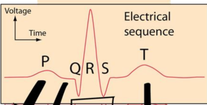
electrical conduction of heart pt 2

waveforms
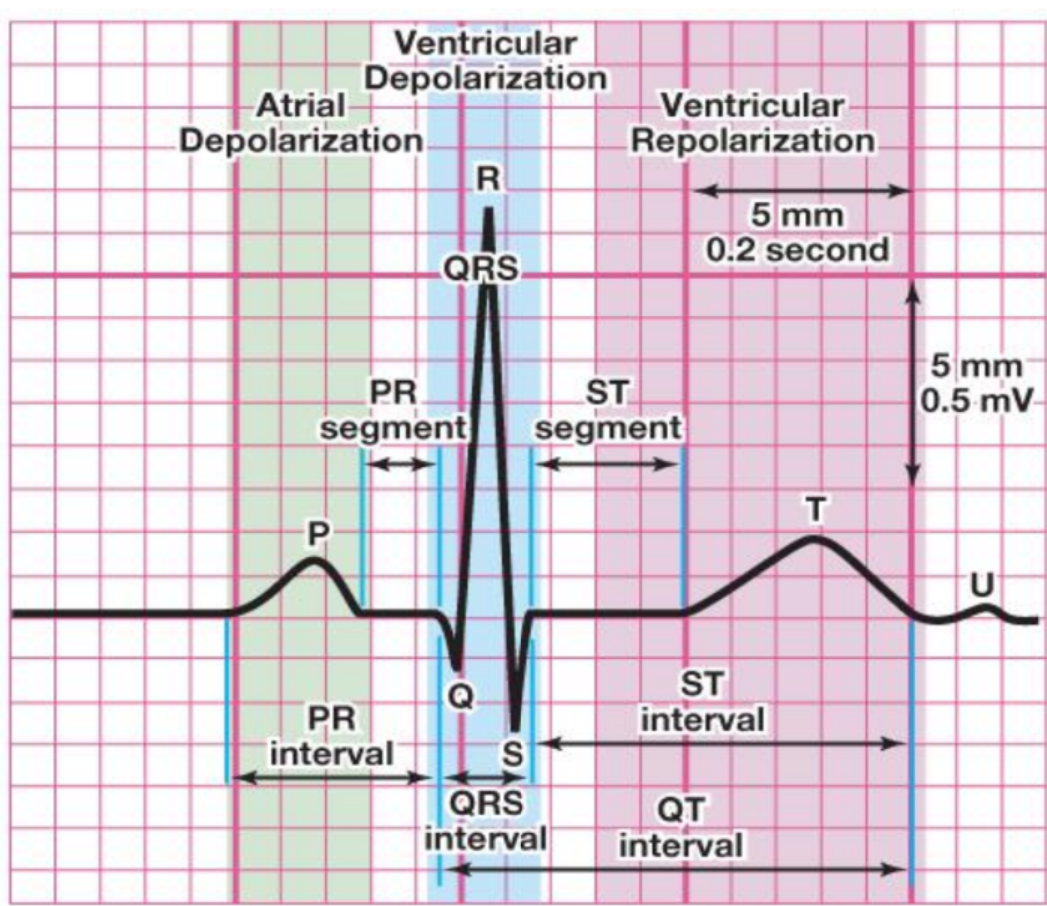
normal PR interval?
0.12-0.20 sec
normal QRS complex?
0.06-0.12 sec
why isnt there a standard QT interval range
it changes with heart rates?
sinus rhythm
Rate: 60-100
rhythm: even, P-P even and R-R even
measurement: PR: 0.2 or less sec (less than or equal to 5 boxes), QRS: 0.12 sec or less (less than or equal to 3 boxes), QT varies
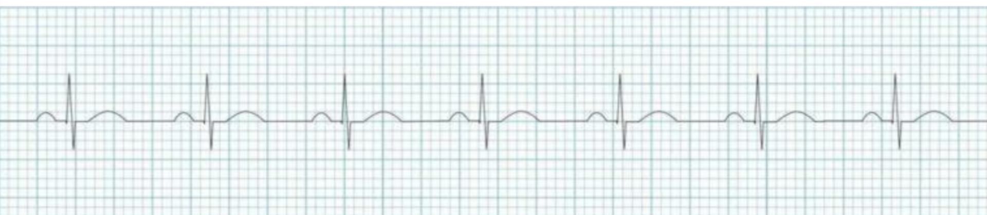
sinus bradycardia
Rate: less than 60
rhythm: even, P-P even and R-R even
measurement: PR: 0.2 or less sec (less than or equal to 5 boxes), QRS: 0.12 sec or less (less than or equal to 3 boxes), QT varies
Not an emergency unless symptomatic
hypotension, delayed cap refill, confusion, weak pulses
tx: atropine, external pace
No it's still in sinus because it has AP wave before the QRS complex
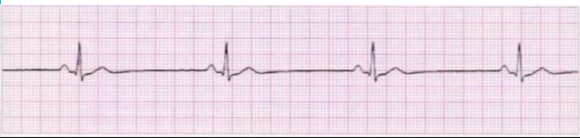
Sinus Tachycardia
Rate: greater than 100
rhythm: even, P-P even and R-R even
measurement: PR: 0.2 or less sec (less than or equal to 5 boxes), QRS: 0.12 sec or less (less than or equal to 3 boxes), QT varies
Prolonged tachycardia will cause the heart to fail
drug is a stimulant, poor cardiac output, stress, secondary tachycardia, hypoperfusion, fever are all causes

sinus dysrhythmia
rate: 60-100
rhythm: not even
measurement: PR: 0.2 or less sec (less than or equal to 5 boxes), QRS: 0.12 sec or less (less than or equal to 3 boxes), QT varies
as client breathes in rate speeds up, as breathes out rate slows down

What does it mean to be symptomatic bradycardia
When being Bradycardic becomes an emergency due to poor profusion
What pharmacology might encourage bradycardia
Negative chronotropes because it slows down the heart rate
Beta blockers and digoxin
What symptoms might the nurse notice if the SA node isn't working
The AV node will take over causing bradycardia
How would the nurse respond if a client was demonstrating bradycardia
Not an emergency unless symptomatic
What are some causes of tachycardia
Anxiety, positive chronotropes, exercise
Chronic tachycardia will fail the heart
premature atrial contraction (PAC)
An over excited AV node Causes an extra cardiac cycle
An early ectopic atrial beat that occurs before the next expected sinus beat
It can be caused by an irritable SA node, stimulant, electrolyte imbalance
rate: underlying is normal, rhythm: irregular due to PAC, and measurement: PR: shortened/ abnormal for PAC, QRS: 0.06-0.12 sec, QT: ~0.40 sec
UNDERLYING RHYTHM IS REGULAR

atrial flutter
rate: atria: 250-350. ventricles: half of atria rate
regular rhythm (fixed block)
measurement: PR and QRS not measurable, QT: ~40 sec
Can be due to electrolytes stress or heart failure
each sawtooth pattern is the SA node firing
Atria contract rapidly in a reentry circuit

atrial fibrillation
rate: tachycardic, A lot of times in RVR
irregularly regular
measurements: hard
Most deadly rhythm we're learning this semester
atria shaking so there is no discernible P wave
There is poor cardiac output causing poor perfusion which can lead to clotting problems and stroke
Can be caused by atrial enlargement or structural heart disease, hypertension, valvular disease, ischemia
An atrial kick is the final atrial contraction that contributes to about 20 to 30%
The goal is to control ventricular rate and rhythm, prevent thromboembolism or stroke and treat the underlying cause (heparin→ start 2nd line) (dont draw labs off of central line with heparin)
monitor PTT every 6 hours
before cardioversion, check for clots using TEE, if cardioversion doesnt work → ablasion (cauterize)
atria is not fully contracting
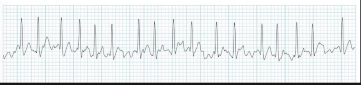
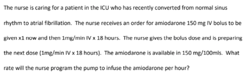
40 mL/hr

an irregular rhythm can allow for blood stasis and clot formation. An embolus can travel to the brain causing an ischemic stroke and deficits
When is bradycardia an emergency
When the patient is symptomatic or it is causing poor perfusion like hypotension ultimate status acute heart failure shock or syncope
normal sinus
HR: 100

Sinus tachycardia
HR: 140
sinus bradycardia
HR: 40
afib w/ RVR
HR: 150

atrial flutter
HR: 80

atrial flutter
HR: 70
normal sinus
HR: 90

Afib
HR: 50
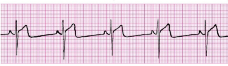
Sinus bradycardia with STEMI
HR: 50

PAC
HR: 70
hemodynamics
Blood Pressure (BP) = Cardiac Output (CO) × Peripheral Vascular Resistance (PVR)
Systolic BP : The pressure when the ventricles contract forcefully and push blood into circulation.
Diastolic BP : The pressure when the ventricles rest and fill with blood from the atria.
S1 : The "LUB" heart sound.
S2 :The "DUB" heart sound.
S3 :Heart sound that occurs early in diastole.
S4 :Heart sound that occurs before systole (pre-systolic).
preload
volume of blood in the ventricles at the end of diastole
afterload
resistance left ventricle must overcome to circulate blood
increased afterload = increased cardiac workload
why should we care (TEST Q)
HTN is a silent killer
leads to peripheral vascular disease affecting:
heart: leads to HF, affecting coronary arteries (clamped down)
brain: stroke, aneurisms
eyes: retina
kidneys: decreased GFR, poor perfusion
HTN classification (TEST Q)
Hypertension (HTN): A condition of persistently elevated blood pressure that increases the risk for cardiovascular and end-organ complications.
Normal BP: Systolic <120 mmHg AND diastolic <80 mmHg.
Elevated BP: Systolic 120–129 mmHg AND diastolic <80 mmHg.
Stage 1 Hypertension: Systolic 130–139 mmHg OR diastolic 80–89 mmHg.
Stage 2 Hypertension: Systolic ≥140 mmHg OR diastolic ≥90 mmHg.
Hypertensive Crisis: Systolic >180 mmHg AND/OR diastolic >120 mmHg.
primary/essesntial HTN: no underlying rhyme or reason
secondary HTN: from something
needs to be on 3 separate occasions (recheck in 10 minutes if not sypmtomatic (exercise, PAIN, cuff size, positioning can affect BP)
RISK factors for HTH
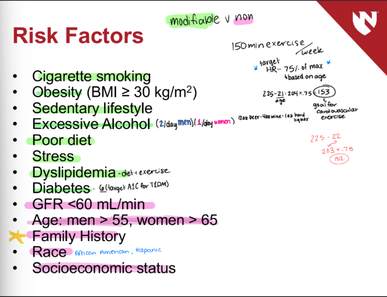
mechanisms that result in HTN
Increased Sodium Intake: ↑ Sodium causes fluid retention, ↑ stroke volume, and ↑ blood pressure. and increased preload
RAAS: Activation of the renin-angiotensin-aldosterone system contributes to hypertension. Angiotensin II A potent vasoconstrictor that increases blood pressure. Angiotensin II and Aldosterone ↑ Angiotensin II causes ↑ aldosterone release.
Aldosterone: Promotes sodium and fluid retention and increases potassium excretion. Low Potassium and Hypertension Low plasma K+ contributes to increased vasoconstriction.
Sympathetic Nervous System: ↑ Sympathetic activity causes ↑ vasoconstriction and ↑ peripheral vascular resistance.
labs and diagnostic testing (TEST Q)
Renal: UA/BUN/ and or creatinine- Protein in the urine; may indicate renal target-organ damage. Elevated BUN and Creatinine May indicate renal target-organ damage.
Endocrine Testing for Hypertension: Serum sodium, potassium, calcium, and TSH.
Metabolic Testing for Hypertension: Fasting blood glucose and lipid profile. Hypertension Metabolic Findings ↑ fasting blood glucose (>100), ↑ LDL, ↑ triglycerides, and ↓ HDL.
Echocardiogram Finding in Hypertension Left ventricular hypertrophy may indicate cardiac target-organ damage.
management of HTN
Assess underlying cause and implement lifestyle modifications and medications as needed.
Lifestyle Modifications for Hypertension
Weight management, diet modification (DASH -dietary approash to stop HTN, decrease sodium, lean meatsm decrease saturated fats, whole foods, decrease sugars, increase fruits and veggies)
limiting alcohol
exercise
stress management
Hypertension Medications
Diuretics (know these)
calcium channel blockers
beta blockers
ACE inhibitors
ARBs
central alpha agonists
diet considerations
DASH Diet Dietary approach used to help manage blood pressure.
Mediterranean Diet A dietary approach recommended as a consideration for hypertension management. (fruits, veggies,, nuts)
Low-Sodium Diet Dietary approach that limits sodium intake to help manage blood pressure.